
- Jul. 14, 2025
- Home
- About Us
- Editorial Board
- Instruction
- Subscription
- Advertisement
- Contact Us
- Chinese
- RSS
Chinese Journal of Magnetic Resonance ›› 2022, Vol. 39 ›› Issue (3): 356-365.doi: 10.11938/cjmr20222999
• Review Articles & Perspectives • Previous Articles Next Articles
Yi ZHANG1,Fei-yang LOU2,Ke FANG3,Gao CHEN1,Xiao-tong ZHANG1,2,4,*( )
)
Received:2022-04-15
Online:2022-09-05
Published:2022-06-13
Contact:
Xiao-tong ZHANG
E-mail:zhangxiaotong@zju.edu.cn
CLC Number:
Yi ZHANG,Fei-yang LOU,Ke FANG,Gao CHEN,Xiao-tong ZHANG. Review of a New Molecular Imaging Method——Deuterium Metabolic Spectroscopy and Imaging[J]. Chinese Journal of Magnetic Resonance, 2022, 39(3): 356-365.

Fig.1
Metabolic process of deuterium-labeled compounds in vivo: includes mainly glycolysis and citric acid cycle. D (deuterium), D/H (deuterium or hydrogen), * indicates the substrates that have been used, and # indicates the products detected so far
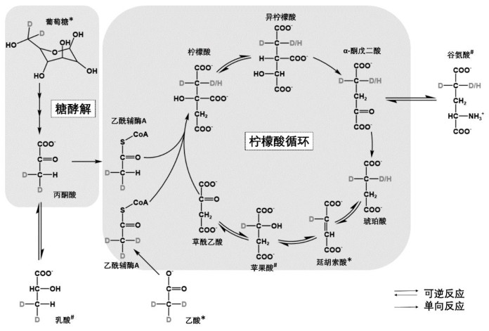
Table 1
A summary of the research on deuterium metabolic spectroscopy and imaging
| 文献 | 氘代底物 | 物种 | 场强/T | 器官组织 | 研究内容及结论 |
| [ | 葡萄糖 | 大鼠 | 11.7 | 脑、肝 | 可检测出脑胶质瘤中糖代谢异常(Warburg效应);但无法区分肝脏中的糖原和葡萄糖 |
| 人 | 4 | ||||
| [ | 葡萄糖 | 大鼠 | 16.4 | 脑 | 可检测脑葡萄糖代谢速率 |
| [ | 葡萄糖 | 小鼠 | 9.4 | 淋巴瘤 | 早期检测肿瘤组织化疗后糖代谢变化 |
| [ | 延胡索酸 | 小鼠 | 7 | 乳腺癌 | 早期检测肿瘤组织化疗后糖代谢变化 |
| [ | 葡萄糖 | 小鼠 | 11.7 | 肝 | 难以检测肝脏中的糖原代谢 |
| [ | 葡萄糖 | 大鼠 | 9.4 | 脑 | 检测中风相关区域糖代谢变化 |
| [ | 葡萄糖 | 大鼠 | 9.4 | 棕色脂肪 | 检测寒冷刺激下棕色脂肪代谢变化 |
| [ | 乙酸盐 | 大鼠 | 16.4 | 心肌 | 不同情况下心肌细胞对能量底物的偏好 |
| [ | 葡萄糖 | 大鼠 | 4.7 | 脑 | 氘与非超极化13C成像的信号强度比较 |
| [ | 葡萄糖 | 小鼠 | 14.1 | 肝细胞 | 氘成像过程中代谢产生的重水含量变化 |
| [ | 葡萄糖 | 大鼠 | 11.7 | 脑 | 检测氘代谢过程中因氢氘化学交换而损失的氘信号 |
| [ | 葡萄糖 | 大鼠 | 9.4 | 脑 | 以氢氘化学交换产生的氢信号差值反映氘代谢物变化 |
| [ | 葡萄糖 | 大鼠 | 11.7 | 脑 | 检测氘信号强度等参数随场强提高而产生的变化 |
| 人 | 7 | ||||
| [ | 葡萄糖 | 人 | 9.4 | 脑 | 硬件提升提高氘成像的时空分辨率 |
| [ | 葡萄糖 | 小鼠 | 9.4 | 黑色素瘤 | 氘波谱相关硬件设计 |
| [ | 葡萄糖 | 小鼠 | 15.2 | 胰腺 | 多回波bbSP序列的信噪比高于CSI |
| [ | 葡萄糖 | 大鼠 | 16.4 | 脑 | 机器学习提高氘成像信噪比 |
| [ | 葡萄糖 | 小鼠 | 14.1 | 脑 | 利用氘波谱成像检测肿瘤组织负荷及其对化疗的反应 |
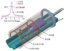
Fig.2
The perspective view of 1H/2H RF coil design and DMS results. Two concentric birdcages whose rungs are azimuthally interleaved with each other for better decoupling are housed in a polycarbonate cylinder. The animal holder, bed, bite stick and ear bar are placed in the coil core for animal support, orientation, and anesthesia. DMS result of control mice and tumor mice is illustrated at the top left. Notice that both heavy water and deuterium-labeled glucose signals can be detected, whereas the deuterium-labeled lactate signal can only be identified in tumor group

| 1 | KUBOTA K . From tumor biology to clinical PET: a review of positron emission tomography (PET) in oncology[J]. Anna Nucl Med, 2001, 15 (6): 471- 486. |
| 2 | LIU T T , WANG J , GUO X Y . Proton magnetic resonance spectroscopy in brain science researches[J]. Chinese J Magn Reson, 2020, 37 (2): 232- 240. |
| 刘涛涛, 王杰, 郭向阳. 脑科学研究中的质子磁共振波谱方法[J]. 波谱学杂志, 2020, 37 (2): 232- 240. | |
| 3 |
XU X , YADAV N N , KNUTSSON L , et al. Dynamic glucose-enhanced (DGE) MRI: translation to human scanning and first results in glioma patients[J]. Tomography, 2015, 1 (2): 105.
doi: 10.18383/j.tom.2015.00175 |
| 4 |
LI H D , ZHANG Z Y , HAN Y Q , et al. Lung MRI using hyperpolarized gases[J]. Chinese J Magn Reson, 2014, 31 (3): 307- 320.
doi: 10.3969/j.issn.1000-4556.2014.03.002 |
|
李海东, 张智颖, 韩叶清, 等. 超极化气体肺部磁共振成像[J]. 波谱学杂志, 2014, 31 (3): 307- 320.
doi: 10.3969/j.issn.1000-4556.2014.03.002 |
|
| 5 | WANG C W , HUANG X , SHI L , et al. Cathepsin B triggered hyperpolarization 129Xe MRI probe for ultra-sensitive lung cancer cells detection[J]. Chinese J Magn Reson, 2021, 38 (3): 336- 344. |
| 王崇武, 黄曦, 石磊, 等. 组织蛋白酶B响应的超极化129Xe MRI探针对肺癌细胞的超灵敏探测[J]. 波谱学杂志, 2021, 38 (3): 336- 344. | |
| 6 | KURHANEWICZ J , VIGNERON D B , ARDENKJAER-LARSEN J H , et al. Hyperpolarized 13C MRI: path to clinical translation in oncology[J]. Neoplasia, 2019, 21 (1): 1- 16. |
| 7 |
DE FEYTER H M , BEHAR K L , CORBIN Z A , et al. Deuterium metabolic imaging (DMI) for MRI-based 3D mapping of metabolism in vivo[J]. Sci Adv, 2018, 4 (8): eaat7314.
doi: 10.1126/sciadv.aat7314 |
| 8 | UREY H C , BRICKWEDDE F G , MURPHY G M . A hydrogen isotope of mass 2 and its concentration[J]. Phys Rev, 1932, 40 (1): 1- 15. |
| 9 | 俎栋林, 高家红. 核磁共振成像——物理原理和方法[M]. 北京: 北京大学出版社, 2014, 1- 19. |
| 10 | LU M , ZHU X H , ZHANG Y , et al. Quantitative assessment of brain glucose metabolic rates using in vivo deuterium magnetic resonance spectroscopy[J]. Journal of Cerebral Blood Flow & Metabolism, 2017, 37 (11): 3518- 3530. |
| 11 | 朱圣庚, 徐长法. 生物化学.下册[M]. 第4版 北京: 高等教育出版社, 2016, 49- 90. |
| 12 | HOUSE S W , WARBURG O , BURK D , et al. On respiratory impairment in cancer cells[J]. Science, 1956, 124 (3215): 267- 272. |
| 13 | KREIS F , WRIGHT A J , HESSE F , et al. Measuring tumor glycolytic flux in vivo by using fast deuterium MRI[J]. Radiology, 2020, 294 (2): 289- 296. |
| 14 | GERWING M , HERRMANN K , HELFEN A , et al. The beginning of the end for conventional RECIST—novel therapies require novel imaging approaches[J]. Nat Rev Clin Oncol, 2019, 16 (7): 442- 458. |
| 15 | GALLAGHER F A , KETTUNEN M I , HU D E , et al. Production of hyperpolarized[1, 4-13C2] malate from[1, 4-13C2] fumarate is a marker of cell necrosis and treatment response in tumors[J]. Proc Natl Acad Sci, 2009, 106 (47): 19801- 19806. |
| 16 | HESSE F, SOMAI V, KREIS F, et al. Monitoring tumor cell death in murine tumor models using deuterium magnetic resonance spectroscopy and spectroscopic imaging[J]. Proc Natl Acad Sci, 2021, 118(12): e2014631118. |
| 17 | DE FEYTER H M , THOMAS M A , BEHAR K L , et al. NMR visibility of deuterium-labeled liver glycogen in vivo[J]. Magn Reson Med, 2021, 86 (1): 62- 68. |
| 18 | TAKASAWA M , MOUSTAFA R R , BARON J C . Applications of nitroimidazole in vivo hypoxia imaging in ischemic stroke[J]. Stroke, 2008, 39 (5): 1629- 1637. |
| 19 | STRAATHOF M , MEERWALDT A E , DE FEYTER H M , et al. Deuterium metabolic imaging of the healthy and diseased brain[J]. Neuroscience, 2021, 474, 94- 99. |
| 20 | RIIS-VESTERGAARD M J , LAUSTSEN C , MARIAGER C Ø , et al. Glucose metabolism in brown adipose tissue determined by deuterium metabolic imaging in rats[J]. Inter J Obesity, 2020, 44 (6): 1417- 1427. |
| 21 | WANG T , ZHU X H , LI H , et al. Noninvasive assessment of myocardial energy metabolism and dynamics using in vivo deuterium MRS imaging[J]. Magn Reson Med, 2021, 86 (6): 2899- 2909. |
| 22 | VON MORZE C , ENGELBACH J A , BLAZEY T , et al. Comparison of hyperpolarized 13C and non-hyperpolarized deuterium MRI approaches for imaging cerebral glucose metabolism at 4.7 T[J]. Magn Reson Med, 2021, 85 (4): 1795- 1804. |
| 23 | MAHAR R , DONABEDIAN P L , MERRITT M E . HDO production from[2H7] glucose quantitatively identifies warburg metabolism[J]. Sci Rep, 2020, 10 (1): 8885. |
| 24 | DE GRAAF R A , THOMAS M A , BEHAR K L , et al. Characterization of kinetic isotope effects and label loss in deuterium-based isotopic labeling studies[J]. ACS Chem Neurosci, 2020, 12 (1): 234- 243. |
| 25 | RICH L J , BAGGA P , WILSON N E , et al. 1H magnetic resonance spectroscopy of 2H-to-1H exchange quantifies the dynamics of cellular metabolism in vivo[J]. Nat Biomed Engineer, 2020, 4 (3): 335- 342. |
| 26 | DE GRAAF R A , HENDRIKS A D , KLOMP D W J , et al. On the magnetic field dependence of deuterium metabolic imaging[J]. NMR Biomed, 2020, 33 (3): e4235. |
| 27 | RUHM L , AVDIEVICH N , ZIEGS T , et al. Deuterium metabolic imaging in the human brain at 9.4 Tesla with high spatial and temporal resolution[J]. NeuroImage, 2021, 244, 118639. |
| 28 | ZHANG Y , GAO Y , ZHANG X T , et al. Proton/deuterium magnetic resonance imaging of rodents at 9.4 T using birdcage coils[J]. Bioelectromagnetics, 2022, 43 (1): 40- 46. |
| 29 | DE FEYTER H M , DE GRAAF R A . Deuterium metabolic imaging–Back to the future[J]. J Magn Reson, 2021, 326, 106932. |
| 30 | PETERS D C , MARKOVIC S , BAO Q , et al. Improving deuterium metabolic imaging (DMI) signal-to-noise ratio by spectroscopic multi-echo bSSFP: A pancreatic cancer investigation[J]. Magn Reson Med, 2021, 86 (5): 2604- 2617. |
| 31 | LI Y D , ZHAO Y B , GUO R , et al. Machine learning-enabled high-resolution dynamic deuterium MR spectroscopic imaging[J]. IEEE T Med Imaging, 2021, 40 (12): 3879- 3890. |
| 32 | NARESSI A , COUTURIER C , DEVOS J M , et al. Java-based graphical user interface for the MRUI quantitation package[J]. Magnetic resonance materials in physics, biology and medicine, 2001, 12 (2): 141- 152. |
| 33 | PROVENCHER S W . Estimation of metabolite concentrations from localized in vivo proton NMR spectra[J]. Magn Reson Med, 1993, 30 (6): 672- 679. |
| 34 | TAGLANG C , BATSIOS G , MUKHERJEE J , et al. Deuterium magnetic resonance spectroscopy enables non-invasive metabolic imaging of tumor burden and response to therapy in low-grade gliomas[J]. Neuro-oncology, 2022, 24 (7): 1101- 1112. |
| 35 | LAM F, CHU J, CHOI J S, et al. Epigenetic MRI: Noninvasive imaging of DNA methylation in the brain[J]. Proc Natl Acad Scie, 2022, 119(10): e2119891119. |
| [1] | Lian-hua LIU,Bin JIANG,Dai-xie CHEN,Chi SU. The Status and Challenge of the Domestic Manufacturing of Superconduct Magnetic Resonance Instruments in China [J]. Chinese Journal of Magnetic Resonance, 2022, 39(3): 345-355. |
| [2] | Xian-xin QIU,Xu HAN,Yao WANG,Wei-na DING,Ya-wen SUN,Yan ZHOU,Hao LEI,Fu-chun LIN. The Alteration of Rich Club in Brain Functional Network in Internet Gaming Disorder [J]. Chinese Journal of Magnetic Resonance, 2022, 39(3): 258-266. |
| [3] | Wen-shan LIAO, Jun-cheng XU, Shou-quan YAO, Jian-qi LI, Yu JIANG. Phase Coherence Technology of Digital MR Console Based on Dual Reference Sources [J]. Chinese Journal of Magnetic Resonance, 2022, 39(3): 327-336. |
| [4] | Yuan-yuan LIU, Yu-xin YANG, Qing-yong ZHU, Zhuo-xu CUI, Jing CHENG, Cong-cong LIU, Dong LIANG, Yan-jie ZHU. Accelerating T1ρ Dispersion Imaging with Multiple Relaxation Signal Compensation [J]. Chinese Journal of Magnetic Resonance, 2022, 39(3): 243-257. |
| [5] | Ying-shan WANG, Ao-qi DENG, Jin-ling MAO, Zhong-qi ZHU, Jie SHI, Guang YANG, Wei-wei MA, Qing LU, Hong-zhi WANG. Automatic Segmentation of Knee Joint Synovial Magnetic Resonance Images Based on 3D VNetTrans [J]. Chinese Journal of Magnetic Resonance, 2022, 39(3): 303-315. |
| [6] | Xiao-ming CHEN, Xiu-chao ZHAO, Xian-ping SUN, Jun-shuai XIE, Hai-dong LI, Ye-qing HAN, Xiao-ling LIU, Qi CHEN, Xin ZHOU. Study on the Automatic Accumulation-thawing Device of Hyperpolarized 129Xe [J]. Chinese Journal of Magnetic Resonance, 2022, 39(3): 316-326. |
| [7] | Han HU,Wei-yu WANG,Jun XU,Feng DENG. 1, 3-Butadienen Hydrogenation on Supported Pd-Sn Bimetallic Catalysts Investigated by Parahydrogen-induced Polarization [J]. Chinese Journal of Magnetic Resonance, 2022, 39(2): 133-143. |
| [8] | Min-xiong ZHOU, Hui-ting ZHANG, Yi-da WANG, Guang YANG, Xu-feng YAO, An-kang GAO, Jing-liang CHENG, Jie BAI, Xu YAN. Evaluation of the Influence of Data Sampling Schemes on Neural Diffusion Models [J]. Chinese Journal of Magnetic Resonance, 2022, 39(2): 220-229. |
| [9] | Jun LUO, Sheng-ping LIU, Xing YANG, Jia-sheng WANG, Ye LI. Design of a 5 T Non-magnetic Magnetic Resonance Radio Frequency Power Amplifier [J]. Chinese Journal of Magnetic Resonance, 2022, 39(2): 163-173. |
| [10] | Yan MA, Cang-ju XING, Liang XIAO. Knee Joint Image Segmentation and Model Construction Based on Cascaded Network [J]. Chinese Journal of Magnetic Resonance, 2022, 39(2): 184-195. |
| [11] |
De-gang TANG,Hong-chuang LI,Xiao-ling LIU,Lei SHI,Hai-dong LI,Chao-hui YE,Xin ZHOU.
A Simulation Study on the Effect of the High Permittivity Materials Geometrical Structure on the Transmit Field |
| [12] | Zhen-yu WANG, Ying-shan WANG, Jin-ling MAO, Wei-wei MA, Qing LU, Jie SHI, Hong-zhi WANG. Magnetic Resonance Images Segmentation of Synovium Based on Dense-UNet++ [J]. Chinese Journal of Magnetic Resonance, 2022, 39(2): 208-219. |
| [13] | Yue QIU, Sheng-dong NIE, Long WEI. Segmentation of Breast Tumors Based on Fully Convolutional Network and Dynamic Contrast Enhanced Magnetic Resonance Image [J]. Chinese Journal of Magnetic Resonance, 2022, 39(2): 196-207. |
| [14] | Shu ZENG, Shu-tao XU, Ying-xu WEI, Zhong-min LIU. Investigation of the Ethanol Dehydration to Ethene Reaction on H-SSZ-13 Molecular Sieve by in situ Solid-state NMR Spectroscopy [J]. Chinese Journal of Magnetic Resonance, 2022, 39(2): 123-132. |
| [15] | Lei CHEN,Hong-bing LIU,Hui-li LIU. Comparison of Different Approaches for Estimation of the Detection Limit of Quantitative NMR [J]. Chinese Journal of Magnetic Resonance, 2022, 39(2): 230-242. |
| Viewed | ||||||||||||||||||||||||||||||||||||||||||||||||||
|
Full text 481
|
|
|||||||||||||||||||||||||||||||||||||||||||||||||
|
Abstract 292
|
|
|||||||||||||||||||||||||||||||||||||||||||||||||