
- Aug. 5, 2025
- Home
- About Us
- Editorial Board
- Instruction
- Subscription
- Advertisement
- Contact Us
- Chinese
- RSS
Chinese Journal of Magnetic Resonance ›› 2022, Vol. 39 ›› Issue (3): 303-315.doi: 10.11938/cjmr20222988
• Articles • Previous Articles Next Articles
Ying-shan WANG1,Ao-qi DENG3,Jin-ling MAO1,Zhong-qi ZHU1,Jie SHI2,*( ),Guang YANG1,Wei-wei MA4,Qing LU4,*(),Hong-zhi WANG1,*()
Received:2022-03-23
Online:2022-09-05
Published:2022-05-11
Contact:
Jie SHI,Qing LU,Hong-zhi WANG
E-mail:ghyyfsk@163.com;drluqingsjtu@163.com;hzwang@phy.ecnu.edu.cn
CLC Number:
Ying-shan WANG, Ao-qi DENG, Jin-ling MAO, Zhong-qi ZHU, Jie SHI, Guang YANG, Wei-wei MA, Qing LU, Hong-zhi WANG. Automatic Segmentation of Knee Joint Synovial Magnetic Resonance Images Based on 3D VNetTrans[J]. Chinese Journal of Magnetic Resonance, 2022, 39(3): 303-315.

Fig.1
Experimental process of this research
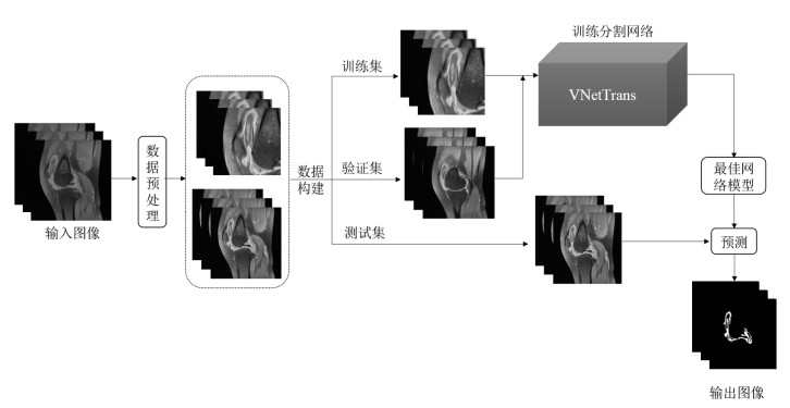
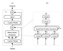
Fig.2
(a) Transformer encoder; (b) Multi-head attention (MHA)[21]
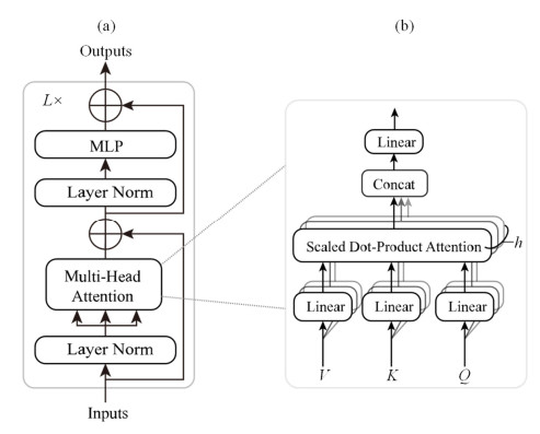
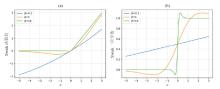
Fig.3
(a) Swish activation function and (b) its first derivatives with different β values[24]
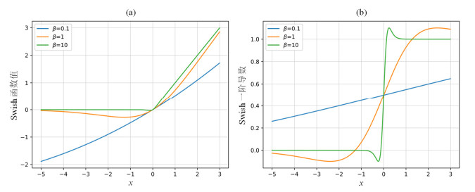
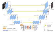
Fig.4
Overview of VNetTrans architecture

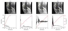
Fig.5
Comparison of the effects of different image contrast enhancement methods. (a) Original image, (b) Contrast stretching, (c) Histogram equalization, (d) CLAHE
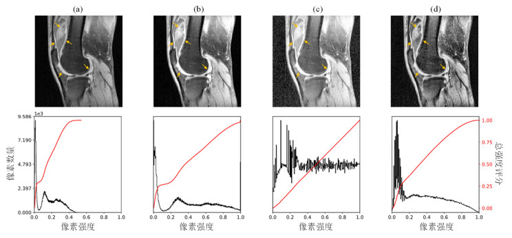
Table 1
2D and 3D segmentation effect comparison
| 模型 | 卷积核维度 | Dice | HD/mm |
| UNet | 2D | 0.6489 | 89.8 |
| VNet | 2D | 0.6640 | 52.5 |
| UNet | 3D | 0.6528 | 72.2 |
| VNet | 3D | 0.6749 | 34.6 |
Table 2
Comparison of results using different activation functions in UNet and VNetTrans
| 模型 | 激活函数 | Dice | HD/mm | 训练耗时 |
| UNet | ReLU | 0.6528 | 72.2 | 23min |
| Swish | 0.6613 | 50.8 | 34min | |
| MemSwish | 0.6670 | 37.4 | 30min | |
| VNetTrans | ReLU | 0.7140 | 33.6 | 5h58min |
| Swish | 0.7505 | 29.0 | 6h36min | |
| MemSwish | 0.7585 | 24.6 | 6h21min |
Table 3
Comparison of different network models
| 模型 | Dice | Sensitivity | Specificity | RVD/% | HD/mm |
| UNet | 0.6528 | 0.7828 | 0.9864 | 33.84 | 72.2 |
| VNet | 0.6749 | 0.7262 | 0.9908 | 9.21 | 34.6 |
| TransBTS | 0.6996 | 0.7186 | 0.9917 | 4.87 | 36.0 |
| UNETR | 0.6355 | 0.6560 | 0.9920 | ?3.31 | 68.8 |
| VNetTrans | 0.7585 | 0.7633 | 0.9943 | ?1.34 | 24.6 |

Fig.6
Training process of knee joint synovial magnetic resonance image segmentation based on different networks. (a) Loss curves of training set; (b) Dice curves of validation set

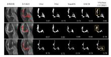
Fig.7
Comparison of segmentation results of synovium of knee joint with different network models
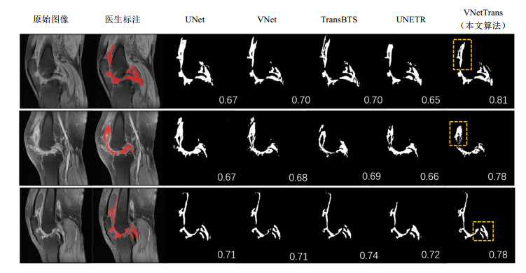
Table 4
Ablation experiment
| 网络模型 | 激活函数 | 是否加入Transformer模块 | Dice | Sensitivity | Specificity | RVD/% | HD/mm |
| VNet | ReLU | 否 | 0.6749 | 0.7262 | 0.9908 | 9.21 | 34.6 |
| VNet + MemSwish | MemSwish | 否 | 0.6962 | 0.7580 | 0.9904 | 14.92 | 35.8 |
| VNet + Transformer | ReLU | 是 | 0.7140 | 0.7527 | 0.9913 | 2.52 | 33.6 |
| This work | MemSwish | 是 | 0.7585 | 0.7633 | 0.9943 | ?1.34 | 24.6 |
| 1 |
SMOLEN J S, ALETAHA D, BARTON A, et alRheumatoid arthritis[J]. Nat Rev Dis Primers, 2018,4 (1): 18001.
doi: 10.1038/nrdp.2018.1 |
| 2 | JIN S Y, LI M T, FANG Y F, et alChinese registry of rheumatoid arthritis (CREDIT): II. prevalence and risk factors of major comorbidities in Chinese patients with rheumatoid arthritis[J]. Arthritis ResTher, 2017,19 (1): 251. |
| 3 |
中华医学会风湿病学分会2018中国类风湿关节炎诊疗指南[J]. 中华内科杂志, 2018,57 (4): 242- 251.
doi: 10.3760/cma.j.issn.0578-1426.2018.04.004 |
|
ASSOCIATION C R2018 Chinese guideline for the diagnosis and treatment of rheumatoid arthritis[J]. Chin J Intern Med, 2018,57 (4): 242- 251.
doi: 10.3760/cma.j.issn.0578-1426.2018.04.004 |
|
| 4 |
SUGIMOTO H, TAKEDA A, KANO SAssessment of disease activity in rheumatoid arthritis using magnetic resonance imaging: quantification of pannus volume in the hands[J]. Bri J Rheumatol, 1998,37 (8): 854- 861.
doi: 10.1093/rheumatology/37.8.854 |
| 5 |
OSTERGAARD MDifferent approaches to synovial membrane volume determination by magnetic resonance imaging: manual versus automated segmentation[J]. Rheumatology, 1997,36 (11): 1166- 1177.
doi: 10.1093/rheumatology/36.11.1166 |
| 6 |
SAKASHITA T, KAMISHIMA T, KOBAYASHI Y, et alAccurate quantitative assessment of synovitis in rheumatoid arthritis using pixel-by-pixel, time-intensity curve shape analysis[J]. Br J Radiol, 2016,89 (1061): 20151000.
doi: 10.1259/bjr.20151000 |
| 7 |
FOTINOS-HOYER A K, GUERMAZI A, JARA H, et alAssessment of synovitis in the osteoarthritic knee: comparison between manual segmentation, semiautomated segmentation, and semiquantitative assessment using contrast-enhanced fat-suppressed T1-weighted MRI[J]. Magn Reson Med, 2010,64 (2): 604- 609.
doi: 10.1002/mrm.22401 |
| 8 |
PERRY T A, GAIT A, O’NEILL T W, et alMeasurement of synovial tissue volume in knee osteoarthritis using a semiautomated MRI-based quantitative approach[J]. Magn Reson Med, 2019,81 (5): 3056- 3064.
doi: 10.1002/mrm.27633 |
| 9 | WANG A, FRANKE A, WESARG S. Semi-automatic segmentation of JIA-induced inflammation in MRI images of ankle joints[C]// Medical Imaging 2019: Image Processing, SPIE, 2019, 10949: 875-881. |
| 10 |
ANDERSEN J K H, PEDERSEN J S, LAURSEN M S, et alNeural networks for automatic scoring of arthritis disease activity on ultrasound images[J]. RMD open, 2019,5 (1): e000891.
doi: 10.1136/rmdopen-2018-000891 |
| 11 |
CHRISTENSEN A B H, JUST S A, ANDERSEN J K H, et alApplying cascaded convolutional neural network design further enhances automatic scoring of arthritis disease activity on ultrasound images from rheumatoid arthritis patients[J]. Ann Rheum Dise, 2020,79 (9): 1189- 1193.
doi: 10.1136/annrheumdis-2019-216636 |
| 12 |
IQBAL I, SHAHZAD G, RAFIQ N, et alDeep learning-based automated detection of human knee joint's synovial fluid from magnetic resonance images with transfer learning[J]. IET Image Processing, 2020,14 (10): 1990- 1998.
doi: 10.1049/iet-ipr.2019.1646 |
| 13 |
WONG L M, SHI L, XIAO F, et alFully automated segmentation of wrist bones on T2-weighted fat-suppressed MR images in early rheumatoid arthritis[J]. Quant Imag Med Surg, 2019,9 (4): 579.
doi: 10.21037/qims.2019.04.03 |
| 14 | 魏小娜, 邢嘉祺, 王振宇, 等基于改进U-Net的关节滑膜磁共振图像的分割[J]. 计算机应用, 2020,40 (11): 3340- 3345. |
| WEI X N, XIN J Q, WANG Z Y, et alMagnetic resonance image segmentation of articular synovium based on improved U-Net[J]. Journal of Computer Applications, 2020,40 (11): 3340- 3345. | |
| 15 | 王振宇, 王颖珊, 毛瑾玲, 等基于Dense-UNet++的关节滑膜磁共振图像分割[J]. 波谱学杂志, 2022,39 (2): 208- 219. |
| WANG Z Y, WANG Y S, MAO J L, et alMagnetic resonance images segmentation of synovium based on Dense-UNet++[J]. Chinese J Magn Reson, 2022,39 (2): 208- 219. | |
| 16 | WANG W X, CHEN C, DING M, et al. TransBTS: Multimodal brain tumor segmentation using transformer[C]// International Conference on Medical Image Computing and Computer-Assisted Intervention, Springer, 2021: 109-119. |
| 17 | LONG J, SHELHAMER E, DARRELL T. Fully convolutional networks for semantic segmentation[C]// Proceedings of the IEEE conference on computer vision and pattern recognition, 2015: 3431-3440. |
| 18 | RONNEBERGER O, FISCHER P, BROX T. U-Net: Convolutional Networks for Biomedical Image Segmentation[C]// International Conference on Medical image computing and computer-assisted intervention, Cham: Springer, 2015: 234-241. |
| 19 | ÇIçEK Ö, ABDULKADIR A, LIENKAMP S S, et al. 3D U-Net: learning dense volumetric segmentation from sparse annotation[C]// International conference on medical image computing and computer-assisted intervention. Springer, 2016: 424-432. |
| 20 | MILLETARI F, NAVAB N, AHMADI S-A. V-net: Fully convolutional neural networks for volumetric medical image segmentation[C]// 2016 fourth international conference on 3D vision (3DV), IEEE, 2016: 565-571. |
| 21 | VASWANI A, SHAZEER N, PARMAR N, et al. Attention is all you need[C]// Proceedings of the Advances in Neural Information Processing Systems, 2017: 6000-6010. |
| 22 | DOSOVITSKIY A, BEYER L, KOLESNIKOV A, et al. An image is worth 16×16 words: Transformers for image recognition at scale[OL]. arXiv preprint arXiv: 2010.11929, 2020. |
| 23 | NAIR V, HINTON G E. Rectified linear units improve restricted boltzmann machines[C]// Proceedings of the 27th International Conference on International Conference on Machine Learning, Haifa, Israel: 2010: 807-814. |
| 24 | RAMACHANDRAN P, ZOPH B, LE Q V. Searching for activation functions[J]. arXiv preprint arXiv: 1710.05941, 2017. |
| 25 | TAN M, LE Q. Efficientnet: Rethinking model scaling for convolutional neural networks[C]// International conference on machine learning, PMLR, 2019: 6105-6114. |
| 26 | YUAN L, CHEN Y, WANG T, et al. Tokens-to-token vit: Training vision transformers from scratch on imagenet[C]// Proceedings of the IEEE/CVF International Conference on Computer Vision, 2021: 558-567. |
| 27 | YUSHKEVICH P A, GAO Y, GERIG G. ITK-SNAP: An interactive tool for semi-automatic segmentation of multi-modality biomedical images[C]// 2016 38th Annual International Conference of the IEEE Engineering in Medicine and Biology Society (EMBC), IEEE, 2016: 3342-3345. |
| 28 | HATAMIZADEH A, TANG Y, NATH V, et al. Unetr: Transformers for 3d medical image segmentation[C]// Proceedings of the IEEE/CVF Winter Conference on Applications of Computer Vision, 2022: 574-584. |
| [1] | Qin ZHOU, Yuan-jun WANG. Groupwise Registration for Magnetic Resonance Image Based on Variational Inference [J]. Chinese Journal of Magnetic Resonance, 2022, 39(3): 291-302. |
| [2] | Xiao CHANG,Xin CAI,Guang YANG,Sheng-dong NIE. Applications of Generative Adversarial Networks in Medical Image Translation [J]. Chinese Journal of Magnetic Resonance, 2022, 39(3): 366-380. |
| [3] | Meng CHEN, Chen GENG, Yu-xin LI, Dao-ying GENG, Yi-fang BAO, Ya-kang DAI. Automatic Detection for Cerebral Aneurysms in TOF-MRA Images Based on Fuzzy Label and Deep Learning [J]. Chinese Journal of Magnetic Resonance, 2022, 39(3): 267-277. |
| [4] | Zhen-yu WANG, Ying-shan WANG, Jin-ling MAO, Wei-wei MA, Qing LU, Jie SHI, Hong-zhi WANG. Magnetic Resonance Images Segmentation of Synovium Based on Dense-UNet++ [J]. Chinese Journal of Magnetic Resonance, 2022, 39(2): 208-219. |
| [5] | Jian-sheng LIN,Li-jia WANG. Reconstruction of Displacement Field of Left Ventricle Combined with Biomechanical Model [J]. Chinese Journal of Magnetic Resonance, 2022, 39(1): 72-86. |
| [6] | Lu HUO,Xiao-xin HU,Qin XIAO,Ya-jia GU,Xu CHU,Luan JIANG. Automatic Segmentation of Breast and Fibroglandular Tissues in DCE-MR Images Based on nnU-Net [J]. Chinese Journal of Magnetic Resonance, 2021, 38(3): 367-380. |
| [7] | LIU Peng, ZHONG Yu-min, WANG Li-jia. Automatic Segmentation of Right Ventricle in Cine Cardiac Magnetic Resonance Image Based on a Dense and Multi-Scale U-net Method [J]. Chinese Journal of Magnetic Resonance, 2020, 37(4): 456-468. |
| [8] | WANG Wan-ting, SU Shi, JIA Sen, LIANG Dong, WANG Hai-feng. Reconstruction of Simultaneous Multi-Slice MRI Data by Combining Virtual Conjugate Coil Technology and Convolutional Neural Network [J]. Chinese Journal of Magnetic Resonance, 2020, 37(4): 407-421. |
| [9] | ZHAO Shang-yi, WANG Yuan-jun. Classification of Alzheimer's Disease Patients Based on Magnetic Resonance Images and an Improved UNet++ Model [J]. Chinese Journal of Magnetic Resonance, 2020, 37(3): 321-331. |
| [10] | GONG Jin-chang, WANG Yu, WANG Yuan-jun. A Method for Segmentation of Glioma on Multimodal Magnetic Resonance Images Based on Wavelet Fusion and Deep Learning [J]. Chinese Journal of Magnetic Resonance, 2020, 37(2): 131-143. |
| [11] | CHENG Hui-tao, WANG Shan-shan, KE Zi-wen, JIA Sen, CHENG Jing, QIU Zhi-lang, ZHENG Hai-rong, LIANG Dong. A Deep Recursive Cascaded Convolutional Network for Parallel MRI [J]. Chinese Journal of Magnetic Resonance, 2019, 36(4): 437-445. |
| [12] | SU Xin-yu, WANG Li-jia, NIE Sheng-dong, HU Li-wei, ZHONG Yu-min. Progress of Right Ventricle Segmentation from Short-Axis Images Acquired with Cardiac Cine MRI [J]. Chinese Journal of Magnetic Resonance, 2019, 36(3): 377-391. |
| [13] | SUN Jing-wen, YAN Shi-ju, HAN Yong-sen, SONG Cheng-li. Classifying the Course of Alzheimer's Disease with Brain MR Images and a Method Based on Three-Dimensional Local Pattern Transformation [J]. Chinese Journal of Magnetic Resonance, 2019, 36(3): 268-277. |
| [14] | SHAO Dan-dan, WANG Xue-xue, PAN Zi-lai, CHEN Ke-min, ZHANG Zhong-shuai, YUAN Li-li, XU Zi-yue, CHEN Lei, WANG Jin-hong. Imaging Hippocampus of Mental Patients with BLADE Technique [J]. Chinese Journal of Magnetic Resonance, 2019, 36(3): 261-267. |
| [15] | XU Jia-wen, XU Jian, ZHOU Xiao-dong, ZHANG Cong, CHEN Qun. Multi-GPU Distributed Magnetic Resonance Image Reconstruction Based on Gadgetron [J]. Chinese Journal of Magnetic Resonance, 2018, 35(3): 303-317. |
| Viewed | ||||||||||||||||||||||||||||||||||||||||||||||||||
|
Full text 313
|
|
|||||||||||||||||||||||||||||||||||||||||||||||||
|
Abstract 301
|
|
|||||||||||||||||||||||||||||||||||||||||||||||||