- Jul. 14, 2025
- Home
- About Us
- Editorial Board
- Instruction
- Subscription
- Advertisement
- Contact Us
- Chinese
- RSS

Chinese Journal of Magnetic Resonance ›› 2022, Vol. 39 ›› Issue (2): 220-229.doi: 10.11938/cjmr20202870
• Review Articles & Perspectives • Previous Articles Next Articles
Min-xiong ZHOU1,Hui-ting ZHANG2,Yi-da WANG3,Guang YANG3,Xu-feng YAO1,An-kang GAO4,Jing-liang CHENG4,Jie BAI4,Xu YAN2,*( )
)
Received:2020-11-05
Online:2022-06-05
Published:2022-05-28
Contact:
Xu YAN
E-mail:maxwell4444@hotmail.com
CLC Number:
Min-xiong ZHOU, Hui-ting ZHANG, Yi-da WANG, Guang YANG, Xu-feng YAO, An-kang GAO, Jing-liang CHENG, Jie BAI, Xu YAN. Evaluation of the Influence of Data Sampling Schemes on Neural Diffusion Models[J]. Chinese Journal of Magnetic Resonance, 2022, 39(2): 220-229.
Table 1
Meanings of regular parameters used in diffusion models
| 模型 | 参数 | 全名(英文/中文) | 物理含义 |
| DTI | FA | fractional anisotropy各向异性分数 | 反映扩散系数在空间上分布不均匀的程度. |
| MD\AD\RD | mean\axial\radial diffusivity平均\轴向\径向扩散系数 | 反映平均、轴向(神经纤维方向)和垂直于轴向平面的扩散速率. | |
| DKI | MD\AD\RD | mean\axial\radial diffusivity 平均\轴向\径向扩散系数 | 与DTI的MD\AD\RD参数类似,但为校正后的扩散速率. |
| MK\AK\RK | mean\axial\radial kurtosis 平均\轴向\径向扩散峰度 | 反映平均、轴向(神经纤维方向)和垂直于轴向平面的扩散偏离正态分布的程度,与扩散受限和多组织成分混杂有关. | |
| NODDI | ICVF | intra-cellular volume fraction 细胞内容积比 | 为细胞内信号占总扩散信号的比例,与神经突密度相关. |
| ISOVF | isotropic volume fraction 各向同性容积比 | 为各向同性信号占总扩散信号的比例,通常反映脑脊液信号 | |
| ODI | orientation dispersion index 方向分散指数 | 量化神经轴突方向角度的不一致性,其值在单方向的神经组织中趋近于0,在各向同性组织趋近于1. | |
| MAP | RTOP | return-to-the-origin probability 返回原点概率 | 水分子在扩散过程中不发生净位移的概率,反映扩散受限程度. |
| MSD | mean squared displacement 平均平方位移 | 单位时间内水分子的均方位移,反映扩散速率,与DTI/DKI的MD参数接近. | |
| QIV | Q space inverse variance Q空间逆方差 | Q空间信号几何平均值的逆方差 | |
| NG | no-Gaussianity 非高斯性 | 含义与DKI中的MK接近,反映扩散偏离正态分布的程度. |

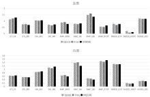
Fig.1
Based on the mean values in ROIs, the influence of different sampling schemes on the quantitative parameters of various diffusion models was compared. The sampling schemes included QGrid, Free and MDDW. The diffusion models included DTI, DKI, NODDI and MAP. ROIs were selected in gray matter and white matter regions respectively (Each parameter is in different value range, thus a rescale is applied to display them together)

Table 2
The coefficients of variation of quantitative diffusion parameters among 3 sampling schemes, namely QGrid, Free, MDDW
| DTI | DKI | MAP | NODDI | |||||||||||
| FA | MD | MD | MK | MSD | NG | QIV | RTOP | ICVF | ISOVF | ODI | ||||
| 灰质 | 0.06 | 0.04 | 0.01 | 0.05 | 0.09 | 0.04 | 0.08 | 0.11 | 0.07 | 0.47 | 0.01 | |||
| 白质 | 0.02 | 0.01 | 0.03 | 0.04 | 0.06 | 0.08 | 0.09 | 0.03 | 0.01 | 0.11 | 0.04 | |||
| 1 |
BASSER P J , MATTIELLO J , LEBIHAN D . MR diffusion tensor spectroscopy and imaging[J]. Biophys J, 1994, 66 (1): 259- 267.
doi: 10.1016/S0006-3495(94)80775-1 |
| 2 | JIANG F , WANG Y J . A review on interpolation methods for diffusion tensor images[J]. Chinese J Magn Reson, 2019, 36 (3): 392- 407. |
| 蒋帆, 王远军. 扩散张量图像的插值方法综述[J]. 波谱学杂志, 2019, 36 (3): 392- 407. | |
| 3 | XU Y H , GAO S C , HAO X F . An improved spectral quaternion interpolation method of diffusion tensor imaging[J]. J Biomed Eng, 2016, 33 (2): 362- 367. |
| 徐永红, 高上策, 郝小飞. 一种改进的扩散张量成像谱四元数插值方法[J]. 生物医学工程学杂志, 2016, 33 (2): 362- 367. | |
| 4 |
JENSEN J H , HELPERN J A , RAMANI A , et al. Diffusional kurtosis imaging: the quantification of non-Gaussian water diffusion by means of magnetic resonance imaging[J]. Magn Reson Med, 2005, 53 (6): 1432- 1440.
doi: 10.1002/mrm.20508 |
| 5 |
ZHANG H , SCHNEIDER T , WHEELER-KINGSHOTT C A , et al. NODDI: practical in vivo neurite orientation dispersion and density imaging of the human brain[J]. Neuroimage, 2012, 61 (4): 1000- 1016.
doi: 10.1016/j.neuroimage.2012.03.072 |
| 6 | ÖZARSLANE, KOAYC G, SHEPHERDT M, 等. Mean apparent propagator (MAP) MRI: A novel diffusion imaging method for mapping tissue microstructure[J]. Neuroimage, 2013, 78, 16- 32. |
| 7 | FICK R, DAIANU M, PIZZOLATO M, et al. Comparison of biomarkers in transgenic alzheimer rats using multi-shell diffusion MRI[C] //MICCAI, 2017: 187-199. |
| 8 |
PINES A R , CIESLAK M , LARSEN B , et al. Leveraging multi-shell diffusion for studies of brain development in youth and young adulthood[J]. Dev Cogn Neurosci, 2020, 43, 100788.
doi: 10.1016/j.dcn.2020.100788 |
| 9 |
MA K R , ZHANG X N , ZHANG H T , et al. Mean apparent propagator-MRI: A new diffusion model which improves temporal lobe epilepsy lateralization[J]. Eur J Radiol, 2020, 126, 108914.
doi: 10.1016/j.ejrad.2020.108914 |
| 10 |
LE H B , ZENG W K , ZHANG H H , et al. Mean apparent propagator MRI is better than conventional diffusion tensor imaging for the Evaluation of Parkinson's disease: a prospective pilot study[J]. Front Aging Neurosci, 2020, 12, 563595.
doi: 10.3389/fnagi.2020.563595 |
| 11 |
WEDEEN V J , HAGMANN P , TSENG W Y I , et al. Mapping complex tissue architecture with diffusion spectrum magnetic resonance imaging[J]. Magn Reson Med, 2005, 54 (6): 1377- 1386.
doi: 10.1002/mrm.20642 |
| 12 |
CARUYER E , LENGLET C , SAPIRO G , et al. Design of multishell sampling schemes with uniform coverage in diffusion MRI[J]. Magn Reson Med, 2013, 69 (6): 1534- 1540.
doi: 10.1002/mrm.24736 |
| 13 |
JONES D K , HORSFIELD M A , SIMMONS A . Optimal strategies for measuring diffusion in anisotropic systems by magnetic resonance imaging[J]. Magn Reson Med, 1999, 42 (3): 515- 525.
doi: 10.1002/(SICI)1522-2594(199909)42:3<515::AID-MRM14>3.0.CO;2-Q |
| 14 |
PAPADAKIS N G , MURRILLS C D , HALL L D , et al. Minimal gradient encoding for robust estimation of diffusion anisotropy[J]. Magn Reson Imaging, 2000, 18 (6): 671- 679.
doi: 10.1016/S0730-725X(00)00151-X |
| 15 |
TABESH A , JENSEN J H , ARDEKANI B A , et al. Estimation of tensors and tensor-derived measures in diffusional kurtosis imaging[J]. Magn Reson Med, 2011, 65 (3): 823- 836.
doi: 10.1002/mrm.22655 |
| 16 |
WU Y C , ALEXANDER A L . Hybrid diffusion imaging[J]. Neuroimage, 2007, 36 (3): 617- 629.
doi: 10.1016/j.neuroimage.2007.02.050 |
| 17 |
HUTCHINSON E B , AVRAM A V , IRFANOGLU M O , et al. Analysis of the effects of noise, DWI sampling, and value of assumed parameters in diffusion MRI models[J]. Magn Reson Med, 2017, 78 (5): 1767- 1780.
doi: 10.1002/mrm.26575 |
| 18 |
ANDICA C , KAMAGATA K , HATANO T , et al. MR biomarkers of degenerative brain disorders derived from diffusion imaging[J]. J Magn Reson Imaging, 2020, 52 (6): 1620- 1636.
doi: 10.1002/jmri.27019 |
| 19 |
FIGINI M , RIVA M , GRAHAM M , et al. Prediction of isocitrate dehydrogenase genotype in brain gliomas with MRI: Single-shell versus multishell diffusion models[J]. Radiology, 2018, 289 (3): 788- 796.
doi: 10.1148/radiol.2018180054 |
| 20 |
SOTIROPOULOS S N , JBABDI S , XU J Q , et al. Advances in diffusion MRI acquisition and processing in the Human Connectome Project[J]. Neuroimage, 2013, 80, 125- 143.
doi: 10.1016/j.neuroimage.2013.05.057 |
| 21 |
JENKINSON M , BECKMANN C F , BEHRENS T E , et al. FSL[J]. Neuroimage, 2012, 62 (2): 782- 790.
doi: 10.1016/j.neuroimage.2011.09.015 |
| 22 |
XIE S M , CHEN L F , ZUO N M , et al. DiffusionKit: A light one-stop solution for diffusion MRI data analysis[J]. J Neurosci Methods, 2016, 273, 107- 119.
doi: 10.1016/j.jneumeth.2016.08.011 |
| 23 | GARYFALLIDIS E , BRETT M , AMIRBEKIAN B , et al. Dipy, a library for the analysis of diffusion MRI data[J]. Front Neuroinform, 2014, 8, 8. |
| 24 |
DADUCCI A , CANALES-RODRÍGUEZ E J , ZHANG H , et al. Accelerated microstructure imaging via convex optimization (AMICO) from diffusion MRI data[J]. Neuroimage, 2015, 105, 32- 44.
doi: 10.1016/j.neuroimage.2014.10.026 |
| 25 |
KLEIN S , STARING M , MURPHY K , et al. elastix: a toolbox for intensity-based medical image registration[J]. IEEE Trans Med Imaging, 2010, 29 (1): 196- 205.
doi: 10.1109/TMI.2009.2035616 |
| 26 |
LE BIHAN D , TURNER R . Intravoxel incoherent motion imaging using spin echoes[J]. Magn Reson Med, 1991, 19 (2): 221- 227.
doi: 10.1002/mrm.1910190206 |
| 27 |
BENNETT K M , SCHMAINDA K M , BENNETT R T , et al. Characterization of continuously distributed cortical water diffusion rates with a stretched-exponential model[J]. Magn Reson Med, 2003, 50 (4): 727- 734.
doi: 10.1002/mrm.10581 |
| 28 | WANG Y , WANG Q , HALDAR J P , et al. Quantification of increased cellularity during inflammatory demyelination[J]. Brain, 2011, 134 (Pt 12): 3590- 3601. |
| 29 |
PARK J E , KIM H S , PARK S Y , et al. Prediction of core signaling pathway by using diffusionand perfusion-based MRI radiomics and next-generation sequencing in isocitrate dehydrogenase wild-type glioblastoma[J]. Radiology, 2020, 294 (2): 388- 397.
doi: 10.1148/radiol.2019190913 |
| [1] | Yi-feng YANG,Zhang-xuan QI,Sheng-dong NIE. Differentiation of Benign and Malignant Breast Lesions Based on Multimodal MRI and Deep Learning [J]. Chinese Journal of Magnetic Resonance, 2022, 39(4): 401-412. |
| [2] | Lan DENG,Yuan-jun WANG. DTI Brain Template Construction Based on Gaussian Averaging [J]. Chinese Journal of Magnetic Resonance, 2022, 39(4): 413-427. |
| [3] | Xiao-ming CHEN, Xiu-chao ZHAO, Xian-ping SUN, Jun-shuai XIE, Hai-dong LI, Ye-qing HAN, Xiao-ling LIU, Qi CHEN, Xin ZHOU. Study on the Automatic Accumulation-thawing Device of Hyperpolarized 129Xe [J]. Chinese Journal of Magnetic Resonance, 2022, 39(3): 316-326. |
| [4] | Xian-xin QIU,Xu HAN,Yao WANG,Wei-na DING,Ya-wen SUN,Yan ZHOU,Hao LEI,Fu-chun LIN. The Alteration of Rich Club in Brain Functional Network in Internet Gaming Disorder [J]. Chinese Journal of Magnetic Resonance, 2022, 39(3): 258-266. |
| [5] | Yan MA, Cang-ju XING, Liang XIAO. Knee Joint Image Segmentation and Model Construction Based on Cascaded Network [J]. Chinese Journal of Magnetic Resonance, 2022, 39(2): 184-195. |
| [6] | Jun LUO, Sheng-ping LIU, Xing YANG, Jia-sheng WANG, Ye LI. Design of a 5 T Non-magnetic Magnetic Resonance Radio Frequency Power Amplifier [J]. Chinese Journal of Magnetic Resonance, 2022, 39(2): 163-173. |
| [7] | Zhen-yu WANG, Ying-shan WANG, Jin-ling MAO, Wei-wei MA, Qing LU, Jie SHI, Hong-zhi WANG. Magnetic Resonance Images Segmentation of Synovium Based on Dense-UNet++ [J]. Chinese Journal of Magnetic Resonance, 2022, 39(2): 208-219. |
| [8] | Yue QIU, Sheng-dong NIE, Long WEI. Segmentation of Breast Tumors Based on Fully Convolutional Network and Dynamic Contrast Enhanced Magnetic Resonance Image [J]. Chinese Journal of Magnetic Resonance, 2022, 39(2): 196-207. |
| [9] |
De-gang TANG,Hong-chuang LI,Xiao-ling LIU,Lei SHI,Hai-dong LI,Chao-hui YE,Xin ZHOU.
A Simulation Study on the Effect of the High Permittivity Materials Geometrical Structure on the Transmit Field    |
| [10] | Zhi-chao WANG,Ji-lei ZHANG,Yu ZHAO,Ting HUA,Guang-yu TANG,Jian-qi LI. CEST Imaging of the Abdomen with Neural Network Fitting [J]. Chinese Journal of Magnetic Resonance, 2022, 39(1): 33-42. |
| [11] | Yan-yan LI,Lv LI,Xue-song LI,Hua GUO. 3D Dynamic MRI with Homotopic l0 Minimization Reconstruction [J]. Chinese Journal of Magnetic Resonance, 2022, 39(1): 20-32. |
| [12] | Yang-yang CUI,Huai-bin LIANG,Qian ZHU,Wei TANG,Ting-ting GAO,Jian-ren LIU,Xiao-xia DU. A Study on the Alteration of Spontaneous Brain Activity in Somatic Symptoms Disorder Combining Regional Homogeneity and Amplitude of Low-frequency Fluctuation [J]. Chinese Journal of Magnetic Resonance, 2022, 39(1): 64-71. |
| [13] | Han-wei WANG,Hao WU,Jing TIAN,Jun-feng ZHANG,Peng ZHONG,Li-zhao CHEN,Shu-nan WANG. The Diagnostic Value of Quantitative Parameters of T2/FLAIR Mismatch Sign in Evaluating the Molecular Typing of Lower-grade Gliomas [J]. Chinese Journal of Magnetic Resonance, 2022, 39(1): 56-63. |
| [14] | Ju-min ZHANG,Shi-zhen CHEN,Xin ZHOU. Dual-modal MRI T1-T2 Contrast Agent Based on Dynamic Organic Gadolinium Nanoparticles [J]. Chinese Journal of Magnetic Resonance, 2022, 39(1): 11-19. |
| [15] | Nan WANG,Yuan-jun WANG,Peng LIAN. Prediction of Preoperative T Staging of Rectal Cancer Based on Radiomics [J]. Chinese Journal of Magnetic Resonance, 2022, 39(1): 43-55. |
| Viewed | ||||||||||||||||||||||||||||||||||||||||||||||||||
|
Full text 271
|
|
|||||||||||||||||||||||||||||||||||||||||||||||||
|
Abstract 140
|
|
|||||||||||||||||||||||||||||||||||||||||||||||||