
- Jun. 9, 2025
- Home
- About Us
- Editorial Board
- Instruction
- Subscription
- Advertisement
- Contact Us
- Chinese
- RSS
Chinese Journal of Magnetic Resonance ›› 2022, Vol. 39 ›› Issue (1): 43-55.doi: 10.11938/cjmr20212908
• Articles • Previous Articles Next Articles
Nan WANG1,Yuan-jun WANG1,*( ),Peng LIAN2,*()
Received:2021-04-15
Online:2022-03-05
Published:2021-07-14
Contact:
Yuan-jun WANG,Peng LIAN
E-mail:yjusst@126.com;lianpeng_crcc@163.com
CLC Number:
Nan WANG,Yuan-jun WANG,Peng LIAN. Prediction of Preoperative T Staging of Rectal Cancer Based on Radiomics[J]. Chinese Journal of Magnetic Resonance, 2022, 39(1): 43-55.

Fig.1
Flow chart of radiomics research

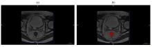
Fig.2
(a) T2WI image (b) and the region of interest of one patient


Fig.3
Support vector machine algorithm derivation diagram
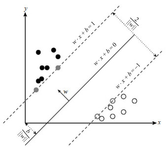
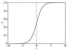
Fig.4
Sigmoid function
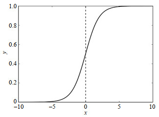
Table 1
Feature selection results
| 特征名称 | 系数 | 特征详情 |
| 灰度相关矩阵高灰度依赖程度(original_gldm_LargeDependenceHighGrayLevelEmphasis) | 0.0318 | 灰度相关矩阵高灰度依赖程度 |
| 伸长率(original_shape_Elongation) | 0.0043 | ROI形状中两个最大的主成分之间的关系 |
| 平面度(original_shape_Flatness) | ?0.0448 | ROI形状中最大和最小主成分之间的关系 |
| 最大2D直径(列)(original_shape_Maximum2DDiameterColumn) | 0.0616 | 冠状平面中肿瘤表面网格顶点之间最大的欧几里得距离 |
| 最大2D直径(切片)(original_shape_Maximum2DDiameterSlice) | 0.0431 | 轴向平面中肿瘤表面网格顶点之间最大的欧几里得距离 |
| 短轴长(original_shape_MinorAxisLength) | 0.1205 | 包围ROI的椭球的第二轴长 |
| 表面积与体积之比(original_shape_SurfaceVolumeRatio) | ?0.0384 | 较低的值表示更紧凑的球形形状 |
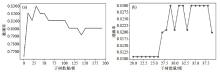
Fig.5
Learning curves with n_estimators of (a) 0~200 and (b) 20~40
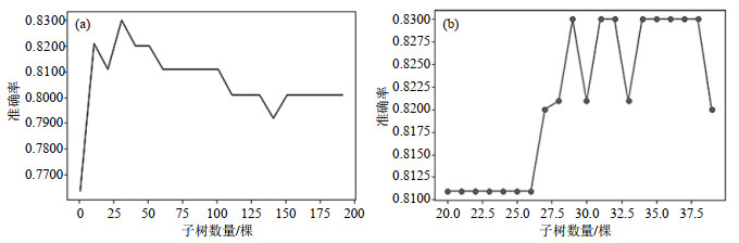
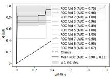
Fig.6
Receiver operating characteristic curve of the prediction model based on random forest
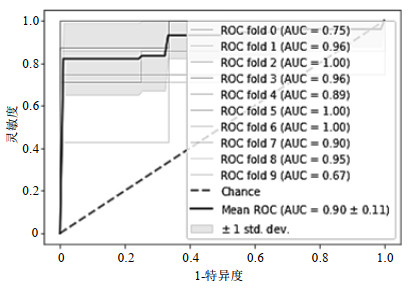
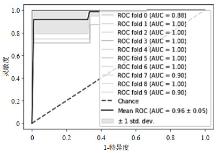
Fig.7
Receiver operation characteristic curve of the prediction model based on SVM
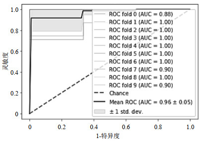
Fig.8
Learning curve of logistic regression

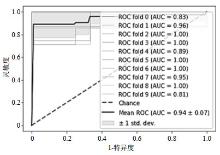
Fig.9
Receiver operation characteristic curve based on logistic regression
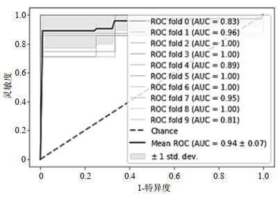
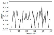
Fig.10
Learning curve of gradient boosting descent tree
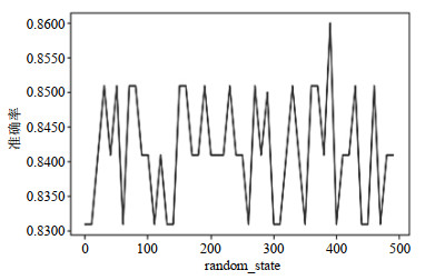
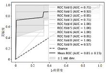
Fig.11
Receiver operation characteristic curve of the prediction model based on GBDT
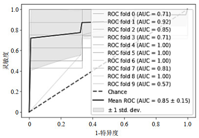
Table 2
The index values predicted by the four models
| 模型 | AUC | 准确率 | 灵敏度 | 特异度 | 测试集准确率 |
| 随机森林 | 0.9015 | 0.8300 | 0.9107 | 0.8690 | 0.8571 |
| 支持向量机 | 0.9685 | 0.8864 | 0.9625 | 0.8992 | 0.9047 |
| 逻辑回归 | 0.9439 | 0.8476 | 0.9057 | 0.8894 | 0.8571 |
| GBDT | 0.8568 | 0.8600 | 0.9250 | 0.8960 | 0.8095 |
| 1 | VUIK F E, NIEUWENBURG S A, BARDOU M, et al. Increasing incidence of colorectal cancer in young adults in Europe over the last 25 years[J]. Gut, 2019, 68(10): gutjnl-2018-317592. |
| 2 | GLOBAL BURDEN OF DISEASE CANCER COLLABORATION . Global, regional, and national cancer incidence, mortality, years of life lost, years lived with disability, and disability-adjusted life-years for 29 cancer groups, 1990 to 2016:a systematic analysis for the global burden of disease study[J]. JAMA On Col, 2018, 4 (11): 1553- 1568. |
| 3 |
CONNELL L C , MOTA J M , BRAGHIROLI M I , et al. The rising incidence of younger patients with colorectal cancer: questions about screening, biology, and treatment[J]. Curr Treat Options Oncol, 2017, 18 (4): 23.
doi: 10.1007/s11864-017-0463-3 |
| 4 |
PETERSE E F P , MEESTER R G S , SIEGEL R L , et al. The impact of the rising colorectal cancer incidence in young adults on the optimal age to start screening: Microsimulation analysis I to inform the American Cancer Society colorectal cancer screening guideline[J]. Cancer, 2018, 124 (14): 2964- 2973.
doi: 10.1002/cncr.31543 |
| 5 |
郑荣寿, 孙可欣, 张思维, 等. 2015年中国恶性肿瘤流行情况分析[J]. 中华肿瘤杂志, 2019, 41 (1): 19- 28.
doi: 10.3760/cma.j.issn.0253-3766.2019.01.005 |
| 6 |
ENGSTROM P F , ARNOLETTI J P , BENSON A B , et al. NCCN clinical practice guidelines in oncology: rectal cancer[J]. J Natl Compr Canc Ne, 2009, 7 (8): 838- 881.
doi: 10.6004/jnccn.2009.0057 |
| 7 |
LAMBIN P , RIOS-VELAZQUEZ E , LEIJENAAR R , et al. Radiomics: Extracting more information from medical images using advanced feature analysis[J]. Eur J Cancer, 2012, 48 (4): 441- 446.
doi: 10.1016/j.ejca.2011.11.036 |
| 8 |
徐从斌. MRI与CT在直肠癌诊断及术前分期中价值探究[J]. 影像研究与医学应用, 2018, 2 (24): 152- 153.
doi: 10.3969/j.issn.2096-3807.2018.24.092 |
| 9 |
CUI S F , WANG X S . The accuracy of MRI in preoperative T staging diagnosis of rectal cancer[J]. Chin J Colorec Dis (Electronic Edition), 2014, 3 (5): 29- 36.
doi: 10.3877/cma.j.issn.2095-3224.2014.05.09 |
|
崔书发, 王锡山. 术前应用MRI评估直肠癌T分期的价值[J]. 中华结直肠疾病电子杂志, 2014, 3 (5): 29- 36.
doi: 10.3877/cma.j.issn.2095-3224.2014.05.09 |
|
| 10 |
LIANG C S , HUANG Y Q , HE L , et al. The development and validation of a CT-based radiomics signature for the preoperative discrimination of stage Ⅰ-Ⅱ and stage Ⅲ-Ⅳ colorectal cancer[J]. Oncotarget, 2016, 7 (21): 31401- 31412.
doi: 10.18632/oncotarget.8919 |
| 11 |
DOU Y F , TANG X F , LIU Y Y , et al. T stage prediction of colorectal tumor based on multiparametric functional images[J]. Transl Cancer Res, 2020, 9 (2): 522- 528.
doi: 10.21037/tcr.2019.11.41 |
| 12 |
KIM J , OH J E , LEE J , et al. Rectal cancer: Toward fully automatic discrimination of T2 and T3 rectal cancers using deep convolutional neural network[J]. Int J Imaging Syst Technol, 2019, 29 (3): 247- 259.
doi: 10.1002/ima.22311 |
| 13 |
XU X P , WANG H J , DU P , et al. A predictive nomogram for individualized recurrence stratification of bladder cancer using multiparametric MRI and clinical risk factors[J]. J Magn Reson Imaging, 2019, 50 (6): 1893- 1904.
doi: 10.1002/jmri.26749 |
| 14 |
YUSHKEVICH P A , PIVEN J , HAZLETT H C , et al. User-guided 3D active contour segmentation of anatomical structures: Significantly improved efficiency and reliability[J]. Neuroimage, 2006, 31 (3): 1116- 1128.
doi: 10.1016/j.neuroimage.2006.01.015 |
| 15 |
BREIMAN L . Random forest[J]. Machine Learning, 2001, 45 (1): 5- 32.
doi: 10.1023/A:1010933404324 |
| 16 | LIAW A , WIENER M . Classification and regression by randomforest[J]. R News, 2002, (2, 3): 18- 22. |
| 17 | 周志华. 机器学习[M]. 北京: 清华大学出版社, 2016. |
| 18 |
VAN GRIETHUYSEN J J M , FEDOROV A , PARMAR C , et al. Computational radiomics system to decode the radiographic phenotype[J]. Cancer Res, 2017, 77 (21): e104- e107.
doi: 10.1158/0008-5472.CAN-17-0339 |
| 19 | KOOPERBERG C , RUCZINSKI I . Identifying interacting SNPs using Monte Carlo logic regression[J]. Genetic Epidemiology, 2010, 28 (2): 157- 170. |
| 20 | 李航. 统计学习方法[M]. 北京: 清华大学出版社, 2012. |
| 21 | SAUNDERS C , STITSON M O , WESTON J , et al. Support vector machine[J]. Computer Ence, 2002, 1 (4): 1- 28. |
| 22 | BAESENS B , VIAENE S , VAN GESTEL T , et al. Least squares support vector machine classifiers: an empirical evaluation[J]. DTEW Research Report 0003, 2000, 1- 16. |
| 23 |
FRIEDMAN J H . Greedy function approximation: A gradient boosting machine[J]. Ann Statist, 2001, 29 (5): 1189- 1232.
doi: 10.1214/aos/1013203450 |
| 24 | FRIEDMAN J H . Stochastic gradient boosting[J]. Computational Statistics & Data Analysis, 2002, 38 (4): 367- 378. |
| 25 | PEDREGOSA F , VAROQUAUX G , GRAMFORT A , et al. Scikit-learn: machine learning in python[J]. J Mach Learn Res, 2011, 2825- 2830. |
| 26 | WANG J , LI Z H , SHEN F , et al. The value of high resolution T2WI-based radiomics in the preoperative staging of rectal cancer[J]. Radiol Practice, 2019, 34 (11): 1251- 1254. |
| 王进, 李智慧, 沈浮, 等. 基于高分辨T2WI的影像组学对直肠癌术前分期的应用价值[J]. 放射学实践, 2019, 34 (11): 1251- 1254. | |
| 27 |
LAMBIN P , LEIJENAAR R T H , DEIST T M , et al. Radiomics: the bridge between medical imaging and personalized medicine[J]. Nat Rev Clin Oncol, 2017, 14, 749- 762.
doi: 10.1038/nrclinonc.2017.141 |
| [1] | Ju-min ZHANG,Shi-zhen CHEN,Xin ZHOU. Dual-modal MRI T1-T2 Contrast Agent Based on Dynamic Organic Gadolinium Nanoparticles [J]. Chinese Journal of Magnetic Resonance, 2022, 39(1): 11-19. |
| [2] | Zhi-chao WANG,Ji-lei ZHANG,Yu ZHAO,Ting HUA,Guang-yu TANG,Jian-qi LI. CEST Imaging of the Abdomen with Neural Network Fitting [J]. Chinese Journal of Magnetic Resonance, 2022, 39(1): 33-42. |
| [3] | Yan-yan LI,Lv LI,Xue-song LI,Hua GUO. 3D Dynamic MRI with Homotopic l0 Minimization Reconstruction [J]. Chinese Journal of Magnetic Resonance, 2022, 39(1): 20-32. |
| [4] | CUI Yang-yang, LIANG Huai-bin, ZHU Qian, TANG Wei, GAO Ting-ting, LIU Jian-ren, DU Xiao-xia. A Study on the Alteration of Spontaneous Brain Activity in Somatic Symptoms Disorder Combining Regional Homogeneity and Amplitude of Low-frequency Fluctuation [J]. Chinese Journal of Magnetic Resonance, 2022, 39(1): 64-71. |
| [5] | WANG Han-wei, WU Hao, TIAN Jing, ZHANG Jun-feng, ZHONG Peng, CHEN Li-zhao, WANG Shu-nan. The Diagnostic Value of Quantitative Parameters of T2/FLAIR Mismatch Sign in Evaluating the Molecular Typing of Lower-grade Gliomas [J]. Chinese Journal of Magnetic Resonance, 2022, 39(1): 56-63. |
| [6] | Long XIAO,Xiao-lei ZHU,Ye-qing HAN,Shi-zhen CHEN,Xin ZHOU. Design and Application of Micellar Magnetic Resonance Imaging Molecular Probe [J]. Chinese Journal of Magnetic Resonance, 2021, 38(4): 474-490. |
| [7] | Shi-ju YAN,Yong-sen HAN,Guang-yu TANG. An Improved Level Set Algorithm for Prostate Region Segmentation [J]. Chinese Journal of Magnetic Resonance, 2021, 38(3): 356-366. |
| [8] | Ying-dan HU,Yue CAI,Xu-xia WANG,Si-jie LIU,Yan KANG,Hao LEI,Fu-chun LIN. Magnetic Resonance Imaging the Brain Structures Involved in Nicotine Susceptibility in Rats [J]. Chinese Journal of Magnetic Resonance, 2021, 38(3): 345-355. |
| [9] | HE Hong-yan, WEI Shu-feng, WANG Hui-xian, YANG Wen-hui. Matrix Gradient Coil: Current Research Status and Perspectives [J]. Chinese Journal of Magnetic Resonance, 2021, 38(1): 140-153. |
| [10] | MA Cong-wei, YANG Hong-yi, ZHONG Kai. Research Progresses of High-Field MRI 1H/31P Dual-Tuned Radio Frequency Coil [J]. Chinese Journal of Magnetic Resonance, 2021, 38(1): 118-139. |
| [11] | XIN Hong-tao, WU Guang-yao, WEN Zhi, LEI Hao, LIN Fu-chun. Effects of Antiretroviral Therapy on Brain Gray Matter Volumes in AIDS Patients [J]. Chinese Journal of Magnetic Resonance, 2021, 38(1): 69-79. |
| [12] | JIA Jia-ying, KUANG Li-qun, XIONG Feng-guang, HAN Xie. Analysis of Dynamic Evolution of Complex Brain Networks Based on Persistent Homology [J]. Chinese Journal of Magnetic Resonance, 2021, 38(1): 80-91. |
| [13] | HU Ge-li, DENG Ye-hui, WANG Kun, JIANG Tian-zi. A New MRI System Architecture Based on 5G Remote Control and Processing [J]. Chinese Journal of Magnetic Resonance, 2020, 37(4): 490-495. |
| [14] | YUE Qing, WANG Yuan-jun. A Fiber Tracking Algorithm Based on Non-Local Constrained Spherical Deconvolution [J]. Chinese Journal of Magnetic Resonance, 2020, 37(4): 422-433. |
| [15] | WU Ming-di, FENG Jie, JIA Hui-hui, WU Ji-zhi, ZHANG Xin, CHANG Yan, YANG Xiao-dong, SHENG Mao. MRI-Based Morphological Quantification of Developmental Dysplasia of the Hip in Children [J]. Chinese Journal of Magnetic Resonance, 2020, 37(4): 434-446. |
| Viewed | ||||||||||||||||||||||||||||||||||||||||||||||||||
|
Full text 367
|
|
|||||||||||||||||||||||||||||||||||||||||||||||||
|
Abstract 304
|
|
|||||||||||||||||||||||||||||||||||||||||||||||||