
- Nov. 14, 2025
- Home
- About Us
- Editorial Board
- Instruction
- Subscription
- Advertisement
- Contact Us
- Chinese
- RSS
Chinese Journal of Magnetic Resonance ›› 2022, Vol. 39 ›› Issue (1): 72-86.doi: 10.11938/cjmr20212898
• Articles • Previous Articles Next Articles
Jian-sheng LIN,Li-jia WANG*( )
)
Received:2021-03-22
Published:2022-03-05
Online:2021-07-05
Contact:
Li-jia WANG
E-mail:lijiawangmri@163.com
CLC Number:
Jian-sheng LIN,Li-jia WANG. Reconstruction of Displacement Field of Left Ventricle Combined with Biomechanical Model[J]. Chinese Journal of Magnetic Resonance, 2022, 39(1): 72-86.
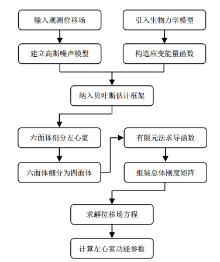
Fig.1
Reconstruction for displacement field of left ventricle
Fig.2
Procedures of motion tracking of left ventricle
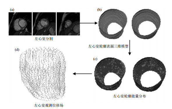
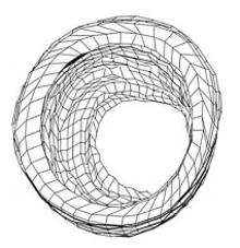
Fig.3
Hexahedron dissection of left ventricle

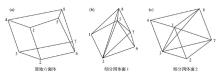
Fig.4
Numbered hexahedral mesh and division of tetrahedral mesh
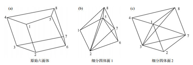
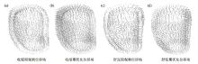
Fig.5
Comparison of observed and optimized displacement fields
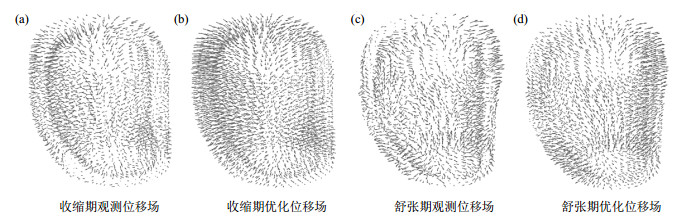
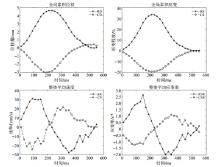
Fig.6
Functional parameters curves of left ventricle
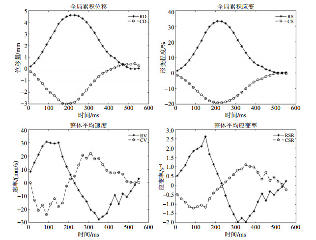
Table 1
Comparison of functional parameters of LV between normal EF group and weak EF group
| 左心室功能参数 | 径向方向(峰值) | 圆周方向(峰值) | |||||||
| RD/mm | RS/% | RV/(mm/s) | RSR/s?1 | CD/mm | CS/% | CV/(mm/s) | CSR/s?1 | ||
| 射血正常 | 4.86±1.10 | 20.30±6.50 | 28.15±8.28 | 1.40±0.68 | ?3.09±0.71 | ?19.43±5.61 | ?18.19±5.34 | ?1.22±0.52 | |
| 射血无力 | 3.29±0.92 | 11.62±5.65 | 19.69±5.60 | 0.78±0.32 | ?2.05±0.61 | ?10.86±4.63 | ?12.62±3.58 | ?0.63±0.24 | |
| p值 | 1.46e-11 | 2.04e-10 | 5.24e-08 | 1.45e-07 | 7.67e-12 | 6.40e-13 | 2.86e-08 | 2.06e-10 | |
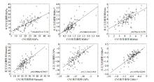
Fig.7
The correlation between the functional parameters of LV obtained by the proposed method and CVI software
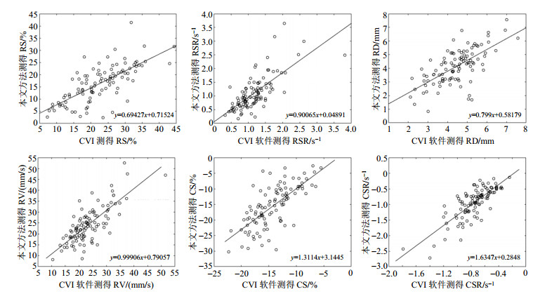
Fig.8
Left ventricular function parameter curves of objects with different LVEF values
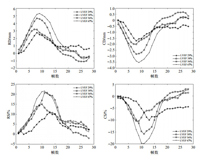
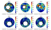
Fig.9
Distribution of left ventricle strain of objects with different LVEF values
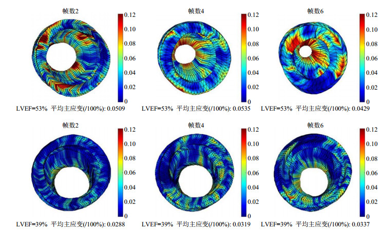
Table 2
Comparison of functional parameters of LV under different constraints
| 约束条件 | 径向方向(峰值) | 圆周方向(峰值) | |||||||
| RD/mm | RS/% | RV/(mm/s) | RSR/s?1 | CD/mm | CS/% | CV/(mm/s) | CSR/s?1 | ||
| 无约束 | 4.07±0.97 | 21.03±6.89 | 24.83±6.45 | 1.35±0.49 | ?2.31±0.56 | ?13.06±6.56 | ?14.07±3.63 | ?1.19±0.88 | |
| 平滑约束 | 4.23±0.96 | 18.76±5.05 | 25.72±6.48 | 1.10±0.35 | ?2.28±0.58 | ?10.51±3.68 | ?14.25±3.58 | ?0.76±0.33 | |
| 模型约束 | 4.08±1.01 | 15.96±6.08 | 23.92±6.94 | 1.09±0.50 | ?2.57±0.66 | ?15.15±5.12 | ?15.40±4.46 | ?0.93±0.38 | |
| 1 |
DEMARINIS S . Cancer overtakes cardiovascular disease as leading cause of death in wealthy nations[J]. Explore, 2020, 16 (1): 6- 7.
doi: 10.1016/j.explore.2019.11.003 |
| 2 |
PANG Y , LYU J , YU C , et al. Risk factors for cardiovascular disease in the Chinese population: recent progress and implications[J]. Glo Hea J, 2020, 4 (3): 65- 71.
doi: 10.1016/j.glohj.2020.08.004 |
| 3 |
FRANGI A F , NIESSEN W J , VIERGEVER M A . Three-dimensional modeling for functional analysis of cardiac images: a review[J]. IEEE Trans Med Imaging, 2001, 20 (1): 2- 5.
doi: 10.1109/42.906421 |
| 4 |
WANG H , AMINI A A . Cardiac motion and deformation recovery from MRI: A Review[J]. IEEE Trans Med Imaging, 2012, 31 (2): 487- 503.
doi: 10.1109/TMI.2011.2171706 |
| 5 |
ZERHOUNI E A , PARISH D M , ROGERS W J , et al. Human heart: Tagging with MR imaging-A method for noninvasive assessment of myocardial motion[J]. Radiology, 1988, 169 (1): 59- 63.
doi: 10.1148/radiology.169.1.3420283 |
| 6 | CHEN T , WANG X , CHUNG S , et al. Automated 3D motion tracking using Gabor filter bank, robust point matching, and deformable models[J]. IEEE Trans Med Imaging, 2009, 29 (1): 1- 11. |
| 7 |
LI M , GUPTA H , LLOYD S G , et al. A graph theoretic approach for computing 3D+ time biventricular cardiac strain from tagged MRI data[J]. Med Image Anal, 2017, 35, 46- 57.
doi: 10.1016/j.media.2016.06.006 |
| 8 |
YU Y , ZHANG S T , LI K , et al. Deformable models with sparsity constraints for cardiac motion analysis[J]. Med Image Anal, 2014, 18 (6): 927- 937.
doi: 10.1016/j.media.2014.03.002 |
| 9 |
TSADOK Y , FRIEDMAN Z , HALUSKA B A , et al. Myocardial strain assessment by cine cardiac magnetic resonance imaging using non-rigid registration[J]. Magn Reson Imaging, 2016, 34 (4): 381- 390.
doi: 10.1016/j.mri.2015.12.035 |
| 10 |
SHI P , SINUSAS A J , CONSTABLE R T , et al. Volumetric deformation analysis using mechanics-based data fusion: applications in cardiac motion recovery[J]. Int J Comput Vision, 1999, 35 (1): 87- 107.
doi: 10.1023/A:1008163112590 |
| 11 |
REMME E W , AUGENSTEIN K F , YOUNG A A , et al. Parameter distribution models for estimation of population based left ventricular deformation using sparse fiducial markers[J]. IEEE Trans Med Imaging, 2005, 24 (3): 381- 388.
doi: 10.1109/TMI.2004.842458 |
| 12 |
VERESS A I , GULLBERG G T , WEISS J A . Measurement of strain in the left ventricle during diastole with cine-MRI and deformable image registration[J]. J Biomech Eng, 2005, 127 (7): 1195- 1207.
doi: 10.1115/1.2073677 |
| 13 |
BISTOQUET A , SKRINJAR O M . Left ventricular deformation recovery from cine MRI using a 4d incompressible model[J]. IEEE Trans Med Imaging, 2007, 26 (9): 1136- 1153.
doi: 10.1109/TMI.2007.903693 |
| 14 |
LIU H F , SHI P C . State-space analysis of cardiac motion with biomechanical constraints[J]. IEEE T Image Process, 2007, 16 (4): 901- 917.
doi: 10.1109/TIP.2007.891773 |
| 15 |
WONG K C L , WANG L W , ZHANG H Y , et al. Physiological fusion of functional and structural images for cardiac deformation recovery[J]. IEEE Trans Med Imaging, 2011, 30 (4): 990- 1000.
doi: 10.1109/TMI.2011.2105274 |
| 16 |
QIAO M Y , WANG Y Y , GUO Y , et al. Temporally coherent cardiac motion tracking from cine MRI: traditional registration method and modern CNN method[J]. Med Phys, 2020, 47 (9): 4189- 4198.
doi: 10.1002/mp.14341 |
| 17 | TANG J Y, GAN Z Y, YANG X. Cardiac motion tracking in short-axis MRI using siamese convolution network[C]//2019 IEEE International Conference on Bioinformatics and Biomedicine (BIBM). IEEE, 2019: 865-870. |
| 18 |
ZHANG N , YANG G , GAO Z F , et al. Deep learning for diagnosis of chronic myocardial infarction on nonenhanced cardiac cine MRI[J]. Radiology, 2019, 291 (3): 606- 617.
doi: 10.1148/radiol.2019182304 |
| 19 |
ZHENG Q , DELINGETTE H , AYACHE N . Explainable cardiac pathology classification on cine MRI with motion characterization by semi-supervised learning of apparent flow[J]. Med Image Anal, 2019, 56, 80- 95.
doi: 10.1016/j.media.2019.06.001 |
| 20 |
MANTILLA J J , PAREDES J L , BELLANGER J J , et al. Discriminative dictionary learning for local LV wall motion classification in cardiac MRI[J]. Expert Syst Appl, 2019, 129, 286- 295.
doi: 10.1016/j.eswa.2019.04.010 |
| 21 |
SEDERBERG T W , PARRY S R . Free-form deformation of solid geometric models[J]. Siggraph, 1986, 20 (4): 151- 160.
doi: 10.1145/15886.15903 |
| 22 |
COOTES T F , TAYLOR C J , COOPER D H , et al. Active shape models-their training and application[J]. Comput Vis Image Underst, 1995, 61 (1): 38- 59.
doi: 10.1006/cviu.1995.1004 |
| 23 | GONG J C , WANG Y , WANG Y J . A method for segmentation of glioma on multimodal magnetic resonance images based on wavelet fusion and deep learning[J]. Chinese J Magn Reson, 2020, 37 (2): 131- 143. |
| 宫进昌, 王宇, 王远军. 结合小波融合和深度学习的脑胶质瘤自动分割[J]. 波谱学杂志, 2020, 37 (2): 131- 143. | |
| 24 | LIU P , ZHONG Y M , WANG L J . Automatic segmentation of right ventricle in cine cardiac magnetic resonance image based on a dense and multi-scale U-net method[J]. Chinese J Magn Reson, 2020, 37 (4): 456- 468. |
| 刘鹏, 钟玉敏, 王丽嘉. 基于密集多尺度U-net网络的电影心脏磁共振图像右心室自动分割[J]. 波谱学杂志, 2020, 37 (4): 456- 468. | |
| 25 | GUCCIONE J M, MCCULLOCH A D. Finite element modeling of ventricular mechanics[M]. Springer New York, 1991. |
| 26 | PAPADEMETRIS X, SHI P C, DIONE D P, et al. Recovery of soft tissue object deformation from 3D image sequences using biomechanical models[C]//Proceedings of the 16th International Conference on Information Processing in Medical Imaging. Berlin, Heidelberg: Springer, 2000. |
| 27 |
SHI P C , SINUSAS A J . Point-tracked quantitative analysis of left ventricular surface motion from 3-D image sequences[J]. IEEE Trans Med Imaging, 2000, 19 (1): 36- 50.
doi: 10.1109/42.832958 |
| 28 |
PAPADEMETRIS , XENOPHON , SINUSAS , et al. Estimation of 3-D left ventricular deformation from medical images using biomechanical models[J]. IEEE Trans Med Imaging, 2002, 21 (7): 786- 800.
doi: 10.1109/TMI.2002.801163 |
| 29 | 杨桂通. 弹性力学[M]. 第2版 高等教育出版社, 2012. |
| 30 | GEMAN S , GERMAN D . Stochastic relaxation, gibbs distributions and the Bayesian restoration of images[J]. IEEE Trans Pattern Anal Mach Intell, 1984, 6, 721- 741. |
| 31 | ZHANG H Y , GAO Z , XU L , et al. A meshfree representation for cardiac medical image computing[J]. IEEE J Transl Eng He, 2018, 6, 1- 12. |
| 32 | 李亚智. 有限元法基础与程序设计[M]. 科学出版社, 2004. |
| [1] | LI Haodong, WANG Yuanjun. A Fiber Tracking Algorithm with Seed Point Clustering and Orientation Correction [J]. Chinese Journal of Magnetic Resonance, 2024, 41(4): 430-442. |
| [2] | CHANG Bo, SUN Haoyun, GAO Qingyu, WANG Lijia. Research Progress on Cardiac Segmentation in Different Modal Medical Images by Traditional Methods and Deep Learning [J]. Chinese Journal of Magnetic Resonance, 2024, 41(2): 224-244. |
| [3] | Li Yijie, YANG Xinyu, YANG Xiaomei. Magnetic Resonance Image Reconstruction of Multi-scale Residual Unet Fused with Attention Mechanism [J]. Chinese Journal of Magnetic Resonance, 2023, 40(3): 307-319. |
| [4] | ZHAO Xin, ZHANG Xin, LI Xinjie, WANG Hongkai. Multimodal Glioma Segmentation with Fusion of Multiple Self-attention and Deformable Convolutions [J]. Chinese Journal of Magnetic Resonance, 2023, 40(3): 280-292. |
| [5] | QIAN Chengyi,WANG Yuanjun. Research Progress on Imaging Classification of Alzheimer’s Disease Based on Deep Learning [J]. Chinese Journal of Magnetic Resonance, 2023, 40(2): 220-238. |
| [6] | SHI Weicheng,JIN Zhaoyang,YE Zheng. Fast Multi-channel Magnetic Resonance Imaging Based on PCAU-Net [J]. Chinese Journal of Magnetic Resonance, 2023, 40(1): 39-51. |
| [7] | Lu HUO,Xiao-xin HU,Qin XIAO,Ya-jia GU,Xu CHU,Luan JIANG. Automatic Segmentation of Breast and Fibroglandular Tissues in DCE-MR Images Based on nnU-Net [J]. Chinese Journal of Magnetic Resonance, 2021, 38(3): 367-380. |
| [8] | Qin-yi SHI,Fang YAN,Yang YANG,Yue-fu CHEN,Xiao-lang LIN,Yuan-jun WANG. Image Segmentation of Tooth and Alveolar Bone with the Level Set Model [J]. Chinese Journal of Magnetic Resonance, 2021, 38(2): 182-193. |
| [9] | YUE Qing, WANG Yuan-jun. A Fiber Tracking Algorithm Based on Non-Local Constrained Spherical Deconvolution [J]. Chinese Journal of Magnetic Resonance, 2020, 37(4): 422-433. |
| [10] | LOU Yun-zhong, LIU Ying, JIANG Hua, ZHANG Hao-wei. A Deep Learning Algorithm for Classifying Meningioma and Auditory Neuroma in the Cerebellopontine Angle from Magnetic Resonance Images [J]. Chinese Journal of Magnetic Resonance, 2020, 37(3): 300-310. |
| [11] | ZHAO Shang-yi, WANG Yuan-jun. Classification of Alzheimer's Disease Patients Based on Magnetic Resonance Images and an Improved UNet++ Model [J]. Chinese Journal of Magnetic Resonance, 2020, 37(3): 321-331. |
| [12] | CAI Wen-qin, WANG Yuan-jun. Advances in Construction of Human Brain Atlases from Magnetic Resonance Images [J]. Chinese Journal of Magnetic Resonance, 2020, 37(2): 241-253. |
| [13] | GONG Jin-chang, WANG Yu, WANG Yuan-jun. A Method for Segmentation of Glioma on Multimodal Magnetic Resonance Images Based on Wavelet Fusion and Deep Learning [J]. Chinese Journal of Magnetic Resonance, 2020, 37(2): 131-143. |
| [14] | JIANG Fan, WANG Yuan-jun. Construction of Human Brain Templates with Diffusion Tensor Imaging Data: A Review [J]. Chinese Journal of Magnetic Resonance, 2018, 35(4): 520-530. |
| [15] | YAN Zhi-yu, CHEN Zhi-wei. Implementation of Graphical Pulse Sequence Design Using Eclipse Graphical Modeling Framework [J]. Chinese Journal of Magnetic Resonance, 2017, 34(2): 175-182. |
| Viewed | ||||||
|
Full text |
|
|||||
|
Abstract |
|
|||||