Interpretation of report on cardiovascular health and diseases in China 2022
1
2023
... 根据《中国心血管疾病与健康报告2022》要点解读[1]统计,我国现存心血管疾病(Cardiovascular Disease,CVD)患者达到3.3亿.随着人口老龄化加剧,CVD在我国的影响日益突出,高死亡率和巨大的经济负担引起了大量研究者的关注.心脏主要由被心肌包裹着的左心室(Left Ventricle,LV)、右心室(Right Ventricle,RV)、左心房(Left Atrium,LA)和右心房(Right Atrium,RA)组成.借助医学成像技术了解心脏结构及功能是临床常规诊断方式.目前常用的成像技术主要有X射线计算机断层扫描(Computed Tomography,CT)、心脏磁共振成像(Cardiac Magnetic Resonance Imaging,CMRI)和超声心动图(Ultrasonic Cardiogram,UCG).CT通过利用计算机重建X射线投影数据进行断层成像,速度快、空间分辨率高,此外,CTA(CT Angiography)将CT增强技术与薄层、大范围、快速扫描技术相结合,清晰显示全身各部位血管细节,但X射线会对人体产生电离辐射,不适于检查身体较差的CVD患者.CMRI基于核磁共振原理,配合不同的梯度磁场可从任意方位成像,软组织对比度高.其中,短轴电影CMRI心室内部血液与心肌组织对比度高,可动态地展示心室甚至心房在心动周期内不同时相的变化,常用于心脏分割及功能分析. 例如,通过分割心室心肌可以计算不同时相血液体积变化,从而计算射血分数和充盈曲线,进一步追踪心肌运动以及分析心肌应力变化等;通过分割心房则可以计算不同时相心房体积,以评估心房负荷以及诊断是否患有房颤.UCG可实现心脏实时成像,能够捕捉心脏的运动细节,便于心脏异常功能的检查以及指导心脏手术和介入治疗过程. ...
《中国心血管健康与疾病报告2022》要点解读
1
2023
... 根据《中国心血管疾病与健康报告2022》要点解读[1]统计,我国现存心血管疾病(Cardiovascular Disease,CVD)患者达到3.3亿.随着人口老龄化加剧,CVD在我国的影响日益突出,高死亡率和巨大的经济负担引起了大量研究者的关注.心脏主要由被心肌包裹着的左心室(Left Ventricle,LV)、右心室(Right Ventricle,RV)、左心房(Left Atrium,LA)和右心房(Right Atrium,RA)组成.借助医学成像技术了解心脏结构及功能是临床常规诊断方式.目前常用的成像技术主要有X射线计算机断层扫描(Computed Tomography,CT)、心脏磁共振成像(Cardiac Magnetic Resonance Imaging,CMRI)和超声心动图(Ultrasonic Cardiogram,UCG).CT通过利用计算机重建X射线投影数据进行断层成像,速度快、空间分辨率高,此外,CTA(CT Angiography)将CT增强技术与薄层、大范围、快速扫描技术相结合,清晰显示全身各部位血管细节,但X射线会对人体产生电离辐射,不适于检查身体较差的CVD患者.CMRI基于核磁共振原理,配合不同的梯度磁场可从任意方位成像,软组织对比度高.其中,短轴电影CMRI心室内部血液与心肌组织对比度高,可动态地展示心室甚至心房在心动周期内不同时相的变化,常用于心脏分割及功能分析. 例如,通过分割心室心肌可以计算不同时相血液体积变化,从而计算射血分数和充盈曲线,进一步追踪心肌运动以及分析心肌应力变化等;通过分割心房则可以计算不同时相心房体积,以评估心房负荷以及诊断是否患有房颤.UCG可实现心脏实时成像,能够捕捉心脏的运动细节,便于心脏异常功能的检查以及指导心脏手术和介入治疗过程. ...
Image segmentation by using thresholding techniques for medical images
1
2016
... 传统医学图像分割方法主要基于图像自身的阈值、边缘和区域.其中,基于阈值的分割算法通过设定灰度阈值将图像划分为不同区域,可按照任务需求制定约束准则以实现最佳阈值的确定,常采用自适应阈值分割法、直方图双峰法以及迭代阈值分割法,这类算法简单易用,适于二值化处理[2],但对噪声敏感且图像质量要求高.基于边缘的分割算法可以处理不规则轮廓的图像,主要依据颜色、灰度和纹理等特征的显著变化,通过一阶导数的极值和二阶导数的过零点来确定边缘,常用水平集、边缘增强、Canny算子等,在进行肝脏和肺等大器官分割时效果较好[3⇓⇓-6].基于区域的分割算法根据像素相似性、利用不同的统计方法进行像素聚集、边缘确定,主要有区域生长、活动轮廓模型、聚类、图谱等算法[7⇓-9].总结这些方式在图像处理中的应用,传统分割方法运行速率高,不需要额外的数据标签进行监督;但是缺乏对图像特征的自动提取能力,并且在分割过程中需要手动介入且采用多个算法集成的方式优化轮廓,鲁棒性较差. ...
Efficient liver segmentation using a level-set method with optimal detection of the initial liver boundary from level-set speed images
1
2007
... 传统医学图像分割方法主要基于图像自身的阈值、边缘和区域.其中,基于阈值的分割算法通过设定灰度阈值将图像划分为不同区域,可按照任务需求制定约束准则以实现最佳阈值的确定,常采用自适应阈值分割法、直方图双峰法以及迭代阈值分割法,这类算法简单易用,适于二值化处理[2],但对噪声敏感且图像质量要求高.基于边缘的分割算法可以处理不规则轮廓的图像,主要依据颜色、灰度和纹理等特征的显著变化,通过一阶导数的极值和二阶导数的过零点来确定边缘,常用水平集、边缘增强、Canny算子等,在进行肝脏和肺等大器官分割时效果较好[3⇓⇓-6].基于区域的分割算法根据像素相似性、利用不同的统计方法进行像素聚集、边缘确定,主要有区域生长、活动轮廓模型、聚类、图谱等算法[7⇓-9].总结这些方式在图像处理中的应用,传统分割方法运行速率高,不需要额外的数据标签进行监督;但是缺乏对图像特征的自动提取能力,并且在分割过程中需要手动介入且采用多个算法集成的方式优化轮廓,鲁棒性较差. ...
Liver segmentation based on region growing and level set active contour model with new signed pressure force function
1
2020
... 传统医学图像分割方法主要基于图像自身的阈值、边缘和区域.其中,基于阈值的分割算法通过设定灰度阈值将图像划分为不同区域,可按照任务需求制定约束准则以实现最佳阈值的确定,常采用自适应阈值分割法、直方图双峰法以及迭代阈值分割法,这类算法简单易用,适于二值化处理[2],但对噪声敏感且图像质量要求高.基于边缘的分割算法可以处理不规则轮廓的图像,主要依据颜色、灰度和纹理等特征的显著变化,通过一阶导数的极值和二阶导数的过零点来确定边缘,常用水平集、边缘增强、Canny算子等,在进行肝脏和肺等大器官分割时效果较好[3⇓⇓-6].基于区域的分割算法根据像素相似性、利用不同的统计方法进行像素聚集、边缘确定,主要有区域生长、活动轮廓模型、聚类、图谱等算法[7⇓-9].总结这些方式在图像处理中的应用,传统分割方法运行速率高,不需要额外的数据标签进行监督;但是缺乏对图像特征的自动提取能力,并且在分割过程中需要手动介入且采用多个算法集成的方式优化轮廓,鲁棒性较差. ...
A novel approach for lung nodules segmentation in chest CT using level sets
1
2013
... 传统医学图像分割方法主要基于图像自身的阈值、边缘和区域.其中,基于阈值的分割算法通过设定灰度阈值将图像划分为不同区域,可按照任务需求制定约束准则以实现最佳阈值的确定,常采用自适应阈值分割法、直方图双峰法以及迭代阈值分割法,这类算法简单易用,适于二值化处理[2],但对噪声敏感且图像质量要求高.基于边缘的分割算法可以处理不规则轮廓的图像,主要依据颜色、灰度和纹理等特征的显著变化,通过一阶导数的极值和二阶导数的过零点来确定边缘,常用水平集、边缘增强、Canny算子等,在进行肝脏和肺等大器官分割时效果较好[3⇓⇓-6].基于区域的分割算法根据像素相似性、利用不同的统计方法进行像素聚集、边缘确定,主要有区域生长、活动轮廓模型、聚类、图谱等算法[7⇓-9].总结这些方式在图像处理中的应用,传统分割方法运行速率高,不需要额外的数据标签进行监督;但是缺乏对图像特征的自动提取能力,并且在分割过程中需要手动介入且采用多个算法集成的方式优化轮廓,鲁棒性较差. ...
A level-set approach to joint image segmentation and registration with application to CT lung imaging
1
2018
... 传统医学图像分割方法主要基于图像自身的阈值、边缘和区域.其中,基于阈值的分割算法通过设定灰度阈值将图像划分为不同区域,可按照任务需求制定约束准则以实现最佳阈值的确定,常采用自适应阈值分割法、直方图双峰法以及迭代阈值分割法,这类算法简单易用,适于二值化处理[2],但对噪声敏感且图像质量要求高.基于边缘的分割算法可以处理不规则轮廓的图像,主要依据颜色、灰度和纹理等特征的显著变化,通过一阶导数的极值和二阶导数的过零点来确定边缘,常用水平集、边缘增强、Canny算子等,在进行肝脏和肺等大器官分割时效果较好[3⇓⇓-6].基于区域的分割算法根据像素相似性、利用不同的统计方法进行像素聚集、边缘确定,主要有区域生长、活动轮廓模型、聚类、图谱等算法[7⇓-9].总结这些方式在图像处理中的应用,传统分割方法运行速率高,不需要额外的数据标签进行监督;但是缺乏对图像特征的自动提取能力,并且在分割过程中需要手动介入且采用多个算法集成的方式优化轮廓,鲁棒性较差. ...
Automatic seeded region growing image segmentation for medical image segmentation: a brief review
1
2020
... 传统医学图像分割方法主要基于图像自身的阈值、边缘和区域.其中,基于阈值的分割算法通过设定灰度阈值将图像划分为不同区域,可按照任务需求制定约束准则以实现最佳阈值的确定,常采用自适应阈值分割法、直方图双峰法以及迭代阈值分割法,这类算法简单易用,适于二值化处理[2],但对噪声敏感且图像质量要求高.基于边缘的分割算法可以处理不规则轮廓的图像,主要依据颜色、灰度和纹理等特征的显著变化,通过一阶导数的极值和二阶导数的过零点来确定边缘,常用水平集、边缘增强、Canny算子等,在进行肝脏和肺等大器官分割时效果较好[3⇓⇓-6].基于区域的分割算法根据像素相似性、利用不同的统计方法进行像素聚集、边缘确定,主要有区域生长、活动轮廓模型、聚类、图谱等算法[7⇓-9].总结这些方式在图像处理中的应用,传统分割方法运行速率高,不需要额外的数据标签进行监督;但是缺乏对图像特征的自动提取能力,并且在分割过程中需要手动介入且采用多个算法集成的方式优化轮廓,鲁棒性较差. ...
Active contour model based on local and global intensity information for medical image segmentation
1
2016
... 传统医学图像分割方法主要基于图像自身的阈值、边缘和区域.其中,基于阈值的分割算法通过设定灰度阈值将图像划分为不同区域,可按照任务需求制定约束准则以实现最佳阈值的确定,常采用自适应阈值分割法、直方图双峰法以及迭代阈值分割法,这类算法简单易用,适于二值化处理[2],但对噪声敏感且图像质量要求高.基于边缘的分割算法可以处理不规则轮廓的图像,主要依据颜色、灰度和纹理等特征的显著变化,通过一阶导数的极值和二阶导数的过零点来确定边缘,常用水平集、边缘增强、Canny算子等,在进行肝脏和肺等大器官分割时效果较好[3⇓⇓-6].基于区域的分割算法根据像素相似性、利用不同的统计方法进行像素聚集、边缘确定,主要有区域生长、活动轮廓模型、聚类、图谱等算法[7⇓-9].总结这些方式在图像处理中的应用,传统分割方法运行速率高,不需要额外的数据标签进行监督;但是缺乏对图像特征的自动提取能力,并且在分割过程中需要手动介入且采用多个算法集成的方式优化轮廓,鲁棒性较差. ...
Multi-atlas segmentation of biomedical images: a survey
1
2015
... 传统医学图像分割方法主要基于图像自身的阈值、边缘和区域.其中,基于阈值的分割算法通过设定灰度阈值将图像划分为不同区域,可按照任务需求制定约束准则以实现最佳阈值的确定,常采用自适应阈值分割法、直方图双峰法以及迭代阈值分割法,这类算法简单易用,适于二值化处理[2],但对噪声敏感且图像质量要求高.基于边缘的分割算法可以处理不规则轮廓的图像,主要依据颜色、灰度和纹理等特征的显著变化,通过一阶导数的极值和二阶导数的过零点来确定边缘,常用水平集、边缘增强、Canny算子等,在进行肝脏和肺等大器官分割时效果较好[3⇓⇓-6].基于区域的分割算法根据像素相似性、利用不同的统计方法进行像素聚集、边缘确定,主要有区域生长、活动轮廓模型、聚类、图谱等算法[7⇓-9].总结这些方式在图像处理中的应用,传统分割方法运行速率高,不需要额外的数据标签进行监督;但是缺乏对图像特征的自动提取能力,并且在分割过程中需要手动介入且采用多个算法集成的方式优化轮廓,鲁棒性较差. ...
GVFOM: a novel external force for active contour based image segmentation
1
2020
... 应用于CMRI分割的传统算法主要有:主动轮廓[10]、水平集[11]、动态规划[12]以及基于图谱[13,14]的模型.其中,主动轮廓模型(Active Contour Model,ACM)是最有影响力的计算机视觉技术之一,已成功应用于分割等各种图像分析任务,该算法首先在图像中初始化一个封闭轮廓并定义能量函数,其中内部能量惩罚轮廓弯曲程度,外部能量驱动轮廓向目标收敛,基于能量最小化原理,优化能量函数来找到图像中目标对象的边界. 在ACM变体中,引入概率统计和先验轮廓等方式可以优化迭代过程动态演化,但由于ACM基于几何特征和物理原理,初始轮廓、能量泛函参数以及图像特征都对分割结果影响很大.Chan等[15]提出新的轮廓模型,使用具有图像去噪和边缘保持的停止项代替传统的图像梯度,在处理弱边缘图像时更具鲁棒性;Saini等[16]在此基础上使用Newton-Raphson方法代替Chan提出的停止项,实现LV的快速分割.Liu等[17]构造了ACM广义法向有偏梯度矢量流外力模型,该模型具有更大捕捉范围和更强抗噪能力,在解决弱边界泄露问题上性能突出,最后引入圆形约束能量项克服乳头肌导致的局部极小问题. ...
Left ventricle segmentation via two-layer level sets with circular shape constraint
3
2017
... 应用于CMRI分割的传统算法主要有:主动轮廓[10]、水平集[11]、动态规划[12]以及基于图谱[13,14]的模型.其中,主动轮廓模型(Active Contour Model,ACM)是最有影响力的计算机视觉技术之一,已成功应用于分割等各种图像分析任务,该算法首先在图像中初始化一个封闭轮廓并定义能量函数,其中内部能量惩罚轮廓弯曲程度,外部能量驱动轮廓向目标收敛,基于能量最小化原理,优化能量函数来找到图像中目标对象的边界. 在ACM变体中,引入概率统计和先验轮廓等方式可以优化迭代过程动态演化,但由于ACM基于几何特征和物理原理,初始轮廓、能量泛函参数以及图像特征都对分割结果影响很大.Chan等[15]提出新的轮廓模型,使用具有图像去噪和边缘保持的停止项代替传统的图像梯度,在处理弱边缘图像时更具鲁棒性;Saini等[16]在此基础上使用Newton-Raphson方法代替Chan提出的停止项,实现LV的快速分割.Liu等[17]构造了ACM广义法向有偏梯度矢量流外力模型,该模型具有更大捕捉范围和更强抗噪能力,在解决弱边界泄露问题上性能突出,最后引入圆形约束能量项克服乳头肌导致的局部极小问题. ...
... 水平集算法将图像分割演化过程用偏微分方程来描述,通过对方程求解来实现曲线的演化.在求解方程时,通常采用迭代法、有限元等方式.Yang等[11]与Zhao等[18]实现两层水平集分别提取LV内外膜.Yang等[11]利用距离正则化优化解剖几何形状,利用圆拟合项来检测心内膜,通过对演变轮廓施加惩罚,解决了基底切片中存在边缘流出以及小梁肌和心肌之间强度重叠的问题;Zhao等[18]优化LV能量函数和正则化函数以更好地约束外轮廓演化,在加快曲线演化过程的同时避免出现震荡,其中外膜分割对比距离正则化水平集(Distance Regularized Level Set Evolution,DRLSE)模型精度显著提升.基于图谱的算法[13,14]是利用多幅图像与目标图像配准的仿射变换并结合图像融合实现分割.Bai等[13]将灰度、梯度和背景信息合并成一个增强特征向量以提供更全面的图像特征描述,并融合支持向量机优化标签,避免传统图谱分割只关注局部特征而导致的边缘不连续,从而提高左心肌分割精度.Nuñez-Garcia等[14]基于钆增强磁共振图像根据LA形状对图谱进行分类,最后与目标图像进行配准分割,相比随机选取图谱,该方式选取最优图谱集,减少形状差异从而提高配准的精度;而Qiao等[19]则将钆增强磁共振图像转化为概率图,选择恰当的图谱进行标签融合,最后利用水平集修正边缘.Wang等[20]提出COLLATE(Consensus Level Labeler Accuracy and Truth Estimation)融合多图谱的RV分割方法,在对目标图像和图谱集进行B样条配准获得粗分割结果的基础上采用COLLATE进行融合,最后采用区域生长修正数据得到最优分割结果,在计算射血分数时具有更高的相关性和一致性,在此基础上,Su等[21]使用仿射传播算法获取图谱集,依次采用仿射变换和Diffeomorphic demons算法对目标图像进行配准,对30例临床数据进行回顾性分析,相比卷积神经网络(Convolutional Neural Network,CNN),在收缩末期精度更接近专家手动分割结果. ...
... [11]利用距离正则化优化解剖几何形状,利用圆拟合项来检测心内膜,通过对演变轮廓施加惩罚,解决了基底切片中存在边缘流出以及小梁肌和心肌之间强度重叠的问题;Zhao等[18]优化LV能量函数和正则化函数以更好地约束外轮廓演化,在加快曲线演化过程的同时避免出现震荡,其中外膜分割对比距离正则化水平集(Distance Regularized Level Set Evolution,DRLSE)模型精度显著提升.基于图谱的算法[13,14]是利用多幅图像与目标图像配准的仿射变换并结合图像融合实现分割.Bai等[13]将灰度、梯度和背景信息合并成一个增强特征向量以提供更全面的图像特征描述,并融合支持向量机优化标签,避免传统图谱分割只关注局部特征而导致的边缘不连续,从而提高左心肌分割精度.Nuñez-Garcia等[14]基于钆增强磁共振图像根据LA形状对图谱进行分类,最后与目标图像进行配准分割,相比随机选取图谱,该方式选取最优图谱集,减少形状差异从而提高配准的精度;而Qiao等[19]则将钆增强磁共振图像转化为概率图,选择恰当的图谱进行标签融合,最后利用水平集修正边缘.Wang等[20]提出COLLATE(Consensus Level Labeler Accuracy and Truth Estimation)融合多图谱的RV分割方法,在对目标图像和图谱集进行B样条配准获得粗分割结果的基础上采用COLLATE进行融合,最后采用区域生长修正数据得到最优分割结果,在计算射血分数时具有更高的相关性和一致性,在此基础上,Su等[21]使用仿射传播算法获取图谱集,依次采用仿射变换和Diffeomorphic demons算法对目标图像进行配准,对30例临床数据进行回顾性分析,相比卷积神经网络(Convolutional Neural Network,CNN),在收缩末期精度更接近专家手动分割结果. ...
Hybrid segmentation of left ventricle in cardiac MRI using gaussian-mixture model and region restricted dynamic programming
1
2013
... 应用于CMRI分割的传统算法主要有:主动轮廓[10]、水平集[11]、动态规划[12]以及基于图谱[13,14]的模型.其中,主动轮廓模型(Active Contour Model,ACM)是最有影响力的计算机视觉技术之一,已成功应用于分割等各种图像分析任务,该算法首先在图像中初始化一个封闭轮廓并定义能量函数,其中内部能量惩罚轮廓弯曲程度,外部能量驱动轮廓向目标收敛,基于能量最小化原理,优化能量函数来找到图像中目标对象的边界. 在ACM变体中,引入概率统计和先验轮廓等方式可以优化迭代过程动态演化,但由于ACM基于几何特征和物理原理,初始轮廓、能量泛函参数以及图像特征都对分割结果影响很大.Chan等[15]提出新的轮廓模型,使用具有图像去噪和边缘保持的停止项代替传统的图像梯度,在处理弱边缘图像时更具鲁棒性;Saini等[16]在此基础上使用Newton-Raphson方法代替Chan提出的停止项,实现LV的快速分割.Liu等[17]构造了ACM广义法向有偏梯度矢量流外力模型,该模型具有更大捕捉范围和更强抗噪能力,在解决弱边界泄露问题上性能突出,最后引入圆形约束能量项克服乳头肌导致的局部极小问题. ...
Multi-atlas segmentation with augmented features for cardiac MR images
3
2015
... 应用于CMRI分割的传统算法主要有:主动轮廓[10]、水平集[11]、动态规划[12]以及基于图谱[13,14]的模型.其中,主动轮廓模型(Active Contour Model,ACM)是最有影响力的计算机视觉技术之一,已成功应用于分割等各种图像分析任务,该算法首先在图像中初始化一个封闭轮廓并定义能量函数,其中内部能量惩罚轮廓弯曲程度,外部能量驱动轮廓向目标收敛,基于能量最小化原理,优化能量函数来找到图像中目标对象的边界. 在ACM变体中,引入概率统计和先验轮廓等方式可以优化迭代过程动态演化,但由于ACM基于几何特征和物理原理,初始轮廓、能量泛函参数以及图像特征都对分割结果影响很大.Chan等[15]提出新的轮廓模型,使用具有图像去噪和边缘保持的停止项代替传统的图像梯度,在处理弱边缘图像时更具鲁棒性;Saini等[16]在此基础上使用Newton-Raphson方法代替Chan提出的停止项,实现LV的快速分割.Liu等[17]构造了ACM广义法向有偏梯度矢量流外力模型,该模型具有更大捕捉范围和更强抗噪能力,在解决弱边界泄露问题上性能突出,最后引入圆形约束能量项克服乳头肌导致的局部极小问题. ...
... 水平集算法将图像分割演化过程用偏微分方程来描述,通过对方程求解来实现曲线的演化.在求解方程时,通常采用迭代法、有限元等方式.Yang等[11]与Zhao等[18]实现两层水平集分别提取LV内外膜.Yang等[11]利用距离正则化优化解剖几何形状,利用圆拟合项来检测心内膜,通过对演变轮廓施加惩罚,解决了基底切片中存在边缘流出以及小梁肌和心肌之间强度重叠的问题;Zhao等[18]优化LV能量函数和正则化函数以更好地约束外轮廓演化,在加快曲线演化过程的同时避免出现震荡,其中外膜分割对比距离正则化水平集(Distance Regularized Level Set Evolution,DRLSE)模型精度显著提升.基于图谱的算法[13,14]是利用多幅图像与目标图像配准的仿射变换并结合图像融合实现分割.Bai等[13]将灰度、梯度和背景信息合并成一个增强特征向量以提供更全面的图像特征描述,并融合支持向量机优化标签,避免传统图谱分割只关注局部特征而导致的边缘不连续,从而提高左心肌分割精度.Nuñez-Garcia等[14]基于钆增强磁共振图像根据LA形状对图谱进行分类,最后与目标图像进行配准分割,相比随机选取图谱,该方式选取最优图谱集,减少形状差异从而提高配准的精度;而Qiao等[19]则将钆增强磁共振图像转化为概率图,选择恰当的图谱进行标签融合,最后利用水平集修正边缘.Wang等[20]提出COLLATE(Consensus Level Labeler Accuracy and Truth Estimation)融合多图谱的RV分割方法,在对目标图像和图谱集进行B样条配准获得粗分割结果的基础上采用COLLATE进行融合,最后采用区域生长修正数据得到最优分割结果,在计算射血分数时具有更高的相关性和一致性,在此基础上,Su等[21]使用仿射传播算法获取图谱集,依次采用仿射变换和Diffeomorphic demons算法对目标图像进行配准,对30例临床数据进行回顾性分析,相比卷积神经网络(Convolutional Neural Network,CNN),在收缩末期精度更接近专家手动分割结果. ...
... [13]将灰度、梯度和背景信息合并成一个增强特征向量以提供更全面的图像特征描述,并融合支持向量机优化标签,避免传统图谱分割只关注局部特征而导致的边缘不连续,从而提高左心肌分割精度.Nuñez-Garcia等[14]基于钆增强磁共振图像根据LA形状对图谱进行分类,最后与目标图像进行配准分割,相比随机选取图谱,该方式选取最优图谱集,减少形状差异从而提高配准的精度;而Qiao等[19]则将钆增强磁共振图像转化为概率图,选择恰当的图谱进行标签融合,最后利用水平集修正边缘.Wang等[20]提出COLLATE(Consensus Level Labeler Accuracy and Truth Estimation)融合多图谱的RV分割方法,在对目标图像和图谱集进行B样条配准获得粗分割结果的基础上采用COLLATE进行融合,最后采用区域生长修正数据得到最优分割结果,在计算射血分数时具有更高的相关性和一致性,在此基础上,Su等[21]使用仿射传播算法获取图谱集,依次采用仿射变换和Diffeomorphic demons算法对目标图像进行配准,对30例临床数据进行回顾性分析,相比卷积神经网络(Convolutional Neural Network,CNN),在收缩末期精度更接近专家手动分割结果. ...
Left atrial segmentation combining multi-atlas whole heart labeling and shape-based atlas selection
3
2019
... 应用于CMRI分割的传统算法主要有:主动轮廓[10]、水平集[11]、动态规划[12]以及基于图谱[13,14]的模型.其中,主动轮廓模型(Active Contour Model,ACM)是最有影响力的计算机视觉技术之一,已成功应用于分割等各种图像分析任务,该算法首先在图像中初始化一个封闭轮廓并定义能量函数,其中内部能量惩罚轮廓弯曲程度,外部能量驱动轮廓向目标收敛,基于能量最小化原理,优化能量函数来找到图像中目标对象的边界. 在ACM变体中,引入概率统计和先验轮廓等方式可以优化迭代过程动态演化,但由于ACM基于几何特征和物理原理,初始轮廓、能量泛函参数以及图像特征都对分割结果影响很大.Chan等[15]提出新的轮廓模型,使用具有图像去噪和边缘保持的停止项代替传统的图像梯度,在处理弱边缘图像时更具鲁棒性;Saini等[16]在此基础上使用Newton-Raphson方法代替Chan提出的停止项,实现LV的快速分割.Liu等[17]构造了ACM广义法向有偏梯度矢量流外力模型,该模型具有更大捕捉范围和更强抗噪能力,在解决弱边界泄露问题上性能突出,最后引入圆形约束能量项克服乳头肌导致的局部极小问题. ...
... 水平集算法将图像分割演化过程用偏微分方程来描述,通过对方程求解来实现曲线的演化.在求解方程时,通常采用迭代法、有限元等方式.Yang等[11]与Zhao等[18]实现两层水平集分别提取LV内外膜.Yang等[11]利用距离正则化优化解剖几何形状,利用圆拟合项来检测心内膜,通过对演变轮廓施加惩罚,解决了基底切片中存在边缘流出以及小梁肌和心肌之间强度重叠的问题;Zhao等[18]优化LV能量函数和正则化函数以更好地约束外轮廓演化,在加快曲线演化过程的同时避免出现震荡,其中外膜分割对比距离正则化水平集(Distance Regularized Level Set Evolution,DRLSE)模型精度显著提升.基于图谱的算法[13,14]是利用多幅图像与目标图像配准的仿射变换并结合图像融合实现分割.Bai等[13]将灰度、梯度和背景信息合并成一个增强特征向量以提供更全面的图像特征描述,并融合支持向量机优化标签,避免传统图谱分割只关注局部特征而导致的边缘不连续,从而提高左心肌分割精度.Nuñez-Garcia等[14]基于钆增强磁共振图像根据LA形状对图谱进行分类,最后与目标图像进行配准分割,相比随机选取图谱,该方式选取最优图谱集,减少形状差异从而提高配准的精度;而Qiao等[19]则将钆增强磁共振图像转化为概率图,选择恰当的图谱进行标签融合,最后利用水平集修正边缘.Wang等[20]提出COLLATE(Consensus Level Labeler Accuracy and Truth Estimation)融合多图谱的RV分割方法,在对目标图像和图谱集进行B样条配准获得粗分割结果的基础上采用COLLATE进行融合,最后采用区域生长修正数据得到最优分割结果,在计算射血分数时具有更高的相关性和一致性,在此基础上,Su等[21]使用仿射传播算法获取图谱集,依次采用仿射变换和Diffeomorphic demons算法对目标图像进行配准,对30例临床数据进行回顾性分析,相比卷积神经网络(Convolutional Neural Network,CNN),在收缩末期精度更接近专家手动分割结果. ...
... [14]基于钆增强磁共振图像根据LA形状对图谱进行分类,最后与目标图像进行配准分割,相比随机选取图谱,该方式选取最优图谱集,减少形状差异从而提高配准的精度;而Qiao等[19]则将钆增强磁共振图像转化为概率图,选择恰当的图谱进行标签融合,最后利用水平集修正边缘.Wang等[20]提出COLLATE(Consensus Level Labeler Accuracy and Truth Estimation)融合多图谱的RV分割方法,在对目标图像和图谱集进行B样条配准获得粗分割结果的基础上采用COLLATE进行融合,最后采用区域生长修正数据得到最优分割结果,在计算射血分数时具有更高的相关性和一致性,在此基础上,Su等[21]使用仿射传播算法获取图谱集,依次采用仿射变换和Diffeomorphic demons算法对目标图像进行配准,对30例临床数据进行回顾性分析,相比卷积神经网络(Convolutional Neural Network,CNN),在收缩末期精度更接近专家手动分割结果. ...
Active contours without edges
1
2001
... 应用于CMRI分割的传统算法主要有:主动轮廓[10]、水平集[11]、动态规划[12]以及基于图谱[13,14]的模型.其中,主动轮廓模型(Active Contour Model,ACM)是最有影响力的计算机视觉技术之一,已成功应用于分割等各种图像分析任务,该算法首先在图像中初始化一个封闭轮廓并定义能量函数,其中内部能量惩罚轮廓弯曲程度,外部能量驱动轮廓向目标收敛,基于能量最小化原理,优化能量函数来找到图像中目标对象的边界. 在ACM变体中,引入概率统计和先验轮廓等方式可以优化迭代过程动态演化,但由于ACM基于几何特征和物理原理,初始轮廓、能量泛函参数以及图像特征都对分割结果影响很大.Chan等[15]提出新的轮廓模型,使用具有图像去噪和边缘保持的停止项代替传统的图像梯度,在处理弱边缘图像时更具鲁棒性;Saini等[16]在此基础上使用Newton-Raphson方法代替Chan提出的停止项,实现LV的快速分割.Liu等[17]构造了ACM广义法向有偏梯度矢量流外力模型,该模型具有更大捕捉范围和更强抗噪能力,在解决弱边界泄露问题上性能突出,最后引入圆形约束能量项克服乳头肌导致的局部极小问题. ...
A fast region-based active contour model for boundary detection of echocardiographic images
1
2012
... 应用于CMRI分割的传统算法主要有:主动轮廓[10]、水平集[11]、动态规划[12]以及基于图谱[13,14]的模型.其中,主动轮廓模型(Active Contour Model,ACM)是最有影响力的计算机视觉技术之一,已成功应用于分割等各种图像分析任务,该算法首先在图像中初始化一个封闭轮廓并定义能量函数,其中内部能量惩罚轮廓弯曲程度,外部能量驱动轮廓向目标收敛,基于能量最小化原理,优化能量函数来找到图像中目标对象的边界. 在ACM变体中,引入概率统计和先验轮廓等方式可以优化迭代过程动态演化,但由于ACM基于几何特征和物理原理,初始轮廓、能量泛函参数以及图像特征都对分割结果影响很大.Chan等[15]提出新的轮廓模型,使用具有图像去噪和边缘保持的停止项代替传统的图像梯度,在处理弱边缘图像时更具鲁棒性;Saini等[16]在此基础上使用Newton-Raphson方法代替Chan提出的停止项,实现LV的快速分割.Liu等[17]构造了ACM广义法向有偏梯度矢量流外力模型,该模型具有更大捕捉范围和更强抗噪能力,在解决弱边界泄露问题上性能突出,最后引入圆形约束能量项克服乳头肌导致的局部极小问题. ...
A method for segmenting cardiac magnetic resonance images using active contours
1
2012
... 应用于CMRI分割的传统算法主要有:主动轮廓[10]、水平集[11]、动态规划[12]以及基于图谱[13,14]的模型.其中,主动轮廓模型(Active Contour Model,ACM)是最有影响力的计算机视觉技术之一,已成功应用于分割等各种图像分析任务,该算法首先在图像中初始化一个封闭轮廓并定义能量函数,其中内部能量惩罚轮廓弯曲程度,外部能量驱动轮廓向目标收敛,基于能量最小化原理,优化能量函数来找到图像中目标对象的边界. 在ACM变体中,引入概率统计和先验轮廓等方式可以优化迭代过程动态演化,但由于ACM基于几何特征和物理原理,初始轮廓、能量泛函参数以及图像特征都对分割结果影响很大.Chan等[15]提出新的轮廓模型,使用具有图像去噪和边缘保持的停止项代替传统的图像梯度,在处理弱边缘图像时更具鲁棒性;Saini等[16]在此基础上使用Newton-Raphson方法代替Chan提出的停止项,实现LV的快速分割.Liu等[17]构造了ACM广义法向有偏梯度矢量流外力模型,该模型具有更大捕捉范围和更强抗噪能力,在解决弱边界泄露问题上性能突出,最后引入圆形约束能量项克服乳头肌导致的局部极小问题. ...
一种基于主动轮廓模型的心脏核磁共振图像分割方法
1
2012
... 应用于CMRI分割的传统算法主要有:主动轮廓[10]、水平集[11]、动态规划[12]以及基于图谱[13,14]的模型.其中,主动轮廓模型(Active Contour Model,ACM)是最有影响力的计算机视觉技术之一,已成功应用于分割等各种图像分析任务,该算法首先在图像中初始化一个封闭轮廓并定义能量函数,其中内部能量惩罚轮廓弯曲程度,外部能量驱动轮廓向目标收敛,基于能量最小化原理,优化能量函数来找到图像中目标对象的边界. 在ACM变体中,引入概率统计和先验轮廓等方式可以优化迭代过程动态演化,但由于ACM基于几何特征和物理原理,初始轮廓、能量泛函参数以及图像特征都对分割结果影响很大.Chan等[15]提出新的轮廓模型,使用具有图像去噪和边缘保持的停止项代替传统的图像梯度,在处理弱边缘图像时更具鲁棒性;Saini等[16]在此基础上使用Newton-Raphson方法代替Chan提出的停止项,实现LV的快速分割.Liu等[17]构造了ACM广义法向有偏梯度矢量流外力模型,该模型具有更大捕捉范围和更强抗噪能力,在解决弱边界泄露问题上性能突出,最后引入圆形约束能量项克服乳头肌导致的局部极小问题. ...
An improved double level set algorithm for left ventricular segmentation of cardiac MRI images
2
2022
... 水平集算法将图像分割演化过程用偏微分方程来描述,通过对方程求解来实现曲线的演化.在求解方程时,通常采用迭代法、有限元等方式.Yang等[11]与Zhao等[18]实现两层水平集分别提取LV内外膜.Yang等[11]利用距离正则化优化解剖几何形状,利用圆拟合项来检测心内膜,通过对演变轮廓施加惩罚,解决了基底切片中存在边缘流出以及小梁肌和心肌之间强度重叠的问题;Zhao等[18]优化LV能量函数和正则化函数以更好地约束外轮廓演化,在加快曲线演化过程的同时避免出现震荡,其中外膜分割对比距离正则化水平集(Distance Regularized Level Set Evolution,DRLSE)模型精度显著提升.基于图谱的算法[13,14]是利用多幅图像与目标图像配准的仿射变换并结合图像融合实现分割.Bai等[13]将灰度、梯度和背景信息合并成一个增强特征向量以提供更全面的图像特征描述,并融合支持向量机优化标签,避免传统图谱分割只关注局部特征而导致的边缘不连续,从而提高左心肌分割精度.Nuñez-Garcia等[14]基于钆增强磁共振图像根据LA形状对图谱进行分类,最后与目标图像进行配准分割,相比随机选取图谱,该方式选取最优图谱集,减少形状差异从而提高配准的精度;而Qiao等[19]则将钆增强磁共振图像转化为概率图,选择恰当的图谱进行标签融合,最后利用水平集修正边缘.Wang等[20]提出COLLATE(Consensus Level Labeler Accuracy and Truth Estimation)融合多图谱的RV分割方法,在对目标图像和图谱集进行B样条配准获得粗分割结果的基础上采用COLLATE进行融合,最后采用区域生长修正数据得到最优分割结果,在计算射血分数时具有更高的相关性和一致性,在此基础上,Su等[21]使用仿射传播算法获取图谱集,依次采用仿射变换和Diffeomorphic demons算法对目标图像进行配准,对30例临床数据进行回顾性分析,相比卷积神经网络(Convolutional Neural Network,CNN),在收缩末期精度更接近专家手动分割结果. ...
... [18]优化LV能量函数和正则化函数以更好地约束外轮廓演化,在加快曲线演化过程的同时避免出现震荡,其中外膜分割对比距离正则化水平集(Distance Regularized Level Set Evolution,DRLSE)模型精度显著提升.基于图谱的算法[13,14]是利用多幅图像与目标图像配准的仿射变换并结合图像融合实现分割.Bai等[13]将灰度、梯度和背景信息合并成一个增强特征向量以提供更全面的图像特征描述,并融合支持向量机优化标签,避免传统图谱分割只关注局部特征而导致的边缘不连续,从而提高左心肌分割精度.Nuñez-Garcia等[14]基于钆增强磁共振图像根据LA形状对图谱进行分类,最后与目标图像进行配准分割,相比随机选取图谱,该方式选取最优图谱集,减少形状差异从而提高配准的精度;而Qiao等[19]则将钆增强磁共振图像转化为概率图,选择恰当的图谱进行标签融合,最后利用水平集修正边缘.Wang等[20]提出COLLATE(Consensus Level Labeler Accuracy and Truth Estimation)融合多图谱的RV分割方法,在对目标图像和图谱集进行B样条配准获得粗分割结果的基础上采用COLLATE进行融合,最后采用区域生长修正数据得到最优分割结果,在计算射血分数时具有更高的相关性和一致性,在此基础上,Su等[21]使用仿射传播算法获取图谱集,依次采用仿射变换和Diffeomorphic demons算法对目标图像进行配准,对30例临床数据进行回顾性分析,相比卷积神经网络(Convolutional Neural Network,CNN),在收缩末期精度更接近专家手动分割结果. ...
改进的双水平集心脏MRI图像左心室分割算法
2
2022
... 水平集算法将图像分割演化过程用偏微分方程来描述,通过对方程求解来实现曲线的演化.在求解方程时,通常采用迭代法、有限元等方式.Yang等[11]与Zhao等[18]实现两层水平集分别提取LV内外膜.Yang等[11]利用距离正则化优化解剖几何形状,利用圆拟合项来检测心内膜,通过对演变轮廓施加惩罚,解决了基底切片中存在边缘流出以及小梁肌和心肌之间强度重叠的问题;Zhao等[18]优化LV能量函数和正则化函数以更好地约束外轮廓演化,在加快曲线演化过程的同时避免出现震荡,其中外膜分割对比距离正则化水平集(Distance Regularized Level Set Evolution,DRLSE)模型精度显著提升.基于图谱的算法[13,14]是利用多幅图像与目标图像配准的仿射变换并结合图像融合实现分割.Bai等[13]将灰度、梯度和背景信息合并成一个增强特征向量以提供更全面的图像特征描述,并融合支持向量机优化标签,避免传统图谱分割只关注局部特征而导致的边缘不连续,从而提高左心肌分割精度.Nuñez-Garcia等[14]基于钆增强磁共振图像根据LA形状对图谱进行分类,最后与目标图像进行配准分割,相比随机选取图谱,该方式选取最优图谱集,减少形状差异从而提高配准的精度;而Qiao等[19]则将钆增强磁共振图像转化为概率图,选择恰当的图谱进行标签融合,最后利用水平集修正边缘.Wang等[20]提出COLLATE(Consensus Level Labeler Accuracy and Truth Estimation)融合多图谱的RV分割方法,在对目标图像和图谱集进行B样条配准获得粗分割结果的基础上采用COLLATE进行融合,最后采用区域生长修正数据得到最优分割结果,在计算射血分数时具有更高的相关性和一致性,在此基础上,Su等[21]使用仿射传播算法获取图谱集,依次采用仿射变换和Diffeomorphic demons算法对目标图像进行配准,对30例临床数据进行回顾性分析,相比卷积神经网络(Convolutional Neural Network,CNN),在收缩末期精度更接近专家手动分割结果. ...
... [18]优化LV能量函数和正则化函数以更好地约束外轮廓演化,在加快曲线演化过程的同时避免出现震荡,其中外膜分割对比距离正则化水平集(Distance Regularized Level Set Evolution,DRLSE)模型精度显著提升.基于图谱的算法[13,14]是利用多幅图像与目标图像配准的仿射变换并结合图像融合实现分割.Bai等[13]将灰度、梯度和背景信息合并成一个增强特征向量以提供更全面的图像特征描述,并融合支持向量机优化标签,避免传统图谱分割只关注局部特征而导致的边缘不连续,从而提高左心肌分割精度.Nuñez-Garcia等[14]基于钆增强磁共振图像根据LA形状对图谱进行分类,最后与目标图像进行配准分割,相比随机选取图谱,该方式选取最优图谱集,减少形状差异从而提高配准的精度;而Qiao等[19]则将钆增强磁共振图像转化为概率图,选择恰当的图谱进行标签融合,最后利用水平集修正边缘.Wang等[20]提出COLLATE(Consensus Level Labeler Accuracy and Truth Estimation)融合多图谱的RV分割方法,在对目标图像和图谱集进行B样条配准获得粗分割结果的基础上采用COLLATE进行融合,最后采用区域生长修正数据得到最优分割结果,在计算射血分数时具有更高的相关性和一致性,在此基础上,Su等[21]使用仿射传播算法获取图谱集,依次采用仿射变换和Diffeomorphic demons算法对目标图像进行配准,对30例临床数据进行回顾性分析,相比卷积神经网络(Convolutional Neural Network,CNN),在收缩末期精度更接近专家手动分割结果. ...
Fully automated left atrium cavity segmentation from 3D GE-MRI by multi-atlas selection and registration
1
2019
... 水平集算法将图像分割演化过程用偏微分方程来描述,通过对方程求解来实现曲线的演化.在求解方程时,通常采用迭代法、有限元等方式.Yang等[11]与Zhao等[18]实现两层水平集分别提取LV内外膜.Yang等[11]利用距离正则化优化解剖几何形状,利用圆拟合项来检测心内膜,通过对演变轮廓施加惩罚,解决了基底切片中存在边缘流出以及小梁肌和心肌之间强度重叠的问题;Zhao等[18]优化LV能量函数和正则化函数以更好地约束外轮廓演化,在加快曲线演化过程的同时避免出现震荡,其中外膜分割对比距离正则化水平集(Distance Regularized Level Set Evolution,DRLSE)模型精度显著提升.基于图谱的算法[13,14]是利用多幅图像与目标图像配准的仿射变换并结合图像融合实现分割.Bai等[13]将灰度、梯度和背景信息合并成一个增强特征向量以提供更全面的图像特征描述,并融合支持向量机优化标签,避免传统图谱分割只关注局部特征而导致的边缘不连续,从而提高左心肌分割精度.Nuñez-Garcia等[14]基于钆增强磁共振图像根据LA形状对图谱进行分类,最后与目标图像进行配准分割,相比随机选取图谱,该方式选取最优图谱集,减少形状差异从而提高配准的精度;而Qiao等[19]则将钆增强磁共振图像转化为概率图,选择恰当的图谱进行标签融合,最后利用水平集修正边缘.Wang等[20]提出COLLATE(Consensus Level Labeler Accuracy and Truth Estimation)融合多图谱的RV分割方法,在对目标图像和图谱集进行B样条配准获得粗分割结果的基础上采用COLLATE进行融合,最后采用区域生长修正数据得到最优分割结果,在计算射血分数时具有更高的相关性和一致性,在此基础上,Su等[21]使用仿射传播算法获取图谱集,依次采用仿射变换和Diffeomorphic demons算法对目标图像进行配准,对30例临床数据进行回顾性分析,相比卷积神经网络(Convolutional Neural Network,CNN),在收缩末期精度更接近专家手动分割结果. ...
Segmentation of right ventricle in cardiac cine MRI using COLLATE fusion-based multi-atlas
1
2018
... 水平集算法将图像分割演化过程用偏微分方程来描述,通过对方程求解来实现曲线的演化.在求解方程时,通常采用迭代法、有限元等方式.Yang等[11]与Zhao等[18]实现两层水平集分别提取LV内外膜.Yang等[11]利用距离正则化优化解剖几何形状,利用圆拟合项来检测心内膜,通过对演变轮廓施加惩罚,解决了基底切片中存在边缘流出以及小梁肌和心肌之间强度重叠的问题;Zhao等[18]优化LV能量函数和正则化函数以更好地约束外轮廓演化,在加快曲线演化过程的同时避免出现震荡,其中外膜分割对比距离正则化水平集(Distance Regularized Level Set Evolution,DRLSE)模型精度显著提升.基于图谱的算法[13,14]是利用多幅图像与目标图像配准的仿射变换并结合图像融合实现分割.Bai等[13]将灰度、梯度和背景信息合并成一个增强特征向量以提供更全面的图像特征描述,并融合支持向量机优化标签,避免传统图谱分割只关注局部特征而导致的边缘不连续,从而提高左心肌分割精度.Nuñez-Garcia等[14]基于钆增强磁共振图像根据LA形状对图谱进行分类,最后与目标图像进行配准分割,相比随机选取图谱,该方式选取最优图谱集,减少形状差异从而提高配准的精度;而Qiao等[19]则将钆增强磁共振图像转化为概率图,选择恰当的图谱进行标签融合,最后利用水平集修正边缘.Wang等[20]提出COLLATE(Consensus Level Labeler Accuracy and Truth Estimation)融合多图谱的RV分割方法,在对目标图像和图谱集进行B样条配准获得粗分割结果的基础上采用COLLATE进行融合,最后采用区域生长修正数据得到最优分割结果,在计算射血分数时具有更高的相关性和一致性,在此基础上,Su等[21]使用仿射传播算法获取图谱集,依次采用仿射变换和Diffeomorphic demons算法对目标图像进行配准,对30例临床数据进行回顾性分析,相比卷积神经网络(Convolutional Neural Network,CNN),在收缩末期精度更接近专家手动分割结果. ...
基于COLLATE融合多图谱的心脏电影MRI右心室分割
1
2018
... 水平集算法将图像分割演化过程用偏微分方程来描述,通过对方程求解来实现曲线的演化.在求解方程时,通常采用迭代法、有限元等方式.Yang等[11]与Zhao等[18]实现两层水平集分别提取LV内外膜.Yang等[11]利用距离正则化优化解剖几何形状,利用圆拟合项来检测心内膜,通过对演变轮廓施加惩罚,解决了基底切片中存在边缘流出以及小梁肌和心肌之间强度重叠的问题;Zhao等[18]优化LV能量函数和正则化函数以更好地约束外轮廓演化,在加快曲线演化过程的同时避免出现震荡,其中外膜分割对比距离正则化水平集(Distance Regularized Level Set Evolution,DRLSE)模型精度显著提升.基于图谱的算法[13,14]是利用多幅图像与目标图像配准的仿射变换并结合图像融合实现分割.Bai等[13]将灰度、梯度和背景信息合并成一个增强特征向量以提供更全面的图像特征描述,并融合支持向量机优化标签,避免传统图谱分割只关注局部特征而导致的边缘不连续,从而提高左心肌分割精度.Nuñez-Garcia等[14]基于钆增强磁共振图像根据LA形状对图谱进行分类,最后与目标图像进行配准分割,相比随机选取图谱,该方式选取最优图谱集,减少形状差异从而提高配准的精度;而Qiao等[19]则将钆增强磁共振图像转化为概率图,选择恰当的图谱进行标签融合,最后利用水平集修正边缘.Wang等[20]提出COLLATE(Consensus Level Labeler Accuracy and Truth Estimation)融合多图谱的RV分割方法,在对目标图像和图谱集进行B样条配准获得粗分割结果的基础上采用COLLATE进行融合,最后采用区域生长修正数据得到最优分割结果,在计算射血分数时具有更高的相关性和一致性,在此基础上,Su等[21]使用仿射传播算法获取图谱集,依次采用仿射变换和Diffeomorphic demons算法对目标图像进行配准,对30例临床数据进行回顾性分析,相比卷积神经网络(Convolutional Neural Network,CNN),在收缩末期精度更接近专家手动分割结果. ...
A new method of multi- atlas segmentation of right ventricle based on cardiac film magnetic resonance images
1
2019
... 水平集算法将图像分割演化过程用偏微分方程来描述,通过对方程求解来实现曲线的演化.在求解方程时,通常采用迭代法、有限元等方式.Yang等[11]与Zhao等[18]实现两层水平集分别提取LV内外膜.Yang等[11]利用距离正则化优化解剖几何形状,利用圆拟合项来检测心内膜,通过对演变轮廓施加惩罚,解决了基底切片中存在边缘流出以及小梁肌和心肌之间强度重叠的问题;Zhao等[18]优化LV能量函数和正则化函数以更好地约束外轮廓演化,在加快曲线演化过程的同时避免出现震荡,其中外膜分割对比距离正则化水平集(Distance Regularized Level Set Evolution,DRLSE)模型精度显著提升.基于图谱的算法[13,14]是利用多幅图像与目标图像配准的仿射变换并结合图像融合实现分割.Bai等[13]将灰度、梯度和背景信息合并成一个增强特征向量以提供更全面的图像特征描述,并融合支持向量机优化标签,避免传统图谱分割只关注局部特征而导致的边缘不连续,从而提高左心肌分割精度.Nuñez-Garcia等[14]基于钆增强磁共振图像根据LA形状对图谱进行分类,最后与目标图像进行配准分割,相比随机选取图谱,该方式选取最优图谱集,减少形状差异从而提高配准的精度;而Qiao等[19]则将钆增强磁共振图像转化为概率图,选择恰当的图谱进行标签融合,最后利用水平集修正边缘.Wang等[20]提出COLLATE(Consensus Level Labeler Accuracy and Truth Estimation)融合多图谱的RV分割方法,在对目标图像和图谱集进行B样条配准获得粗分割结果的基础上采用COLLATE进行融合,最后采用区域生长修正数据得到最优分割结果,在计算射血分数时具有更高的相关性和一致性,在此基础上,Su等[21]使用仿射传播算法获取图谱集,依次采用仿射变换和Diffeomorphic demons算法对目标图像进行配准,对30例临床数据进行回顾性分析,相比卷积神经网络(Convolutional Neural Network,CNN),在收缩末期精度更接近专家手动分割结果. ...
基于心脏电影磁共振图像的一种新的右心室多图谱分割方法
1
2019
... 水平集算法将图像分割演化过程用偏微分方程来描述,通过对方程求解来实现曲线的演化.在求解方程时,通常采用迭代法、有限元等方式.Yang等[11]与Zhao等[18]实现两层水平集分别提取LV内外膜.Yang等[11]利用距离正则化优化解剖几何形状,利用圆拟合项来检测心内膜,通过对演变轮廓施加惩罚,解决了基底切片中存在边缘流出以及小梁肌和心肌之间强度重叠的问题;Zhao等[18]优化LV能量函数和正则化函数以更好地约束外轮廓演化,在加快曲线演化过程的同时避免出现震荡,其中外膜分割对比距离正则化水平集(Distance Regularized Level Set Evolution,DRLSE)模型精度显著提升.基于图谱的算法[13,14]是利用多幅图像与目标图像配准的仿射变换并结合图像融合实现分割.Bai等[13]将灰度、梯度和背景信息合并成一个增强特征向量以提供更全面的图像特征描述,并融合支持向量机优化标签,避免传统图谱分割只关注局部特征而导致的边缘不连续,从而提高左心肌分割精度.Nuñez-Garcia等[14]基于钆增强磁共振图像根据LA形状对图谱进行分类,最后与目标图像进行配准分割,相比随机选取图谱,该方式选取最优图谱集,减少形状差异从而提高配准的精度;而Qiao等[19]则将钆增强磁共振图像转化为概率图,选择恰当的图谱进行标签融合,最后利用水平集修正边缘.Wang等[20]提出COLLATE(Consensus Level Labeler Accuracy and Truth Estimation)融合多图谱的RV分割方法,在对目标图像和图谱集进行B样条配准获得粗分割结果的基础上采用COLLATE进行融合,最后采用区域生长修正数据得到最优分割结果,在计算射血分数时具有更高的相关性和一致性,在此基础上,Su等[21]使用仿射传播算法获取图谱集,依次采用仿射变换和Diffeomorphic demons算法对目标图像进行配准,对30例临床数据进行回顾性分析,相比卷积神经网络(Convolutional Neural Network,CNN),在收缩末期精度更接近专家手动分割结果. ...
2
2018
... 心脏CT图像分割中,主要采用ACM和基于图谱的方式,最常用的是基于图谱[22,23]的方式.Yang等[22]第一次利用多图谱方法分割心脏各区域;在此基础上,Galisot等[23]为每个解剖结构创建不同的局部概率图谱,包含先验形状和位置信息,能够更精细的提取结构形状,随后使用马尔可夫随机场进行像素分类,将学习到的先验信息与不同像素强度相结合.最后,使用Adaboost分类器修正区域边界上的像素并提高分割精度.Li等[24]在单张CT图像中使用模糊水平集方法进行粗分割,再使用C-V模型水平集迭代100次细化分割后得到最终结果.Wu等[25]采用DRLSE方法提高LV内外膜的分割精度,在DRLSE方法中重新设计距离正则化项和外部能量项,LV内外膜的Dice系数分别达到了0.925 3和0.968 7,但DRLSE算法对初始化太敏感,分割结果与初始图像和参数设置密切相关,对于每个图像必须单独设置参数并初始化.He等[26]采用形态重构和随机行走相结合的方法应用于LA分割,相比单独使用随机行走方式,优化并减少了种子点的选取,有效避免了局部过分割、欠分割. ...
... [22]第一次利用多图谱方法分割心脏各区域;在此基础上,Galisot等[23]为每个解剖结构创建不同的局部概率图谱,包含先验形状和位置信息,能够更精细的提取结构形状,随后使用马尔可夫随机场进行像素分类,将学习到的先验信息与不同像素强度相结合.最后,使用Adaboost分类器修正区域边界上的像素并提高分割精度.Li等[24]在单张CT图像中使用模糊水平集方法进行粗分割,再使用C-V模型水平集迭代100次细化分割后得到最终结果.Wu等[25]采用DRLSE方法提高LV内外膜的分割精度,在DRLSE方法中重新设计距离正则化项和外部能量项,LV内外膜的Dice系数分别达到了0.925 3和0.968 7,但DRLSE算法对初始化太敏感,分割结果与初始图像和参数设置密切相关,对于每个图像必须单独设置参数并初始化.He等[26]采用形态重构和随机行走相结合的方法应用于LA分割,相比单独使用随机行走方式,优化并减少了种子点的选取,有效避免了局部过分割、欠分割. ...
2
2018
... 心脏CT图像分割中,主要采用ACM和基于图谱的方式,最常用的是基于图谱[22,23]的方式.Yang等[22]第一次利用多图谱方法分割心脏各区域;在此基础上,Galisot等[23]为每个解剖结构创建不同的局部概率图谱,包含先验形状和位置信息,能够更精细的提取结构形状,随后使用马尔可夫随机场进行像素分类,将学习到的先验信息与不同像素强度相结合.最后,使用Adaboost分类器修正区域边界上的像素并提高分割精度.Li等[24]在单张CT图像中使用模糊水平集方法进行粗分割,再使用C-V模型水平集迭代100次细化分割后得到最终结果.Wu等[25]采用DRLSE方法提高LV内外膜的分割精度,在DRLSE方法中重新设计距离正则化项和外部能量项,LV内外膜的Dice系数分别达到了0.925 3和0.968 7,但DRLSE算法对初始化太敏感,分割结果与初始图像和参数设置密切相关,对于每个图像必须单独设置参数并初始化.He等[26]采用形态重构和随机行走相结合的方法应用于LA分割,相比单独使用随机行走方式,优化并减少了种子点的选取,有效避免了局部过分割、欠分割. ...
... [23]为每个解剖结构创建不同的局部概率图谱,包含先验形状和位置信息,能够更精细的提取结构形状,随后使用马尔可夫随机场进行像素分类,将学习到的先验信息与不同像素强度相结合.最后,使用Adaboost分类器修正区域边界上的像素并提高分割精度.Li等[24]在单张CT图像中使用模糊水平集方法进行粗分割,再使用C-V模型水平集迭代100次细化分割后得到最终结果.Wu等[25]采用DRLSE方法提高LV内外膜的分割精度,在DRLSE方法中重新设计距离正则化项和外部能量项,LV内外膜的Dice系数分别达到了0.925 3和0.968 7,但DRLSE算法对初始化太敏感,分割结果与初始图像和参数设置密切相关,对于每个图像必须单独设置参数并初始化.He等[26]采用形态重构和随机行走相结合的方法应用于LA分割,相比单独使用随机行走方式,优化并减少了种子点的选取,有效避免了局部过分割、欠分割. ...
Fuzzy level set segmentation method for cardiac CT image sequence
1
2015
... 心脏CT图像分割中,主要采用ACM和基于图谱的方式,最常用的是基于图谱[22,23]的方式.Yang等[22]第一次利用多图谱方法分割心脏各区域;在此基础上,Galisot等[23]为每个解剖结构创建不同的局部概率图谱,包含先验形状和位置信息,能够更精细的提取结构形状,随后使用马尔可夫随机场进行像素分类,将学习到的先验信息与不同像素强度相结合.最后,使用Adaboost分类器修正区域边界上的像素并提高分割精度.Li等[24]在单张CT图像中使用模糊水平集方法进行粗分割,再使用C-V模型水平集迭代100次细化分割后得到最终结果.Wu等[25]采用DRLSE方法提高LV内外膜的分割精度,在DRLSE方法中重新设计距离正则化项和外部能量项,LV内外膜的Dice系数分别达到了0.925 3和0.968 7,但DRLSE算法对初始化太敏感,分割结果与初始图像和参数设置密切相关,对于每个图像必须单独设置参数并初始化.He等[26]采用形态重构和随机行走相结合的方法应用于LA分割,相比单独使用随机行走方式,优化并减少了种子点的选取,有效避免了局部过分割、欠分割. ...
模糊水平集心脏CT图像序列分割方法
1
2015
... 心脏CT图像分割中,主要采用ACM和基于图谱的方式,最常用的是基于图谱[22,23]的方式.Yang等[22]第一次利用多图谱方法分割心脏各区域;在此基础上,Galisot等[23]为每个解剖结构创建不同的局部概率图谱,包含先验形状和位置信息,能够更精细的提取结构形状,随后使用马尔可夫随机场进行像素分类,将学习到的先验信息与不同像素强度相结合.最后,使用Adaboost分类器修正区域边界上的像素并提高分割精度.Li等[24]在单张CT图像中使用模糊水平集方法进行粗分割,再使用C-V模型水平集迭代100次细化分割后得到最终结果.Wu等[25]采用DRLSE方法提高LV内外膜的分割精度,在DRLSE方法中重新设计距离正则化项和外部能量项,LV内外膜的Dice系数分别达到了0.925 3和0.968 7,但DRLSE算法对初始化太敏感,分割结果与初始图像和参数设置密切相关,对于每个图像必须单独设置参数并初始化.He等[26]采用形态重构和随机行走相结合的方法应用于LA分割,相比单独使用随机行走方式,优化并减少了种子点的选取,有效避免了局部过分割、欠分割. ...
Cardiac CT segmentation based on distance regularized level set
2
2022
... 心脏CT图像分割中,主要采用ACM和基于图谱的方式,最常用的是基于图谱[22,23]的方式.Yang等[22]第一次利用多图谱方法分割心脏各区域;在此基础上,Galisot等[23]为每个解剖结构创建不同的局部概率图谱,包含先验形状和位置信息,能够更精细的提取结构形状,随后使用马尔可夫随机场进行像素分类,将学习到的先验信息与不同像素强度相结合.最后,使用Adaboost分类器修正区域边界上的像素并提高分割精度.Li等[24]在单张CT图像中使用模糊水平集方法进行粗分割,再使用C-V模型水平集迭代100次细化分割后得到最终结果.Wu等[25]采用DRLSE方法提高LV内外膜的分割精度,在DRLSE方法中重新设计距离正则化项和外部能量项,LV内外膜的Dice系数分别达到了0.925 3和0.968 7,但DRLSE算法对初始化太敏感,分割结果与初始图像和参数设置密切相关,对于每个图像必须单独设置参数并初始化.He等[26]采用形态重构和随机行走相结合的方法应用于LA分割,相比单独使用随机行走方式,优化并减少了种子点的选取,有效避免了局部过分割、欠分割. ...
... Summary of different segmentation networks based on cardiac CT images
Table 3 | 方法 | 时间 | 学习框架 | 数据集 | Dice系数 | | Hausdorff距离(HD)/mm |
左心室
(LV) | 右心室
(RV) | 心肌
(Myo) | 左心室
(LV) | 右心室
(RV) | 心肌
(Myo) |
| CNN[93] | 2016 | - | 60例临床病例 | 0.85 | - | - | | - | - | - |
Combining faster R-CNN and
U-net[54] | 2018 | PyTorch | MM-WHS2017 | 0.879 | 0.902 | 0.822 | | - | - | - |
| CNN[102] | 2018 | TensorFlow | 11例临床病例 | 0.878 | 0.829 | - | | - | - | - |
| Hybrid loss guided CNN[65] | 2018 | TensorFlow | MM-WHS2017 | 0.8680 | 0.7143 | 0.665 | | - | - | - |
CNN and anatomical label
configurations[94] | 2018 | Caffe | MM-WHS2017 | 0.918 | 0.909 | 0.881 | | - | - | - |
| 3D deeply-supervised U-Net[55] | 2018 | - | MM-WHS2017 | 0.893 | 0.810 | 0.837 | | - | - | - |
| DL and shape context[59] | 2018 | Keras | MM-WHS2017 | 0.935 | 0.825 | 0.879 | | - | - | - |
Multi-planar deep segmentation
networks[99] | 2018 | TensorFlow | MM-WHS2017 | 0.904 | 0.883 | 0.851 | | - | - | - |
| 3D CNN[103] | 2018 | TensorFlow | MM-WHS2017 | 0.923 | 0.857 | 0.856 | | - | - | - |
| Two-stage 3D U-net[56] | 2018 | TensorFlow | MM-WHS2017 | 0.800 | 0.786 | 0.729 | | - | - | - |
| Multi-depth fusion network[58] | 2019 | TensorFlow | MICCAI 2017全心
CT数据集 | 0.944 | 0.895 | 0.889 | | - | - | - |
3D deeply supervised attention
U-net[57] | 2020 | MATLAB | 100例临床病例 | 0.916 | - | - | | 6.840 | - | - |
| DL[66] | 2020 | - | 1100例临床数据 | - | - | 0.883 | | - | - | 13.4 |
| Unet-GAN[98] | 2021 | PyTorch | MM-WHS2017 | 整体平均0.889 | | | | |
Multiple GAN guided by
Self-attention mechanism[97] | 2021 | - | MM-WHS2017 | 0.814 | - | 0.669 | | - | - | - |
| AttU_Net_conv1_5Mffp[62] | 2021 | PyTorch | MM-WHS2017 | 0.907 | 0.842 | 0.906 | | - | - | - |
| PC-Unet[60] | 2021 | - | 20例临床数据 | 0.885 | - | - | | 7.05 | - | - |
Computer graphics imaging
and DL[129] | 2022 | - | 130例临床数据 | - | 0.81~0.95 | - | | - | - | - |
| DRLSE[25] | 2022 | - | 5例临床数据 | 0.9253 | - | - | | 7.874 | - | - |
| 4D contrast-enhanced[104] | 2022 | PyTorch | 1509例临床数据 | 整体平均0.8 | | - | - | - |
| MRDFF[95] | 2022 | - | MM-WHS2017 | 0.899 | 0.823 | - | | - | - | - |
| Transnunet[64] | 2022 | - | MM-WHS2017 | 0.921 | - | - | | - | - | - |
| Self-attention mechanism[45] | 2023 | TensorFlow | 96例临床病例 | - | - | 0.9202 | | - | - | - |
4.3 UCG图像分割方法总结UCG图像分割研究中,常用的几何变形、水平集和活动轮模型基于数学和物理原理,在低清晰度的UCG图像中不能有效进行边界轮廓的检测,而且需要人工介入的初始化.在DL方式中,U-Net依旧是主流网络.此外,UCG应用于胎儿检测,能实现先天性心脏病等的快速筛查,提出的动态CNN能显著加快胎儿心室图像分割和测量.表4总结了不同网络模型应用于UCG图像分割时的环境和精度. ...
Study of left atrium segmentation in dual source CT image with random walks algorithms
1
2016
... 心脏CT图像分割中,主要采用ACM和基于图谱的方式,最常用的是基于图谱[22,23]的方式.Yang等[22]第一次利用多图谱方法分割心脏各区域;在此基础上,Galisot等[23]为每个解剖结构创建不同的局部概率图谱,包含先验形状和位置信息,能够更精细的提取结构形状,随后使用马尔可夫随机场进行像素分类,将学习到的先验信息与不同像素强度相结合.最后,使用Adaboost分类器修正区域边界上的像素并提高分割精度.Li等[24]在单张CT图像中使用模糊水平集方法进行粗分割,再使用C-V模型水平集迭代100次细化分割后得到最终结果.Wu等[25]采用DRLSE方法提高LV内外膜的分割精度,在DRLSE方法中重新设计距离正则化项和外部能量项,LV内外膜的Dice系数分别达到了0.925 3和0.968 7,但DRLSE算法对初始化太敏感,分割结果与初始图像和参数设置密切相关,对于每个图像必须单独设置参数并初始化.He等[26]采用形态重构和随机行走相结合的方法应用于LA分割,相比单独使用随机行走方式,优化并减少了种子点的选取,有效避免了局部过分割、欠分割. ...
基于Random Walks算法的心脏双源CT左心房分割
1
2016
... 心脏CT图像分割中,主要采用ACM和基于图谱的方式,最常用的是基于图谱[22,23]的方式.Yang等[22]第一次利用多图谱方法分割心脏各区域;在此基础上,Galisot等[23]为每个解剖结构创建不同的局部概率图谱,包含先验形状和位置信息,能够更精细的提取结构形状,随后使用马尔可夫随机场进行像素分类,将学习到的先验信息与不同像素强度相结合.最后,使用Adaboost分类器修正区域边界上的像素并提高分割精度.Li等[24]在单张CT图像中使用模糊水平集方法进行粗分割,再使用C-V模型水平集迭代100次细化分割后得到最终结果.Wu等[25]采用DRLSE方法提高LV内外膜的分割精度,在DRLSE方法中重新设计距离正则化项和外部能量项,LV内外膜的Dice系数分别达到了0.925 3和0.968 7,但DRLSE算法对初始化太敏感,分割结果与初始图像和参数设置密切相关,对于每个图像必须单独设置参数并初始化.He等[26]采用形态重构和随机行走相结合的方法应用于LA分割,相比单独使用随机行走方式,优化并减少了种子点的选取,有效避免了局部过分割、欠分割. ...
Automated border detection in three-dimensional echocardiography: principles and promises
1
2010
... 基于UCG图像动态分析整个心动周期的心腔容积对评估左右心室功能有不可替代的优势.在2D和3D UCG图像中分割LV的传统方法主要包括几何形变模型和基于活动轮廓的模型[27,28].但这些算法应用于UCG图像分割时缺乏标准化和公开数据集,限制了模型的评估与比较.Hansegard等[29]提出一种创新算法,该算法将四边形网格的三维主动轮廓模型的参数与Lalman滤波的状态向量相结合,实现空间信息测量与融合.Smistad等[30]采用卡尔曼滤波和边缘检测对每帧网格进行实时自动跟踪,使分割模型的精度有显著提升.Huang等[31]利用椭球模型实现了心腔体积的定量测量,在此基础上,引入涡旋梯度矢量流(Vortical Gradient Vector Flow,VGVF)外力场和贪婪算法对心脏超声图像初始轮廓进行变形处理,与传统Snake模型和GVF Snake模型相比,Huang等提出的模型分割结果更接近于专家手动分割结果. ...
A multiple active contour model for cardiac boundary detection on echocardiographic sequences
1
1996
... 基于UCG图像动态分析整个心动周期的心腔容积对评估左右心室功能有不可替代的优势.在2D和3D UCG图像中分割LV的传统方法主要包括几何形变模型和基于活动轮廓的模型[27,28].但这些算法应用于UCG图像分割时缺乏标准化和公开数据集,限制了模型的评估与比较.Hansegard等[29]提出一种创新算法,该算法将四边形网格的三维主动轮廓模型的参数与Lalman滤波的状态向量相结合,实现空间信息测量与融合.Smistad等[30]采用卡尔曼滤波和边缘检测对每帧网格进行实时自动跟踪,使分割模型的精度有显著提升.Huang等[31]利用椭球模型实现了心腔体积的定量测量,在此基础上,引入涡旋梯度矢量流(Vortical Gradient Vector Flow,VGVF)外力场和贪婪算法对心脏超声图像初始轮廓进行变形处理,与传统Snake模型和GVF Snake模型相比,Huang等提出的模型分割结果更接近于专家手动分割结果. ...
1
2007
... 基于UCG图像动态分析整个心动周期的心腔容积对评估左右心室功能有不可替代的优势.在2D和3D UCG图像中分割LV的传统方法主要包括几何形变模型和基于活动轮廓的模型[27,28].但这些算法应用于UCG图像分割时缺乏标准化和公开数据集,限制了模型的评估与比较.Hansegard等[29]提出一种创新算法,该算法将四边形网格的三维主动轮廓模型的参数与Lalman滤波的状态向量相结合,实现空间信息测量与融合.Smistad等[30]采用卡尔曼滤波和边缘检测对每帧网格进行实时自动跟踪,使分割模型的精度有显著提升.Huang等[31]利用椭球模型实现了心腔体积的定量测量,在此基础上,引入涡旋梯度矢量流(Vortical Gradient Vector Flow,VGVF)外力场和贪婪算法对心脏超声图像初始轮廓进行变形处理,与传统Snake模型和GVF Snake模型相比,Huang等提出的模型分割结果更接近于专家手动分割结果. ...
1
2014
... 基于UCG图像动态分析整个心动周期的心腔容积对评估左右心室功能有不可替代的优势.在2D和3D UCG图像中分割LV的传统方法主要包括几何形变模型和基于活动轮廓的模型[27,28].但这些算法应用于UCG图像分割时缺乏标准化和公开数据集,限制了模型的评估与比较.Hansegard等[29]提出一种创新算法,该算法将四边形网格的三维主动轮廓模型的参数与Lalman滤波的状态向量相结合,实现空间信息测量与融合.Smistad等[30]采用卡尔曼滤波和边缘检测对每帧网格进行实时自动跟踪,使分割模型的精度有显著提升.Huang等[31]利用椭球模型实现了心腔体积的定量测量,在此基础上,引入涡旋梯度矢量流(Vortical Gradient Vector Flow,VGVF)外力场和贪婪算法对心脏超声图像初始轮廓进行变形处理,与传统Snake模型和GVF Snake模型相比,Huang等提出的模型分割结果更接近于专家手动分割结果. ...
Adoption of snake variable model-based method in segmentation and quantitative calculation of cardiac ultrasound medical images
1
2021
... 基于UCG图像动态分析整个心动周期的心腔容积对评估左右心室功能有不可替代的优势.在2D和3D UCG图像中分割LV的传统方法主要包括几何形变模型和基于活动轮廓的模型[27,28].但这些算法应用于UCG图像分割时缺乏标准化和公开数据集,限制了模型的评估与比较.Hansegard等[29]提出一种创新算法,该算法将四边形网格的三维主动轮廓模型的参数与Lalman滤波的状态向量相结合,实现空间信息测量与融合.Smistad等[30]采用卡尔曼滤波和边缘检测对每帧网格进行实时自动跟踪,使分割模型的精度有显著提升.Huang等[31]利用椭球模型实现了心腔体积的定量测量,在此基础上,引入涡旋梯度矢量流(Vortical Gradient Vector Flow,VGVF)外力场和贪婪算法对心脏超声图像初始轮廓进行变形处理,与传统Snake模型和GVF Snake模型相比,Huang等提出的模型分割结果更接近于专家手动分割结果. ...
Gradient-based learning applied to document recognition
1
1998
... 医学图像的获取及使用DL分割图像的基本流程如图2所示.近年来,随着GPU(Graphics Processing Unit)等硬件性能的提升和算法的改进,DL通过自主学习图像特征、捕捉大数据有用信息来提升医学图像的利用率.DL技术的高准确度和鲁棒性,为医学图像分割提供了强有力的工具,同时也帮助医生更快做出诊疗决策,从而提高医疗水平和患者的生存率.目前常用于医学图像分割的DL网络主要有CNN、循环神经网络(Recurrent Neural Network,RNN)、全卷积神经网络(Full Convolution Neural Network,FCN)、U-Net、残差网络(Residual Network,ResNet)和生成对抗网络(Generative Adversarial Network,GAN)等方式.其中,CNN模型最初是Lecun等[32]在1998年提出的用于图像分类的LeNet,由于当时算力有限、数据集不足并且缺乏有效的激活函数,未能实现较好的训练结果,从而限制了CNN的发展.2012年提出的AlexNet模型[33]使用ReLU激活函数、Dropout(在DL训练过程中随机删除部分节点的输出,以减少过拟合)和GPU技术,大幅提升了图像识别准确率,促进了DL分割方法的快速发展,但该网络局限于固定尺寸的图像.为实现任意尺寸的输入输出图像,将输入图像的全局信息进行提取和像素级别的输出预测,2015年Long等提出的FCN[34]是在CNN的基础上将全连接层替换为卷积层;同年,Ronneberger等提出的一种端到端神经网络架构U-Net[35]具有相对简单和固定的结构,广泛应用于小尺寸和多模态数据集.U-Net的主要特征是具有对称的U形结构,包括一个捕捉语义信息的下采样过程(编码器)和一个精准定位的上采样过程(解码器);下采样将图像尺寸逐层减小、以提取颜色、轮廓等图像特征;上采样通过跳跃连接将浅层与深层特征进行融合,同时进行反卷积操作将特征图的尺寸还原为输入图像大小. ...
ImageNet classification with deep convolutional neural networks
1
2017
... 医学图像的获取及使用DL分割图像的基本流程如图2所示.近年来,随着GPU(Graphics Processing Unit)等硬件性能的提升和算法的改进,DL通过自主学习图像特征、捕捉大数据有用信息来提升医学图像的利用率.DL技术的高准确度和鲁棒性,为医学图像分割提供了强有力的工具,同时也帮助医生更快做出诊疗决策,从而提高医疗水平和患者的生存率.目前常用于医学图像分割的DL网络主要有CNN、循环神经网络(Recurrent Neural Network,RNN)、全卷积神经网络(Full Convolution Neural Network,FCN)、U-Net、残差网络(Residual Network,ResNet)和生成对抗网络(Generative Adversarial Network,GAN)等方式.其中,CNN模型最初是Lecun等[32]在1998年提出的用于图像分类的LeNet,由于当时算力有限、数据集不足并且缺乏有效的激活函数,未能实现较好的训练结果,从而限制了CNN的发展.2012年提出的AlexNet模型[33]使用ReLU激活函数、Dropout(在DL训练过程中随机删除部分节点的输出,以减少过拟合)和GPU技术,大幅提升了图像识别准确率,促进了DL分割方法的快速发展,但该网络局限于固定尺寸的图像.为实现任意尺寸的输入输出图像,将输入图像的全局信息进行提取和像素级别的输出预测,2015年Long等提出的FCN[34]是在CNN的基础上将全连接层替换为卷积层;同年,Ronneberger等提出的一种端到端神经网络架构U-Net[35]具有相对简单和固定的结构,广泛应用于小尺寸和多模态数据集.U-Net的主要特征是具有对称的U形结构,包括一个捕捉语义信息的下采样过程(编码器)和一个精准定位的上采样过程(解码器);下采样将图像尺寸逐层减小、以提取颜色、轮廓等图像特征;上采样通过跳跃连接将浅层与深层特征进行融合,同时进行反卷积操作将特征图的尺寸还原为输入图像大小. ...
1
2015
... 医学图像的获取及使用DL分割图像的基本流程如图2所示.近年来,随着GPU(Graphics Processing Unit)等硬件性能的提升和算法的改进,DL通过自主学习图像特征、捕捉大数据有用信息来提升医学图像的利用率.DL技术的高准确度和鲁棒性,为医学图像分割提供了强有力的工具,同时也帮助医生更快做出诊疗决策,从而提高医疗水平和患者的生存率.目前常用于医学图像分割的DL网络主要有CNN、循环神经网络(Recurrent Neural Network,RNN)、全卷积神经网络(Full Convolution Neural Network,FCN)、U-Net、残差网络(Residual Network,ResNet)和生成对抗网络(Generative Adversarial Network,GAN)等方式.其中,CNN模型最初是Lecun等[32]在1998年提出的用于图像分类的LeNet,由于当时算力有限、数据集不足并且缺乏有效的激活函数,未能实现较好的训练结果,从而限制了CNN的发展.2012年提出的AlexNet模型[33]使用ReLU激活函数、Dropout(在DL训练过程中随机删除部分节点的输出,以减少过拟合)和GPU技术,大幅提升了图像识别准确率,促进了DL分割方法的快速发展,但该网络局限于固定尺寸的图像.为实现任意尺寸的输入输出图像,将输入图像的全局信息进行提取和像素级别的输出预测,2015年Long等提出的FCN[34]是在CNN的基础上将全连接层替换为卷积层;同年,Ronneberger等提出的一种端到端神经网络架构U-Net[35]具有相对简单和固定的结构,广泛应用于小尺寸和多模态数据集.U-Net的主要特征是具有对称的U形结构,包括一个捕捉语义信息的下采样过程(编码器)和一个精准定位的上采样过程(解码器);下采样将图像尺寸逐层减小、以提取颜色、轮廓等图像特征;上采样通过跳跃连接将浅层与深层特征进行融合,同时进行反卷积操作将特征图的尺寸还原为输入图像大小. ...
U-net: Convolutional networks for biomedical image segmentation
1
2015
... 医学图像的获取及使用DL分割图像的基本流程如图2所示.近年来,随着GPU(Graphics Processing Unit)等硬件性能的提升和算法的改进,DL通过自主学习图像特征、捕捉大数据有用信息来提升医学图像的利用率.DL技术的高准确度和鲁棒性,为医学图像分割提供了强有力的工具,同时也帮助医生更快做出诊疗决策,从而提高医疗水平和患者的生存率.目前常用于医学图像分割的DL网络主要有CNN、循环神经网络(Recurrent Neural Network,RNN)、全卷积神经网络(Full Convolution Neural Network,FCN)、U-Net、残差网络(Residual Network,ResNet)和生成对抗网络(Generative Adversarial Network,GAN)等方式.其中,CNN模型最初是Lecun等[32]在1998年提出的用于图像分类的LeNet,由于当时算力有限、数据集不足并且缺乏有效的激活函数,未能实现较好的训练结果,从而限制了CNN的发展.2012年提出的AlexNet模型[33]使用ReLU激活函数、Dropout(在DL训练过程中随机删除部分节点的输出,以减少过拟合)和GPU技术,大幅提升了图像识别准确率,促进了DL分割方法的快速发展,但该网络局限于固定尺寸的图像.为实现任意尺寸的输入输出图像,将输入图像的全局信息进行提取和像素级别的输出预测,2015年Long等提出的FCN[34]是在CNN的基础上将全连接层替换为卷积层;同年,Ronneberger等提出的一种端到端神经网络架构U-Net[35]具有相对简单和固定的结构,广泛应用于小尺寸和多模态数据集.U-Net的主要特征是具有对称的U形结构,包括一个捕捉语义信息的下采样过程(编码器)和一个精准定位的上采样过程(解码器);下采样将图像尺寸逐层减小、以提取颜色、轮廓等图像特征;上采样通过跳跃连接将浅层与深层特征进行融合,同时进行反卷积操作将特征图的尺寸还原为输入图像大小. ...
Automated left and right ventricular chamber segmentation in cardiac magnetic resonance images using dense fully convolutional neural network
2
2021
... 在增强跳跃连接方式中,Penso等[36]提出将RV和LV的自动分割采用重新设计跳跃连接的U-Net架构,引入了密集块缓解U-Net编码器和解码器之间的语义鸿沟,将每一层与前一层的特征映射连接起来增加并保留特征信息.Wang等[37]在编码器部分融合了压缩激励模块及残差模块,使得跳跃连接能够在解码器特征融合时收集到更多的边缘细节,降低了小梁肌、乳头肌对分割带来的影响. ...
... Summary of segmentation networks based on CMRI images
Table 2 | 方法 | 时间 | 学习框架 | 数据集 | Dice系数 | | Hausdorff距离(HD)/mm |
左心室
(LV) | 右心室
(RV) | 心肌
(Myo) | 左心室
(LV) | 右心室
(RV) | 心肌
(Myo) |
| Active contour models[87] | 2019 | TensorFlow | ACDC | 0.986 | 0.940 | 0.969 | | 4.73 | 5.95 | 5.42 |
| MSU-Net[42] | 2019 | TensorFlow | ACDC | 0.897 | 0.855 | 0.836 | | - | - | - |
| 3D high resolution[92] | 2019 | TensorFlow | 1912例临床数据 | 0.8792 | - | - | | 3.99 | - | |
Dynamic pixel-wise
weighting-FCN[52] | 2020 | TensorFlow | MICCAI 2013 | - | - | 0.803 | | - | - | - |
FCN for left ventricle
segmentation[128] | 2020 | - | MICCAI2009
33例临床数据 | 0.95 | - | 0.914 | | - | - | - |
CNN incorporating
domain-specific
constraints[38] | 2020 | TensorFlow | ACDC | 0.959 | 0.924 | 0.873 | | - | - | - |
Combined CNN and
U-net[50] | 2020 | PyTorch | MICCAI2009 | 0.951 | - | - | | 3.641 | - | - |
Automatic segmentation
and quantification[46] | 2020 | PyTorch | ACDC、临床数据 | 0.96 | - | 0.88 | | 6.31 | - | 7.11 |
Fully automatic
segmentation of RV
and LV[88] | 2020 | PyTorch | ACDC、5570例
临床数据 | 0.927 | 0.873 | - | | - | - | - |
| Deep CNN[41] | 2020 | TensorFlow | MICCAI2009 | 0.961 | 0.949 | 0.867 | | - | - | - |
| DMU-net[43] | 2020 | Keras | 71例临床数据 | - | - | - | | - | 4.445 | - |
| Semi-supervised[47] | 2021 | PyTorch | M&Ms | 0.909 | 0.879 | 0.845 | | 9.42 | 12.65 | 11.85 |
Active contour
models[82] | 2021 | - | ACDC、LVQuan18 | 0.890
0.805 | - | - | | 12.247
19.717 | - | - |
Deep reinforcement
learning[53] | 2021 | - | ACDC、
Sunnybrook2009 | 0.9502
0.9351 | - | - | | - | - | - |
| Attention guided U-Net[39] | 2021 | TensorFlow | LVSC | - | - | 0.956 | | - | - | 1.456 |
| Dens FCN[36] | 2021 | TensorFlow | 210例临床数据 | 0.944 | 0.908 | 0.851 | | 7.2 | 7.35 | 5.9 |
| SegNet[85] | 2022 | TensorFlow | 1354例临床数据 | 0.878 | - | - | | 10.163 | - | - |
| FCN[51] | 2022 | - | 150例临床数据 | 0.930 | - | - | | - | - | - |
Cascade approach
structures[49] | 2022 | TensorFlow | ACDC | 0.963 | 0.900 | 0.894 | | 8.062 | 14.660 | 7.906 |
| DEU-Net2.0[40] | 2022 | PyTorch | ACDC | 0.970 | 0.949 | 0.904 | | 7.0 | 12.2 | 9.0 |
RNN with Atrous Spatial
pyramid pooling[86] | 2022 | PyTorch | 56例临床数据 | - | - | 0.8543 | | - | - | - |
| OSFNet[90] | 2022 | TensorFlow | ACDC | 0.946 | - | - | | 3.976 | - | - |
| Deep Atlas network[44] | 2023 | TensorFlow | 71例临床数据 | - | 0.902 | - | | - | 4.358 | - |
4.2 心脏CT图像分割方法总结心脏CT图像分割的传统算法中最常用的是基于图谱的方式,但此类算法存在初始化敏感问题.在DL方式中,U-Net依旧是使用最多的基础网络,通过优化损失函数和强化U形结构能显著提升分割性能.此外,还介绍了采用级联和Transformer的改进方式.近两年来,Transformer与其他网络的结合成为研究热点,在参数不增加的条件下提高了分割精度.表3总结对比了用于心脏CT图像分割的不同模型性能. ...
Squeeze-and-excitation residual U-shaped network for left myocardium segmentation based on cine cardiac magnetic resonance images
1
2023
... 在增强跳跃连接方式中,Penso等[36]提出将RV和LV的自动分割采用重新设计跳跃连接的U-Net架构,引入了密集块缓解U-Net编码器和解码器之间的语义鸿沟,将每一层与前一层的特征映射连接起来增加并保留特征信息.Wang等[37]在编码器部分融合了压缩激励模块及残差模块,使得跳跃连接能够在解码器特征融合时收集到更多的边缘细节,降低了小梁肌、乳头肌对分割带来的影响. ...
基于心脏磁共振电影图像的压缩激励残差U形网络左心肌分割
1
2023
... 在增强跳跃连接方式中,Penso等[36]提出将RV和LV的自动分割采用重新设计跳跃连接的U-Net架构,引入了密集块缓解U-Net编码器和解码器之间的语义鸿沟,将每一层与前一层的特征映射连接起来增加并保留特征信息.Wang等[37]在编码器部分融合了压缩激励模块及残差模块,使得跳跃连接能够在解码器特征融合时收集到更多的边缘细节,降低了小梁肌、乳头肌对分割带来的影响. ...
Cardiac MRI segmentation with a dilated CNN incorporating domain-specific constraints
4
2020
... 在改进损失函数方式中,Simantiris等[38]提出新的平滑损失函数减少网络过拟合;相比同样层数其它U-Net,该网络训练较少的批次就能有很好的损失收敛.Cui等[39]引入了Focal Tversky损失函数,该函数增加权重调节因子,通过减少简单样本的权重来增强ROI较小的复杂样本特征学习,有效地解决了心脏图像分割过程中目标和背景之间数据量严重不平衡的问题. ...
... 在增加局部模块方式中.Zhang等[45]在U-Net中引入注意力机制,能够同时分割血池和心肌,并且实现了三维U-Net精细分割LV,最后在96例临床数据中进行网络训练和验证,获得Dice系数为0.944 3.Simantiris等[38]在数据增强的基础上利用膨胀卷积进行语义分割,首先利用3D马尔可夫随机场对心脏进行ROI提取,膨胀卷积能够在较少的参数中提高整个网络层的定位精度.文献[39,40]使用多尺度注意力模块,其中Dong等[40]提出基于增强注意变形网络的多尺度注意力模块来捕捉不同尺度特征之间的长程依赖关系.同时概率噪声校正模块将融合后的特征作为一个分布来量化不确定性.Cui等[39]提出的网络屏蔽背景的同时全自动学习目标结构,并将挤压-激励(Squeeze-and-Excitation,SE)模块[46]与主干网络融合构成SEnet结构,实现不同数据集上的有效训练,提高模型泛化能力.Zhang等[47]以半监督方式利用未标记数据,采用标签传播和迭代细化生成未标记图像的伪标签,再使用人工标签和伪标签进行训练并在人工标签数据上进行微调. ...
... 在计算机硬件性能提高和数据集扩充下,基于DL的分割算法逐渐超过了最先进的传统算法,表2对不同网络的分割性能进行总结.目前常用于CMRI图像分割的网络大多数基于U-Net框架,在此基础上针对不同数据进行模块改良,在保证分割精度的前提下降低训练参数并且加快训练速度.文献[38,39,47,124⇓⇓-127]研究了不同的损失函数,主要有加权交叉熵、加权Dice、深度监督和焦点损失函数,用于提高分割性能.在基于FCN的方法中,大多数方法使用2D网络而不是3D网络,因为CMRI低分辨率和运动伪影,这限制了3D网络的适用性.由于传统算法具有运算速度快,可以优化分割细节的特点,文献[79⇓⇓-82]采用传统算法与DL算法相结合的方式提升分割精度,并取得良好效果. ...
... Summary of segmentation networks based on CMRI images
Table 2 | 方法 | 时间 | 学习框架 | 数据集 | Dice系数 | | Hausdorff距离(HD)/mm |
左心室
(LV) | 右心室
(RV) | 心肌
(Myo) | 左心室
(LV) | 右心室
(RV) | 心肌
(Myo) |
| Active contour models[87] | 2019 | TensorFlow | ACDC | 0.986 | 0.940 | 0.969 | | 4.73 | 5.95 | 5.42 |
| MSU-Net[42] | 2019 | TensorFlow | ACDC | 0.897 | 0.855 | 0.836 | | - | - | - |
| 3D high resolution[92] | 2019 | TensorFlow | 1912例临床数据 | 0.8792 | - | - | | 3.99 | - | |
Dynamic pixel-wise
weighting-FCN[52] | 2020 | TensorFlow | MICCAI 2013 | - | - | 0.803 | | - | - | - |
FCN for left ventricle
segmentation[128] | 2020 | - | MICCAI2009
33例临床数据 | 0.95 | - | 0.914 | | - | - | - |
CNN incorporating
domain-specific
constraints[38] | 2020 | TensorFlow | ACDC | 0.959 | 0.924 | 0.873 | | - | - | - |
Combined CNN and
U-net[50] | 2020 | PyTorch | MICCAI2009 | 0.951 | - | - | | 3.641 | - | - |
Automatic segmentation
and quantification[46] | 2020 | PyTorch | ACDC、临床数据 | 0.96 | - | 0.88 | | 6.31 | - | 7.11 |
Fully automatic
segmentation of RV
and LV[88] | 2020 | PyTorch | ACDC、5570例
临床数据 | 0.927 | 0.873 | - | | - | - | - |
| Deep CNN[41] | 2020 | TensorFlow | MICCAI2009 | 0.961 | 0.949 | 0.867 | | - | - | - |
| DMU-net[43] | 2020 | Keras | 71例临床数据 | - | - | - | | - | 4.445 | - |
| Semi-supervised[47] | 2021 | PyTorch | M&Ms | 0.909 | 0.879 | 0.845 | | 9.42 | 12.65 | 11.85 |
Active contour
models[82] | 2021 | - | ACDC、LVQuan18 | 0.890
0.805 | - | - | | 12.247
19.717 | - | - |
Deep reinforcement
learning[53] | 2021 | - | ACDC、
Sunnybrook2009 | 0.9502
0.9351 | - | - | | - | - | - |
| Attention guided U-Net[39] | 2021 | TensorFlow | LVSC | - | - | 0.956 | | - | - | 1.456 |
| Dens FCN[36] | 2021 | TensorFlow | 210例临床数据 | 0.944 | 0.908 | 0.851 | | 7.2 | 7.35 | 5.9 |
| SegNet[85] | 2022 | TensorFlow | 1354例临床数据 | 0.878 | - | - | | 10.163 | - | - |
| FCN[51] | 2022 | - | 150例临床数据 | 0.930 | - | - | | - | - | - |
Cascade approach
structures[49] | 2022 | TensorFlow | ACDC | 0.963 | 0.900 | 0.894 | | 8.062 | 14.660 | 7.906 |
| DEU-Net2.0[40] | 2022 | PyTorch | ACDC | 0.970 | 0.949 | 0.904 | | 7.0 | 12.2 | 9.0 |
RNN with Atrous Spatial
pyramid pooling[86] | 2022 | PyTorch | 56例临床数据 | - | - | 0.8543 | | - | - | - |
| OSFNet[90] | 2022 | TensorFlow | ACDC | 0.946 | - | - | | 3.976 | - | - |
| Deep Atlas network[44] | 2023 | TensorFlow | 71例临床数据 | - | 0.902 | - | | - | 4.358 | - |
4.2 心脏CT图像分割方法总结心脏CT图像分割的传统算法中最常用的是基于图谱的方式,但此类算法存在初始化敏感问题.在DL方式中,U-Net依旧是使用最多的基础网络,通过优化损失函数和强化U形结构能显著提升分割性能.此外,还介绍了采用级联和Transformer的改进方式.近两年来,Transformer与其他网络的结合成为研究热点,在参数不增加的条件下提高了分割精度.表3总结对比了用于心脏CT图像分割的不同模型性能. ...
Multiscale attention guided U-Net architecture for cardiac segmentation in short-axis MRI images
5
2021
... 在改进损失函数方式中,Simantiris等[38]提出新的平滑损失函数减少网络过拟合;相比同样层数其它U-Net,该网络训练较少的批次就能有很好的损失收敛.Cui等[39]引入了Focal Tversky损失函数,该函数增加权重调节因子,通过减少简单样本的权重来增强ROI较小的复杂样本特征学习,有效地解决了心脏图像分割过程中目标和背景之间数据量严重不平衡的问题. ...
... 在增加局部模块方式中.Zhang等[45]在U-Net中引入注意力机制,能够同时分割血池和心肌,并且实现了三维U-Net精细分割LV,最后在96例临床数据中进行网络训练和验证,获得Dice系数为0.944 3.Simantiris等[38]在数据增强的基础上利用膨胀卷积进行语义分割,首先利用3D马尔可夫随机场对心脏进行ROI提取,膨胀卷积能够在较少的参数中提高整个网络层的定位精度.文献[39,40]使用多尺度注意力模块,其中Dong等[40]提出基于增强注意变形网络的多尺度注意力模块来捕捉不同尺度特征之间的长程依赖关系.同时概率噪声校正模块将融合后的特征作为一个分布来量化不确定性.Cui等[39]提出的网络屏蔽背景的同时全自动学习目标结构,并将挤压-激励(Squeeze-and-Excitation,SE)模块[46]与主干网络融合构成SEnet结构,实现不同数据集上的有效训练,提高模型泛化能力.Zhang等[47]以半监督方式利用未标记数据,采用标签传播和迭代细化生成未标记图像的伪标签,再使用人工标签和伪标签进行训练并在人工标签数据上进行微调. ...
... [39]提出的网络屏蔽背景的同时全自动学习目标结构,并将挤压-激励(Squeeze-and-Excitation,SE)模块[46]与主干网络融合构成SEnet结构,实现不同数据集上的有效训练,提高模型泛化能力.Zhang等[47]以半监督方式利用未标记数据,采用标签传播和迭代细化生成未标记图像的伪标签,再使用人工标签和伪标签进行训练并在人工标签数据上进行微调. ...
... 在计算机硬件性能提高和数据集扩充下,基于DL的分割算法逐渐超过了最先进的传统算法,表2对不同网络的分割性能进行总结.目前常用于CMRI图像分割的网络大多数基于U-Net框架,在此基础上针对不同数据进行模块改良,在保证分割精度的前提下降低训练参数并且加快训练速度.文献[38,39,47,124⇓⇓-127]研究了不同的损失函数,主要有加权交叉熵、加权Dice、深度监督和焦点损失函数,用于提高分割性能.在基于FCN的方法中,大多数方法使用2D网络而不是3D网络,因为CMRI低分辨率和运动伪影,这限制了3D网络的适用性.由于传统算法具有运算速度快,可以优化分割细节的特点,文献[79⇓⇓-82]采用传统算法与DL算法相结合的方式提升分割精度,并取得良好效果. ...
... Summary of segmentation networks based on CMRI images
Table 2 | 方法 | 时间 | 学习框架 | 数据集 | Dice系数 | | Hausdorff距离(HD)/mm |
左心室
(LV) | 右心室
(RV) | 心肌
(Myo) | 左心室
(LV) | 右心室
(RV) | 心肌
(Myo) |
| Active contour models[87] | 2019 | TensorFlow | ACDC | 0.986 | 0.940 | 0.969 | | 4.73 | 5.95 | 5.42 |
| MSU-Net[42] | 2019 | TensorFlow | ACDC | 0.897 | 0.855 | 0.836 | | - | - | - |
| 3D high resolution[92] | 2019 | TensorFlow | 1912例临床数据 | 0.8792 | - | - | | 3.99 | - | |
Dynamic pixel-wise
weighting-FCN[52] | 2020 | TensorFlow | MICCAI 2013 | - | - | 0.803 | | - | - | - |
FCN for left ventricle
segmentation[128] | 2020 | - | MICCAI2009
33例临床数据 | 0.95 | - | 0.914 | | - | - | - |
CNN incorporating
domain-specific
constraints[38] | 2020 | TensorFlow | ACDC | 0.959 | 0.924 | 0.873 | | - | - | - |
Combined CNN and
U-net[50] | 2020 | PyTorch | MICCAI2009 | 0.951 | - | - | | 3.641 | - | - |
Automatic segmentation
and quantification[46] | 2020 | PyTorch | ACDC、临床数据 | 0.96 | - | 0.88 | | 6.31 | - | 7.11 |
Fully automatic
segmentation of RV
and LV[88] | 2020 | PyTorch | ACDC、5570例
临床数据 | 0.927 | 0.873 | - | | - | - | - |
| Deep CNN[41] | 2020 | TensorFlow | MICCAI2009 | 0.961 | 0.949 | 0.867 | | - | - | - |
| DMU-net[43] | 2020 | Keras | 71例临床数据 | - | - | - | | - | 4.445 | - |
| Semi-supervised[47] | 2021 | PyTorch | M&Ms | 0.909 | 0.879 | 0.845 | | 9.42 | 12.65 | 11.85 |
Active contour
models[82] | 2021 | - | ACDC、LVQuan18 | 0.890
0.805 | - | - | | 12.247
19.717 | - | - |
Deep reinforcement
learning[53] | 2021 | - | ACDC、
Sunnybrook2009 | 0.9502
0.9351 | - | - | | - | - | - |
| Attention guided U-Net[39] | 2021 | TensorFlow | LVSC | - | - | 0.956 | | - | - | 1.456 |
| Dens FCN[36] | 2021 | TensorFlow | 210例临床数据 | 0.944 | 0.908 | 0.851 | | 7.2 | 7.35 | 5.9 |
| SegNet[85] | 2022 | TensorFlow | 1354例临床数据 | 0.878 | - | - | | 10.163 | - | - |
| FCN[51] | 2022 | - | 150例临床数据 | 0.930 | - | - | | - | - | - |
Cascade approach
structures[49] | 2022 | TensorFlow | ACDC | 0.963 | 0.900 | 0.894 | | 8.062 | 14.660 | 7.906 |
| DEU-Net2.0[40] | 2022 | PyTorch | ACDC | 0.970 | 0.949 | 0.904 | | 7.0 | 12.2 | 9.0 |
RNN with Atrous Spatial
pyramid pooling[86] | 2022 | PyTorch | 56例临床数据 | - | - | 0.8543 | | - | - | - |
| OSFNet[90] | 2022 | TensorFlow | ACDC | 0.946 | - | - | | 3.976 | - | - |
| Deep Atlas network[44] | 2023 | TensorFlow | 71例临床数据 | - | 0.902 | - | | - | 4.358 | - |
4.2 心脏CT图像分割方法总结心脏CT图像分割的传统算法中最常用的是基于图谱的方式,但此类算法存在初始化敏感问题.在DL方式中,U-Net依旧是使用最多的基础网络,通过优化损失函数和强化U形结构能显著提升分割性能.此外,还介绍了采用级联和Transformer的改进方式.近两年来,Transformer与其他网络的结合成为研究热点,在参数不增加的条件下提高了分割精度.表3总结对比了用于心脏CT图像分割的不同模型性能. ...
DeU-Net 2.0: Enhanced deformable U-Net for 3D cardiac cine MRI segmentation
4
2022
... 在强化U形路径方式中,Dong等[40]提出增强可变形网络用于3D CMRI图像分割,它由时间可变形聚集模块、增强可变形注意力网络和概率噪声校正模块构成.时间可变形聚集模块以连续的CMRI切片作为输入,通过偏移预测网络提取时空信息,生成目标切片的融合特征,然后将融合后的特征输入到增强可变形注意力网络中,生成每层分割图的清晰边界.Dong等[41]提出由两个交互式子网络组成的并行U-Net用于LV分割,提取的特征可以在子网络之间传递,提高了特征的利用率;此外,将多任务学习融入到网络设计中提升了网络泛化能力.文献[42,43]采用多尺度U-Net实现分割,其中Wang等[42]提出了多尺度统计U-Net用于CMRI三维数据分割,输入样本建模为多尺度标准形分布,将多尺度数据采样方法与U-Net相结合,充分利用时空相关性来处理数据,与普通U-Net及改进的GridNet方法相比,该网络实现了268%和237%的加速,Dice系数提高了1.6%和3.6%.Liu等[43]提出密集多尺度U-Net用于RV分割,将数据归一化并增强后采用该网络进行特征提取,相比U-Net在收缩末期精度显著提升,在此基础上,Wang等[44]采用密集多尺度U-Net融合多图谱算法实现分割,其中密集多尺度U-Net提取图谱集和目标图像之间的转换参数,将图谱集标签映射到目标图像且该过程有两个损失函数进行约束,在RV分割评估中优于U-Net++等方式. ...
... 在增加局部模块方式中.Zhang等[45]在U-Net中引入注意力机制,能够同时分割血池和心肌,并且实现了三维U-Net精细分割LV,最后在96例临床数据中进行网络训练和验证,获得Dice系数为0.944 3.Simantiris等[38]在数据增强的基础上利用膨胀卷积进行语义分割,首先利用3D马尔可夫随机场对心脏进行ROI提取,膨胀卷积能够在较少的参数中提高整个网络层的定位精度.文献[39,40]使用多尺度注意力模块,其中Dong等[40]提出基于增强注意变形网络的多尺度注意力模块来捕捉不同尺度特征之间的长程依赖关系.同时概率噪声校正模块将融合后的特征作为一个分布来量化不确定性.Cui等[39]提出的网络屏蔽背景的同时全自动学习目标结构,并将挤压-激励(Squeeze-and-Excitation,SE)模块[46]与主干网络融合构成SEnet结构,实现不同数据集上的有效训练,提高模型泛化能力.Zhang等[47]以半监督方式利用未标记数据,采用标签传播和迭代细化生成未标记图像的伪标签,再使用人工标签和伪标签进行训练并在人工标签数据上进行微调. ...
... [40]提出基于增强注意变形网络的多尺度注意力模块来捕捉不同尺度特征之间的长程依赖关系.同时概率噪声校正模块将融合后的特征作为一个分布来量化不确定性.Cui等[39]提出的网络屏蔽背景的同时全自动学习目标结构,并将挤压-激励(Squeeze-and-Excitation,SE)模块[46]与主干网络融合构成SEnet结构,实现不同数据集上的有效训练,提高模型泛化能力.Zhang等[47]以半监督方式利用未标记数据,采用标签传播和迭代细化生成未标记图像的伪标签,再使用人工标签和伪标签进行训练并在人工标签数据上进行微调. ...
... Summary of segmentation networks based on CMRI images
Table 2 | 方法 | 时间 | 学习框架 | 数据集 | Dice系数 | | Hausdorff距离(HD)/mm |
左心室
(LV) | 右心室
(RV) | 心肌
(Myo) | 左心室
(LV) | 右心室
(RV) | 心肌
(Myo) |
| Active contour models[87] | 2019 | TensorFlow | ACDC | 0.986 | 0.940 | 0.969 | | 4.73 | 5.95 | 5.42 |
| MSU-Net[42] | 2019 | TensorFlow | ACDC | 0.897 | 0.855 | 0.836 | | - | - | - |
| 3D high resolution[92] | 2019 | TensorFlow | 1912例临床数据 | 0.8792 | - | - | | 3.99 | - | |
Dynamic pixel-wise
weighting-FCN[52] | 2020 | TensorFlow | MICCAI 2013 | - | - | 0.803 | | - | - | - |
FCN for left ventricle
segmentation[128] | 2020 | - | MICCAI2009
33例临床数据 | 0.95 | - | 0.914 | | - | - | - |
CNN incorporating
domain-specific
constraints[38] | 2020 | TensorFlow | ACDC | 0.959 | 0.924 | 0.873 | | - | - | - |
Combined CNN and
U-net[50] | 2020 | PyTorch | MICCAI2009 | 0.951 | - | - | | 3.641 | - | - |
Automatic segmentation
and quantification[46] | 2020 | PyTorch | ACDC、临床数据 | 0.96 | - | 0.88 | | 6.31 | - | 7.11 |
Fully automatic
segmentation of RV
and LV[88] | 2020 | PyTorch | ACDC、5570例
临床数据 | 0.927 | 0.873 | - | | - | - | - |
| Deep CNN[41] | 2020 | TensorFlow | MICCAI2009 | 0.961 | 0.949 | 0.867 | | - | - | - |
| DMU-net[43] | 2020 | Keras | 71例临床数据 | - | - | - | | - | 4.445 | - |
| Semi-supervised[47] | 2021 | PyTorch | M&Ms | 0.909 | 0.879 | 0.845 | | 9.42 | 12.65 | 11.85 |
Active contour
models[82] | 2021 | - | ACDC、LVQuan18 | 0.890
0.805 | - | - | | 12.247
19.717 | - | - |
Deep reinforcement
learning[53] | 2021 | - | ACDC、
Sunnybrook2009 | 0.9502
0.9351 | - | - | | - | - | - |
| Attention guided U-Net[39] | 2021 | TensorFlow | LVSC | - | - | 0.956 | | - | - | 1.456 |
| Dens FCN[36] | 2021 | TensorFlow | 210例临床数据 | 0.944 | 0.908 | 0.851 | | 7.2 | 7.35 | 5.9 |
| SegNet[85] | 2022 | TensorFlow | 1354例临床数据 | 0.878 | - | - | | 10.163 | - | - |
| FCN[51] | 2022 | - | 150例临床数据 | 0.930 | - | - | | - | - | - |
Cascade approach
structures[49] | 2022 | TensorFlow | ACDC | 0.963 | 0.900 | 0.894 | | 8.062 | 14.660 | 7.906 |
| DEU-Net2.0[40] | 2022 | PyTorch | ACDC | 0.970 | 0.949 | 0.904 | | 7.0 | 12.2 | 9.0 |
RNN with Atrous Spatial
pyramid pooling[86] | 2022 | PyTorch | 56例临床数据 | - | - | 0.8543 | | - | - | - |
| OSFNet[90] | 2022 | TensorFlow | ACDC | 0.946 | - | - | | 3.976 | - | - |
| Deep Atlas network[44] | 2023 | TensorFlow | 71例临床数据 | - | 0.902 | - | | - | 4.358 | - |
4.2 心脏CT图像分割方法总结心脏CT图像分割的传统算法中最常用的是基于图谱的方式,但此类算法存在初始化敏感问题.在DL方式中,U-Net依旧是使用最多的基础网络,通过优化损失函数和强化U形结构能显著提升分割性能.此外,还介绍了采用级联和Transformer的改进方式.近两年来,Transformer与其他网络的结合成为研究热点,在参数不增加的条件下提高了分割精度.表3总结对比了用于心脏CT图像分割的不同模型性能. ...
Automatic segmentation of left ventricle using parallel end-end deep convolutional neural networks framework
2
2020
... 在强化U形路径方式中,Dong等[40]提出增强可变形网络用于3D CMRI图像分割,它由时间可变形聚集模块、增强可变形注意力网络和概率噪声校正模块构成.时间可变形聚集模块以连续的CMRI切片作为输入,通过偏移预测网络提取时空信息,生成目标切片的融合特征,然后将融合后的特征输入到增强可变形注意力网络中,生成每层分割图的清晰边界.Dong等[41]提出由两个交互式子网络组成的并行U-Net用于LV分割,提取的特征可以在子网络之间传递,提高了特征的利用率;此外,将多任务学习融入到网络设计中提升了网络泛化能力.文献[42,43]采用多尺度U-Net实现分割,其中Wang等[42]提出了多尺度统计U-Net用于CMRI三维数据分割,输入样本建模为多尺度标准形分布,将多尺度数据采样方法与U-Net相结合,充分利用时空相关性来处理数据,与普通U-Net及改进的GridNet方法相比,该网络实现了268%和237%的加速,Dice系数提高了1.6%和3.6%.Liu等[43]提出密集多尺度U-Net用于RV分割,将数据归一化并增强后采用该网络进行特征提取,相比U-Net在收缩末期精度显著提升,在此基础上,Wang等[44]采用密集多尺度U-Net融合多图谱算法实现分割,其中密集多尺度U-Net提取图谱集和目标图像之间的转换参数,将图谱集标签映射到目标图像且该过程有两个损失函数进行约束,在RV分割评估中优于U-Net++等方式. ...
... Summary of segmentation networks based on CMRI images
Table 2 | 方法 | 时间 | 学习框架 | 数据集 | Dice系数 | | Hausdorff距离(HD)/mm |
左心室
(LV) | 右心室
(RV) | 心肌
(Myo) | 左心室
(LV) | 右心室
(RV) | 心肌
(Myo) |
| Active contour models[87] | 2019 | TensorFlow | ACDC | 0.986 | 0.940 | 0.969 | | 4.73 | 5.95 | 5.42 |
| MSU-Net[42] | 2019 | TensorFlow | ACDC | 0.897 | 0.855 | 0.836 | | - | - | - |
| 3D high resolution[92] | 2019 | TensorFlow | 1912例临床数据 | 0.8792 | - | - | | 3.99 | - | |
Dynamic pixel-wise
weighting-FCN[52] | 2020 | TensorFlow | MICCAI 2013 | - | - | 0.803 | | - | - | - |
FCN for left ventricle
segmentation[128] | 2020 | - | MICCAI2009
33例临床数据 | 0.95 | - | 0.914 | | - | - | - |
CNN incorporating
domain-specific
constraints[38] | 2020 | TensorFlow | ACDC | 0.959 | 0.924 | 0.873 | | - | - | - |
Combined CNN and
U-net[50] | 2020 | PyTorch | MICCAI2009 | 0.951 | - | - | | 3.641 | - | - |
Automatic segmentation
and quantification[46] | 2020 | PyTorch | ACDC、临床数据 | 0.96 | - | 0.88 | | 6.31 | - | 7.11 |
Fully automatic
segmentation of RV
and LV[88] | 2020 | PyTorch | ACDC、5570例
临床数据 | 0.927 | 0.873 | - | | - | - | - |
| Deep CNN[41] | 2020 | TensorFlow | MICCAI2009 | 0.961 | 0.949 | 0.867 | | - | - | - |
| DMU-net[43] | 2020 | Keras | 71例临床数据 | - | - | - | | - | 4.445 | - |
| Semi-supervised[47] | 2021 | PyTorch | M&Ms | 0.909 | 0.879 | 0.845 | | 9.42 | 12.65 | 11.85 |
Active contour
models[82] | 2021 | - | ACDC、LVQuan18 | 0.890
0.805 | - | - | | 12.247
19.717 | - | - |
Deep reinforcement
learning[53] | 2021 | - | ACDC、
Sunnybrook2009 | 0.9502
0.9351 | - | - | | - | - | - |
| Attention guided U-Net[39] | 2021 | TensorFlow | LVSC | - | - | 0.956 | | - | - | 1.456 |
| Dens FCN[36] | 2021 | TensorFlow | 210例临床数据 | 0.944 | 0.908 | 0.851 | | 7.2 | 7.35 | 5.9 |
| SegNet[85] | 2022 | TensorFlow | 1354例临床数据 | 0.878 | - | - | | 10.163 | - | - |
| FCN[51] | 2022 | - | 150例临床数据 | 0.930 | - | - | | - | - | - |
Cascade approach
structures[49] | 2022 | TensorFlow | ACDC | 0.963 | 0.900 | 0.894 | | 8.062 | 14.660 | 7.906 |
| DEU-Net2.0[40] | 2022 | PyTorch | ACDC | 0.970 | 0.949 | 0.904 | | 7.0 | 12.2 | 9.0 |
RNN with Atrous Spatial
pyramid pooling[86] | 2022 | PyTorch | 56例临床数据 | - | - | 0.8543 | | - | - | - |
| OSFNet[90] | 2022 | TensorFlow | ACDC | 0.946 | - | - | | 3.976 | - | - |
| Deep Atlas network[44] | 2023 | TensorFlow | 71例临床数据 | - | 0.902 | - | | - | 4.358 | - |
4.2 心脏CT图像分割方法总结心脏CT图像分割的传统算法中最常用的是基于图谱的方式,但此类算法存在初始化敏感问题.在DL方式中,U-Net依旧是使用最多的基础网络,通过优化损失函数和强化U形结构能显著提升分割性能.此外,还介绍了采用级联和Transformer的改进方式.近两年来,Transformer与其他网络的结合成为研究热点,在参数不增加的条件下提高了分割精度.表3总结对比了用于心脏CT图像分割的不同模型性能. ...
3
2019
... 在强化U形路径方式中,Dong等[40]提出增强可变形网络用于3D CMRI图像分割,它由时间可变形聚集模块、增强可变形注意力网络和概率噪声校正模块构成.时间可变形聚集模块以连续的CMRI切片作为输入,通过偏移预测网络提取时空信息,生成目标切片的融合特征,然后将融合后的特征输入到增强可变形注意力网络中,生成每层分割图的清晰边界.Dong等[41]提出由两个交互式子网络组成的并行U-Net用于LV分割,提取的特征可以在子网络之间传递,提高了特征的利用率;此外,将多任务学习融入到网络设计中提升了网络泛化能力.文献[42,43]采用多尺度U-Net实现分割,其中Wang等[42]提出了多尺度统计U-Net用于CMRI三维数据分割,输入样本建模为多尺度标准形分布,将多尺度数据采样方法与U-Net相结合,充分利用时空相关性来处理数据,与普通U-Net及改进的GridNet方法相比,该网络实现了268%和237%的加速,Dice系数提高了1.6%和3.6%.Liu等[43]提出密集多尺度U-Net用于RV分割,将数据归一化并增强后采用该网络进行特征提取,相比U-Net在收缩末期精度显著提升,在此基础上,Wang等[44]采用密集多尺度U-Net融合多图谱算法实现分割,其中密集多尺度U-Net提取图谱集和目标图像之间的转换参数,将图谱集标签映射到目标图像且该过程有两个损失函数进行约束,在RV分割评估中优于U-Net++等方式. ...
... [42]提出了多尺度统计U-Net用于CMRI三维数据分割,输入样本建模为多尺度标准形分布,将多尺度数据采样方法与U-Net相结合,充分利用时空相关性来处理数据,与普通U-Net及改进的GridNet方法相比,该网络实现了268%和237%的加速,Dice系数提高了1.6%和3.6%.Liu等[43]提出密集多尺度U-Net用于RV分割,将数据归一化并增强后采用该网络进行特征提取,相比U-Net在收缩末期精度显著提升,在此基础上,Wang等[44]采用密集多尺度U-Net融合多图谱算法实现分割,其中密集多尺度U-Net提取图谱集和目标图像之间的转换参数,将图谱集标签映射到目标图像且该过程有两个损失函数进行约束,在RV分割评估中优于U-Net++等方式. ...
... Summary of segmentation networks based on CMRI images
Table 2 | 方法 | 时间 | 学习框架 | 数据集 | Dice系数 | | Hausdorff距离(HD)/mm |
左心室
(LV) | 右心室
(RV) | 心肌
(Myo) | 左心室
(LV) | 右心室
(RV) | 心肌
(Myo) |
| Active contour models[87] | 2019 | TensorFlow | ACDC | 0.986 | 0.940 | 0.969 | | 4.73 | 5.95 | 5.42 |
| MSU-Net[42] | 2019 | TensorFlow | ACDC | 0.897 | 0.855 | 0.836 | | - | - | - |
| 3D high resolution[92] | 2019 | TensorFlow | 1912例临床数据 | 0.8792 | - | - | | 3.99 | - | |
Dynamic pixel-wise
weighting-FCN[52] | 2020 | TensorFlow | MICCAI 2013 | - | - | 0.803 | | - | - | - |
FCN for left ventricle
segmentation[128] | 2020 | - | MICCAI2009
33例临床数据 | 0.95 | - | 0.914 | | - | - | - |
CNN incorporating
domain-specific
constraints[38] | 2020 | TensorFlow | ACDC | 0.959 | 0.924 | 0.873 | | - | - | - |
Combined CNN and
U-net[50] | 2020 | PyTorch | MICCAI2009 | 0.951 | - | - | | 3.641 | - | - |
Automatic segmentation
and quantification[46] | 2020 | PyTorch | ACDC、临床数据 | 0.96 | - | 0.88 | | 6.31 | - | 7.11 |
Fully automatic
segmentation of RV
and LV[88] | 2020 | PyTorch | ACDC、5570例
临床数据 | 0.927 | 0.873 | - | | - | - | - |
| Deep CNN[41] | 2020 | TensorFlow | MICCAI2009 | 0.961 | 0.949 | 0.867 | | - | - | - |
| DMU-net[43] | 2020 | Keras | 71例临床数据 | - | - | - | | - | 4.445 | - |
| Semi-supervised[47] | 2021 | PyTorch | M&Ms | 0.909 | 0.879 | 0.845 | | 9.42 | 12.65 | 11.85 |
Active contour
models[82] | 2021 | - | ACDC、LVQuan18 | 0.890
0.805 | - | - | | 12.247
19.717 | - | - |
Deep reinforcement
learning[53] | 2021 | - | ACDC、
Sunnybrook2009 | 0.9502
0.9351 | - | - | | - | - | - |
| Attention guided U-Net[39] | 2021 | TensorFlow | LVSC | - | - | 0.956 | | - | - | 1.456 |
| Dens FCN[36] | 2021 | TensorFlow | 210例临床数据 | 0.944 | 0.908 | 0.851 | | 7.2 | 7.35 | 5.9 |
| SegNet[85] | 2022 | TensorFlow | 1354例临床数据 | 0.878 | - | - | | 10.163 | - | - |
| FCN[51] | 2022 | - | 150例临床数据 | 0.930 | - | - | | - | - | - |
Cascade approach
structures[49] | 2022 | TensorFlow | ACDC | 0.963 | 0.900 | 0.894 | | 8.062 | 14.660 | 7.906 |
| DEU-Net2.0[40] | 2022 | PyTorch | ACDC | 0.970 | 0.949 | 0.904 | | 7.0 | 12.2 | 9.0 |
RNN with Atrous Spatial
pyramid pooling[86] | 2022 | PyTorch | 56例临床数据 | - | - | 0.8543 | | - | - | - |
| OSFNet[90] | 2022 | TensorFlow | ACDC | 0.946 | - | - | | 3.976 | - | - |
| Deep Atlas network[44] | 2023 | TensorFlow | 71例临床数据 | - | 0.902 | - | | - | 4.358 | - |
4.2 心脏CT图像分割方法总结心脏CT图像分割的传统算法中最常用的是基于图谱的方式,但此类算法存在初始化敏感问题.在DL方式中,U-Net依旧是使用最多的基础网络,通过优化损失函数和强化U形结构能显著提升分割性能.此外,还介绍了采用级联和Transformer的改进方式.近两年来,Transformer与其他网络的结合成为研究热点,在参数不增加的条件下提高了分割精度.表3总结对比了用于心脏CT图像分割的不同模型性能. ...
Automatic segmentation of right ventricle in cine cardiac magnetic resonance image based on a dense and multi-scale U-net method
3
2020
... 在强化U形路径方式中,Dong等[40]提出增强可变形网络用于3D CMRI图像分割,它由时间可变形聚集模块、增强可变形注意力网络和概率噪声校正模块构成.时间可变形聚集模块以连续的CMRI切片作为输入,通过偏移预测网络提取时空信息,生成目标切片的融合特征,然后将融合后的特征输入到增强可变形注意力网络中,生成每层分割图的清晰边界.Dong等[41]提出由两个交互式子网络组成的并行U-Net用于LV分割,提取的特征可以在子网络之间传递,提高了特征的利用率;此外,将多任务学习融入到网络设计中提升了网络泛化能力.文献[42,43]采用多尺度U-Net实现分割,其中Wang等[42]提出了多尺度统计U-Net用于CMRI三维数据分割,输入样本建模为多尺度标准形分布,将多尺度数据采样方法与U-Net相结合,充分利用时空相关性来处理数据,与普通U-Net及改进的GridNet方法相比,该网络实现了268%和237%的加速,Dice系数提高了1.6%和3.6%.Liu等[43]提出密集多尺度U-Net用于RV分割,将数据归一化并增强后采用该网络进行特征提取,相比U-Net在收缩末期精度显著提升,在此基础上,Wang等[44]采用密集多尺度U-Net融合多图谱算法实现分割,其中密集多尺度U-Net提取图谱集和目标图像之间的转换参数,将图谱集标签映射到目标图像且该过程有两个损失函数进行约束,在RV分割评估中优于U-Net++等方式. ...
... [43]提出密集多尺度U-Net用于RV分割,将数据归一化并增强后采用该网络进行特征提取,相比U-Net在收缩末期精度显著提升,在此基础上,Wang等[44]采用密集多尺度U-Net融合多图谱算法实现分割,其中密集多尺度U-Net提取图谱集和目标图像之间的转换参数,将图谱集标签映射到目标图像且该过程有两个损失函数进行约束,在RV分割评估中优于U-Net++等方式. ...
... Summary of segmentation networks based on CMRI images
Table 2 | 方法 | 时间 | 学习框架 | 数据集 | Dice系数 | | Hausdorff距离(HD)/mm |
左心室
(LV) | 右心室
(RV) | 心肌
(Myo) | 左心室
(LV) | 右心室
(RV) | 心肌
(Myo) |
| Active contour models[87] | 2019 | TensorFlow | ACDC | 0.986 | 0.940 | 0.969 | | 4.73 | 5.95 | 5.42 |
| MSU-Net[42] | 2019 | TensorFlow | ACDC | 0.897 | 0.855 | 0.836 | | - | - | - |
| 3D high resolution[92] | 2019 | TensorFlow | 1912例临床数据 | 0.8792 | - | - | | 3.99 | - | |
Dynamic pixel-wise
weighting-FCN[52] | 2020 | TensorFlow | MICCAI 2013 | - | - | 0.803 | | - | - | - |
FCN for left ventricle
segmentation[128] | 2020 | - | MICCAI2009
33例临床数据 | 0.95 | - | 0.914 | | - | - | - |
CNN incorporating
domain-specific
constraints[38] | 2020 | TensorFlow | ACDC | 0.959 | 0.924 | 0.873 | | - | - | - |
Combined CNN and
U-net[50] | 2020 | PyTorch | MICCAI2009 | 0.951 | - | - | | 3.641 | - | - |
Automatic segmentation
and quantification[46] | 2020 | PyTorch | ACDC、临床数据 | 0.96 | - | 0.88 | | 6.31 | - | 7.11 |
Fully automatic
segmentation of RV
and LV[88] | 2020 | PyTorch | ACDC、5570例
临床数据 | 0.927 | 0.873 | - | | - | - | - |
| Deep CNN[41] | 2020 | TensorFlow | MICCAI2009 | 0.961 | 0.949 | 0.867 | | - | - | - |
| DMU-net[43] | 2020 | Keras | 71例临床数据 | - | - | - | | - | 4.445 | - |
| Semi-supervised[47] | 2021 | PyTorch | M&Ms | 0.909 | 0.879 | 0.845 | | 9.42 | 12.65 | 11.85 |
Active contour
models[82] | 2021 | - | ACDC、LVQuan18 | 0.890
0.805 | - | - | | 12.247
19.717 | - | - |
Deep reinforcement
learning[53] | 2021 | - | ACDC、
Sunnybrook2009 | 0.9502
0.9351 | - | - | | - | - | - |
| Attention guided U-Net[39] | 2021 | TensorFlow | LVSC | - | - | 0.956 | | - | - | 1.456 |
| Dens FCN[36] | 2021 | TensorFlow | 210例临床数据 | 0.944 | 0.908 | 0.851 | | 7.2 | 7.35 | 5.9 |
| SegNet[85] | 2022 | TensorFlow | 1354例临床数据 | 0.878 | - | - | | 10.163 | - | - |
| FCN[51] | 2022 | - | 150例临床数据 | 0.930 | - | - | | - | - | - |
Cascade approach
structures[49] | 2022 | TensorFlow | ACDC | 0.963 | 0.900 | 0.894 | | 8.062 | 14.660 | 7.906 |
| DEU-Net2.0[40] | 2022 | PyTorch | ACDC | 0.970 | 0.949 | 0.904 | | 7.0 | 12.2 | 9.0 |
RNN with Atrous Spatial
pyramid pooling[86] | 2022 | PyTorch | 56例临床数据 | - | - | 0.8543 | | - | - | - |
| OSFNet[90] | 2022 | TensorFlow | ACDC | 0.946 | - | - | | 3.976 | - | - |
| Deep Atlas network[44] | 2023 | TensorFlow | 71例临床数据 | - | 0.902 | - | | - | 4.358 | - |
4.2 心脏CT图像分割方法总结心脏CT图像分割的传统算法中最常用的是基于图谱的方式,但此类算法存在初始化敏感问题.在DL方式中,U-Net依旧是使用最多的基础网络,通过优化损失函数和强化U形结构能显著提升分割性能.此外,还介绍了采用级联和Transformer的改进方式.近两年来,Transformer与其他网络的结合成为研究热点,在参数不增加的条件下提高了分割精度.表3总结对比了用于心脏CT图像分割的不同模型性能. ...
基于密集多尺度U-net网络的电影心脏磁共振图像右心室自动分割
3
2020
... 在强化U形路径方式中,Dong等[40]提出增强可变形网络用于3D CMRI图像分割,它由时间可变形聚集模块、增强可变形注意力网络和概率噪声校正模块构成.时间可变形聚集模块以连续的CMRI切片作为输入,通过偏移预测网络提取时空信息,生成目标切片的融合特征,然后将融合后的特征输入到增强可变形注意力网络中,生成每层分割图的清晰边界.Dong等[41]提出由两个交互式子网络组成的并行U-Net用于LV分割,提取的特征可以在子网络之间传递,提高了特征的利用率;此外,将多任务学习融入到网络设计中提升了网络泛化能力.文献[42,43]采用多尺度U-Net实现分割,其中Wang等[42]提出了多尺度统计U-Net用于CMRI三维数据分割,输入样本建模为多尺度标准形分布,将多尺度数据采样方法与U-Net相结合,充分利用时空相关性来处理数据,与普通U-Net及改进的GridNet方法相比,该网络实现了268%和237%的加速,Dice系数提高了1.6%和3.6%.Liu等[43]提出密集多尺度U-Net用于RV分割,将数据归一化并增强后采用该网络进行特征提取,相比U-Net在收缩末期精度显著提升,在此基础上,Wang等[44]采用密集多尺度U-Net融合多图谱算法实现分割,其中密集多尺度U-Net提取图谱集和目标图像之间的转换参数,将图谱集标签映射到目标图像且该过程有两个损失函数进行约束,在RV分割评估中优于U-Net++等方式. ...
... [43]提出密集多尺度U-Net用于RV分割,将数据归一化并增强后采用该网络进行特征提取,相比U-Net在收缩末期精度显著提升,在此基础上,Wang等[44]采用密集多尺度U-Net融合多图谱算法实现分割,其中密集多尺度U-Net提取图谱集和目标图像之间的转换参数,将图谱集标签映射到目标图像且该过程有两个损失函数进行约束,在RV分割评估中优于U-Net++等方式. ...
... Summary of segmentation networks based on CMRI images
Table 2 | 方法 | 时间 | 学习框架 | 数据集 | Dice系数 | | Hausdorff距离(HD)/mm |
左心室
(LV) | 右心室
(RV) | 心肌
(Myo) | 左心室
(LV) | 右心室
(RV) | 心肌
(Myo) |
| Active contour models[87] | 2019 | TensorFlow | ACDC | 0.986 | 0.940 | 0.969 | | 4.73 | 5.95 | 5.42 |
| MSU-Net[42] | 2019 | TensorFlow | ACDC | 0.897 | 0.855 | 0.836 | | - | - | - |
| 3D high resolution[92] | 2019 | TensorFlow | 1912例临床数据 | 0.8792 | - | - | | 3.99 | - | |
Dynamic pixel-wise
weighting-FCN[52] | 2020 | TensorFlow | MICCAI 2013 | - | - | 0.803 | | - | - | - |
FCN for left ventricle
segmentation[128] | 2020 | - | MICCAI2009
33例临床数据 | 0.95 | - | 0.914 | | - | - | - |
CNN incorporating
domain-specific
constraints[38] | 2020 | TensorFlow | ACDC | 0.959 | 0.924 | 0.873 | | - | - | - |
Combined CNN and
U-net[50] | 2020 | PyTorch | MICCAI2009 | 0.951 | - | - | | 3.641 | - | - |
Automatic segmentation
and quantification[46] | 2020 | PyTorch | ACDC、临床数据 | 0.96 | - | 0.88 | | 6.31 | - | 7.11 |
Fully automatic
segmentation of RV
and LV[88] | 2020 | PyTorch | ACDC、5570例
临床数据 | 0.927 | 0.873 | - | | - | - | - |
| Deep CNN[41] | 2020 | TensorFlow | MICCAI2009 | 0.961 | 0.949 | 0.867 | | - | - | - |
| DMU-net[43] | 2020 | Keras | 71例临床数据 | - | - | - | | - | 4.445 | - |
| Semi-supervised[47] | 2021 | PyTorch | M&Ms | 0.909 | 0.879 | 0.845 | | 9.42 | 12.65 | 11.85 |
Active contour
models[82] | 2021 | - | ACDC、LVQuan18 | 0.890
0.805 | - | - | | 12.247
19.717 | - | - |
Deep reinforcement
learning[53] | 2021 | - | ACDC、
Sunnybrook2009 | 0.9502
0.9351 | - | - | | - | - | - |
| Attention guided U-Net[39] | 2021 | TensorFlow | LVSC | - | - | 0.956 | | - | - | 1.456 |
| Dens FCN[36] | 2021 | TensorFlow | 210例临床数据 | 0.944 | 0.908 | 0.851 | | 7.2 | 7.35 | 5.9 |
| SegNet[85] | 2022 | TensorFlow | 1354例临床数据 | 0.878 | - | - | | 10.163 | - | - |
| FCN[51] | 2022 | - | 150例临床数据 | 0.930 | - | - | | - | - | - |
Cascade approach
structures[49] | 2022 | TensorFlow | ACDC | 0.963 | 0.900 | 0.894 | | 8.062 | 14.660 | 7.906 |
| DEU-Net2.0[40] | 2022 | PyTorch | ACDC | 0.970 | 0.949 | 0.904 | | 7.0 | 12.2 | 9.0 |
RNN with Atrous Spatial
pyramid pooling[86] | 2022 | PyTorch | 56例临床数据 | - | - | 0.8543 | | - | - | - |
| OSFNet[90] | 2022 | TensorFlow | ACDC | 0.946 | - | - | | 3.976 | - | - |
| Deep Atlas network[44] | 2023 | TensorFlow | 71例临床数据 | - | 0.902 | - | | - | 4.358 | - |
4.2 心脏CT图像分割方法总结心脏CT图像分割的传统算法中最常用的是基于图谱的方式,但此类算法存在初始化敏感问题.在DL方式中,U-Net依旧是使用最多的基础网络,通过优化损失函数和强化U形结构能显著提升分割性能.此外,还介绍了采用级联和Transformer的改进方式.近两年来,Transformer与其他网络的结合成为研究热点,在参数不增加的条件下提高了分割精度.表3总结对比了用于心脏CT图像分割的不同模型性能. ...
Automatic right ventricular segmentation for cine cardiac magnetic resonance images based on a new deep atlas network
2
2023
... 在强化U形路径方式中,Dong等[40]提出增强可变形网络用于3D CMRI图像分割,它由时间可变形聚集模块、增强可变形注意力网络和概率噪声校正模块构成.时间可变形聚集模块以连续的CMRI切片作为输入,通过偏移预测网络提取时空信息,生成目标切片的融合特征,然后将融合后的特征输入到增强可变形注意力网络中,生成每层分割图的清晰边界.Dong等[41]提出由两个交互式子网络组成的并行U-Net用于LV分割,提取的特征可以在子网络之间传递,提高了特征的利用率;此外,将多任务学习融入到网络设计中提升了网络泛化能力.文献[42,43]采用多尺度U-Net实现分割,其中Wang等[42]提出了多尺度统计U-Net用于CMRI三维数据分割,输入样本建模为多尺度标准形分布,将多尺度数据采样方法与U-Net相结合,充分利用时空相关性来处理数据,与普通U-Net及改进的GridNet方法相比,该网络实现了268%和237%的加速,Dice系数提高了1.6%和3.6%.Liu等[43]提出密集多尺度U-Net用于RV分割,将数据归一化并增强后采用该网络进行特征提取,相比U-Net在收缩末期精度显著提升,在此基础上,Wang等[44]采用密集多尺度U-Net融合多图谱算法实现分割,其中密集多尺度U-Net提取图谱集和目标图像之间的转换参数,将图谱集标签映射到目标图像且该过程有两个损失函数进行约束,在RV分割评估中优于U-Net++等方式. ...
... Summary of segmentation networks based on CMRI images
Table 2 | 方法 | 时间 | 学习框架 | 数据集 | Dice系数 | | Hausdorff距离(HD)/mm |
左心室
(LV) | 右心室
(RV) | 心肌
(Myo) | 左心室
(LV) | 右心室
(RV) | 心肌
(Myo) |
| Active contour models[87] | 2019 | TensorFlow | ACDC | 0.986 | 0.940 | 0.969 | | 4.73 | 5.95 | 5.42 |
| MSU-Net[42] | 2019 | TensorFlow | ACDC | 0.897 | 0.855 | 0.836 | | - | - | - |
| 3D high resolution[92] | 2019 | TensorFlow | 1912例临床数据 | 0.8792 | - | - | | 3.99 | - | |
Dynamic pixel-wise
weighting-FCN[52] | 2020 | TensorFlow | MICCAI 2013 | - | - | 0.803 | | - | - | - |
FCN for left ventricle
segmentation[128] | 2020 | - | MICCAI2009
33例临床数据 | 0.95 | - | 0.914 | | - | - | - |
CNN incorporating
domain-specific
constraints[38] | 2020 | TensorFlow | ACDC | 0.959 | 0.924 | 0.873 | | - | - | - |
Combined CNN and
U-net[50] | 2020 | PyTorch | MICCAI2009 | 0.951 | - | - | | 3.641 | - | - |
Automatic segmentation
and quantification[46] | 2020 | PyTorch | ACDC、临床数据 | 0.96 | - | 0.88 | | 6.31 | - | 7.11 |
Fully automatic
segmentation of RV
and LV[88] | 2020 | PyTorch | ACDC、5570例
临床数据 | 0.927 | 0.873 | - | | - | - | - |
| Deep CNN[41] | 2020 | TensorFlow | MICCAI2009 | 0.961 | 0.949 | 0.867 | | - | - | - |
| DMU-net[43] | 2020 | Keras | 71例临床数据 | - | - | - | | - | 4.445 | - |
| Semi-supervised[47] | 2021 | PyTorch | M&Ms | 0.909 | 0.879 | 0.845 | | 9.42 | 12.65 | 11.85 |
Active contour
models[82] | 2021 | - | ACDC、LVQuan18 | 0.890
0.805 | - | - | | 12.247
19.717 | - | - |
Deep reinforcement
learning[53] | 2021 | - | ACDC、
Sunnybrook2009 | 0.9502
0.9351 | - | - | | - | - | - |
| Attention guided U-Net[39] | 2021 | TensorFlow | LVSC | - | - | 0.956 | | - | - | 1.456 |
| Dens FCN[36] | 2021 | TensorFlow | 210例临床数据 | 0.944 | 0.908 | 0.851 | | 7.2 | 7.35 | 5.9 |
| SegNet[85] | 2022 | TensorFlow | 1354例临床数据 | 0.878 | - | - | | 10.163 | - | - |
| FCN[51] | 2022 | - | 150例临床数据 | 0.930 | - | - | | - | - | - |
Cascade approach
structures[49] | 2022 | TensorFlow | ACDC | 0.963 | 0.900 | 0.894 | | 8.062 | 14.660 | 7.906 |
| DEU-Net2.0[40] | 2022 | PyTorch | ACDC | 0.970 | 0.949 | 0.904 | | 7.0 | 12.2 | 9.0 |
RNN with Atrous Spatial
pyramid pooling[86] | 2022 | PyTorch | 56例临床数据 | - | - | 0.8543 | | - | - | - |
| OSFNet[90] | 2022 | TensorFlow | ACDC | 0.946 | - | - | | 3.976 | - | - |
| Deep Atlas network[44] | 2023 | TensorFlow | 71例临床数据 | - | 0.902 | - | | - | 4.358 | - |
4.2 心脏CT图像分割方法总结心脏CT图像分割的传统算法中最常用的是基于图谱的方式,但此类算法存在初始化敏感问题.在DL方式中,U-Net依旧是使用最多的基础网络,通过优化损失函数和强化U形结构能显著提升分割性能.此外,还介绍了采用级联和Transformer的改进方式.近两年来,Transformer与其他网络的结合成为研究热点,在参数不增加的条件下提高了分割精度.表3总结对比了用于心脏CT图像分割的不同模型性能. ...
An automatic segmentation method with self-attention mechanism on left ventricle in gated PET/CT myocardial perfusion imaging
2
2023
... 在增加局部模块方式中.Zhang等[45]在U-Net中引入注意力机制,能够同时分割血池和心肌,并且实现了三维U-Net精细分割LV,最后在96例临床数据中进行网络训练和验证,获得Dice系数为0.944 3.Simantiris等[38]在数据增强的基础上利用膨胀卷积进行语义分割,首先利用3D马尔可夫随机场对心脏进行ROI提取,膨胀卷积能够在较少的参数中提高整个网络层的定位精度.文献[39,40]使用多尺度注意力模块,其中Dong等[40]提出基于增强注意变形网络的多尺度注意力模块来捕捉不同尺度特征之间的长程依赖关系.同时概率噪声校正模块将融合后的特征作为一个分布来量化不确定性.Cui等[39]提出的网络屏蔽背景的同时全自动学习目标结构,并将挤压-激励(Squeeze-and-Excitation,SE)模块[46]与主干网络融合构成SEnet结构,实现不同数据集上的有效训练,提高模型泛化能力.Zhang等[47]以半监督方式利用未标记数据,采用标签传播和迭代细化生成未标记图像的伪标签,再使用人工标签和伪标签进行训练并在人工标签数据上进行微调. ...
... Summary of different segmentation networks based on cardiac CT images
Table 3 | 方法 | 时间 | 学习框架 | 数据集 | Dice系数 | | Hausdorff距离(HD)/mm |
左心室
(LV) | 右心室
(RV) | 心肌
(Myo) | 左心室
(LV) | 右心室
(RV) | 心肌
(Myo) |
| CNN[93] | 2016 | - | 60例临床病例 | 0.85 | - | - | | - | - | - |
Combining faster R-CNN and
U-net[54] | 2018 | PyTorch | MM-WHS2017 | 0.879 | 0.902 | 0.822 | | - | - | - |
| CNN[102] | 2018 | TensorFlow | 11例临床病例 | 0.878 | 0.829 | - | | - | - | - |
| Hybrid loss guided CNN[65] | 2018 | TensorFlow | MM-WHS2017 | 0.8680 | 0.7143 | 0.665 | | - | - | - |
CNN and anatomical label
configurations[94] | 2018 | Caffe | MM-WHS2017 | 0.918 | 0.909 | 0.881 | | - | - | - |
| 3D deeply-supervised U-Net[55] | 2018 | - | MM-WHS2017 | 0.893 | 0.810 | 0.837 | | - | - | - |
| DL and shape context[59] | 2018 | Keras | MM-WHS2017 | 0.935 | 0.825 | 0.879 | | - | - | - |
Multi-planar deep segmentation
networks[99] | 2018 | TensorFlow | MM-WHS2017 | 0.904 | 0.883 | 0.851 | | - | - | - |
| 3D CNN[103] | 2018 | TensorFlow | MM-WHS2017 | 0.923 | 0.857 | 0.856 | | - | - | - |
| Two-stage 3D U-net[56] | 2018 | TensorFlow | MM-WHS2017 | 0.800 | 0.786 | 0.729 | | - | - | - |
| Multi-depth fusion network[58] | 2019 | TensorFlow | MICCAI 2017全心
CT数据集 | 0.944 | 0.895 | 0.889 | | - | - | - |
3D deeply supervised attention
U-net[57] | 2020 | MATLAB | 100例临床病例 | 0.916 | - | - | | 6.840 | - | - |
| DL[66] | 2020 | - | 1100例临床数据 | - | - | 0.883 | | - | - | 13.4 |
| Unet-GAN[98] | 2021 | PyTorch | MM-WHS2017 | 整体平均0.889 | | | | |
Multiple GAN guided by
Self-attention mechanism[97] | 2021 | - | MM-WHS2017 | 0.814 | - | 0.669 | | - | - | - |
| AttU_Net_conv1_5Mffp[62] | 2021 | PyTorch | MM-WHS2017 | 0.907 | 0.842 | 0.906 | | - | - | - |
| PC-Unet[60] | 2021 | - | 20例临床数据 | 0.885 | - | - | | 7.05 | - | - |
Computer graphics imaging
and DL[129] | 2022 | - | 130例临床数据 | - | 0.81~0.95 | - | | - | - | - |
| DRLSE[25] | 2022 | - | 5例临床数据 | 0.9253 | - | - | | 7.874 | - | - |
| 4D contrast-enhanced[104] | 2022 | PyTorch | 1509例临床数据 | 整体平均0.8 | | - | - | - |
| MRDFF[95] | 2022 | - | MM-WHS2017 | 0.899 | 0.823 | - | | - | - | - |
| Transnunet[64] | 2022 | - | MM-WHS2017 | 0.921 | - | - | | - | - | - |
| Self-attention mechanism[45] | 2023 | TensorFlow | 96例临床病例 | - | - | 0.9202 | | - | - | - |
4.3 UCG图像分割方法总结UCG图像分割研究中,常用的几何变形、水平集和活动轮模型基于数学和物理原理,在低清晰度的UCG图像中不能有效进行边界轮廓的检测,而且需要人工介入的初始化.在DL方式中,U-Net依旧是主流网络.此外,UCG应用于胎儿检测,能实现先天性心脏病等的快速筛查,提出的动态CNN能显著加快胎儿心室图像分割和测量.表4总结了不同网络模型应用于UCG图像分割时的环境和精度. ...
A deep learning-based approach for automatic segmentation and quantification of the left ventricle from cardiac cine MR images
4
2020
... 在增加局部模块方式中.Zhang等[45]在U-Net中引入注意力机制,能够同时分割血池和心肌,并且实现了三维U-Net精细分割LV,最后在96例临床数据中进行网络训练和验证,获得Dice系数为0.944 3.Simantiris等[38]在数据增强的基础上利用膨胀卷积进行语义分割,首先利用3D马尔可夫随机场对心脏进行ROI提取,膨胀卷积能够在较少的参数中提高整个网络层的定位精度.文献[39,40]使用多尺度注意力模块,其中Dong等[40]提出基于增强注意变形网络的多尺度注意力模块来捕捉不同尺度特征之间的长程依赖关系.同时概率噪声校正模块将融合后的特征作为一个分布来量化不确定性.Cui等[39]提出的网络屏蔽背景的同时全自动学习目标结构,并将挤压-激励(Squeeze-and-Excitation,SE)模块[46]与主干网络融合构成SEnet结构,实现不同数据集上的有效训练,提高模型泛化能力.Zhang等[47]以半监督方式利用未标记数据,采用标签传播和迭代细化生成未标记图像的伪标签,再使用人工标签和伪标签进行训练并在人工标签数据上进行微调. ...
... FCN中的编码器-解码器结构,使得FCN可以接受任意大小的输入并产生相同大小的输出.Tran等[48]第一次将FCN结构应用于CMRI心室的语义分割.文献[46,49,50]采用级联的方式先提取感兴趣区域(Region of Interest,ROI),再进行分割任务,其中Da Silva等[49]提出的分割网络在收缩路径中采用B3模块,扩展路径中使用跳跃连接和注意力模块,最后采用U-Net对分割图像进行后处理,优化分割结果.Abdeltawab等[46]在提取ROI的基础上提出新的径向损失函数改进的FCN实现心室与心肌的分割,在更少参数训练下使LV的预测轮廓与真实轮廓之间的距离最小.Wu等[50]使用FCN进行ROI区域的确定后使用U-Net模型进行LV分割并在MICCAI 2009数据集上进行测试. ...
... [46]在提取ROI的基础上提出新的径向损失函数改进的FCN实现心室与心肌的分割,在更少参数训练下使LV的预测轮廓与真实轮廓之间的距离最小.Wu等[50]使用FCN进行ROI区域的确定后使用U-Net模型进行LV分割并在MICCAI 2009数据集上进行测试. ...
... Summary of segmentation networks based on CMRI images
Table 2 | 方法 | 时间 | 学习框架 | 数据集 | Dice系数 | | Hausdorff距离(HD)/mm |
左心室
(LV) | 右心室
(RV) | 心肌
(Myo) | 左心室
(LV) | 右心室
(RV) | 心肌
(Myo) |
| Active contour models[87] | 2019 | TensorFlow | ACDC | 0.986 | 0.940 | 0.969 | | 4.73 | 5.95 | 5.42 |
| MSU-Net[42] | 2019 | TensorFlow | ACDC | 0.897 | 0.855 | 0.836 | | - | - | - |
| 3D high resolution[92] | 2019 | TensorFlow | 1912例临床数据 | 0.8792 | - | - | | 3.99 | - | |
Dynamic pixel-wise
weighting-FCN[52] | 2020 | TensorFlow | MICCAI 2013 | - | - | 0.803 | | - | - | - |
FCN for left ventricle
segmentation[128] | 2020 | - | MICCAI2009
33例临床数据 | 0.95 | - | 0.914 | | - | - | - |
CNN incorporating
domain-specific
constraints[38] | 2020 | TensorFlow | ACDC | 0.959 | 0.924 | 0.873 | | - | - | - |
Combined CNN and
U-net[50] | 2020 | PyTorch | MICCAI2009 | 0.951 | - | - | | 3.641 | - | - |
Automatic segmentation
and quantification[46] | 2020 | PyTorch | ACDC、临床数据 | 0.96 | - | 0.88 | | 6.31 | - | 7.11 |
Fully automatic
segmentation of RV
and LV[88] | 2020 | PyTorch | ACDC、5570例
临床数据 | 0.927 | 0.873 | - | | - | - | - |
| Deep CNN[41] | 2020 | TensorFlow | MICCAI2009 | 0.961 | 0.949 | 0.867 | | - | - | - |
| DMU-net[43] | 2020 | Keras | 71例临床数据 | - | - | - | | - | 4.445 | - |
| Semi-supervised[47] | 2021 | PyTorch | M&Ms | 0.909 | 0.879 | 0.845 | | 9.42 | 12.65 | 11.85 |
Active contour
models[82] | 2021 | - | ACDC、LVQuan18 | 0.890
0.805 | - | - | | 12.247
19.717 | - | - |
Deep reinforcement
learning[53] | 2021 | - | ACDC、
Sunnybrook2009 | 0.9502
0.9351 | - | - | | - | - | - |
| Attention guided U-Net[39] | 2021 | TensorFlow | LVSC | - | - | 0.956 | | - | - | 1.456 |
| Dens FCN[36] | 2021 | TensorFlow | 210例临床数据 | 0.944 | 0.908 | 0.851 | | 7.2 | 7.35 | 5.9 |
| SegNet[85] | 2022 | TensorFlow | 1354例临床数据 | 0.878 | - | - | | 10.163 | - | - |
| FCN[51] | 2022 | - | 150例临床数据 | 0.930 | - | - | | - | - | - |
Cascade approach
structures[49] | 2022 | TensorFlow | ACDC | 0.963 | 0.900 | 0.894 | | 8.062 | 14.660 | 7.906 |
| DEU-Net2.0[40] | 2022 | PyTorch | ACDC | 0.970 | 0.949 | 0.904 | | 7.0 | 12.2 | 9.0 |
RNN with Atrous Spatial
pyramid pooling[86] | 2022 | PyTorch | 56例临床数据 | - | - | 0.8543 | | - | - | - |
| OSFNet[90] | 2022 | TensorFlow | ACDC | 0.946 | - | - | | 3.976 | - | - |
| Deep Atlas network[44] | 2023 | TensorFlow | 71例临床数据 | - | 0.902 | - | | - | 4.358 | - |
4.2 心脏CT图像分割方法总结心脏CT图像分割的传统算法中最常用的是基于图谱的方式,但此类算法存在初始化敏感问题.在DL方式中,U-Net依旧是使用最多的基础网络,通过优化损失函数和强化U形结构能显著提升分割性能.此外,还介绍了采用级联和Transformer的改进方式.近两年来,Transformer与其他网络的结合成为研究热点,在参数不增加的条件下提高了分割精度.表3总结对比了用于心脏CT图像分割的不同模型性能. ...
3
2021
... 在增加局部模块方式中.Zhang等[45]在U-Net中引入注意力机制,能够同时分割血池和心肌,并且实现了三维U-Net精细分割LV,最后在96例临床数据中进行网络训练和验证,获得Dice系数为0.944 3.Simantiris等[38]在数据增强的基础上利用膨胀卷积进行语义分割,首先利用3D马尔可夫随机场对心脏进行ROI提取,膨胀卷积能够在较少的参数中提高整个网络层的定位精度.文献[39,40]使用多尺度注意力模块,其中Dong等[40]提出基于增强注意变形网络的多尺度注意力模块来捕捉不同尺度特征之间的长程依赖关系.同时概率噪声校正模块将融合后的特征作为一个分布来量化不确定性.Cui等[39]提出的网络屏蔽背景的同时全自动学习目标结构,并将挤压-激励(Squeeze-and-Excitation,SE)模块[46]与主干网络融合构成SEnet结构,实现不同数据集上的有效训练,提高模型泛化能力.Zhang等[47]以半监督方式利用未标记数据,采用标签传播和迭代细化生成未标记图像的伪标签,再使用人工标签和伪标签进行训练并在人工标签数据上进行微调. ...
... 在计算机硬件性能提高和数据集扩充下,基于DL的分割算法逐渐超过了最先进的传统算法,表2对不同网络的分割性能进行总结.目前常用于CMRI图像分割的网络大多数基于U-Net框架,在此基础上针对不同数据进行模块改良,在保证分割精度的前提下降低训练参数并且加快训练速度.文献[38,39,47,124⇓⇓-127]研究了不同的损失函数,主要有加权交叉熵、加权Dice、深度监督和焦点损失函数,用于提高分割性能.在基于FCN的方法中,大多数方法使用2D网络而不是3D网络,因为CMRI低分辨率和运动伪影,这限制了3D网络的适用性.由于传统算法具有运算速度快,可以优化分割细节的特点,文献[79⇓⇓-82]采用传统算法与DL算法相结合的方式提升分割精度,并取得良好效果. ...
... Summary of segmentation networks based on CMRI images
Table 2 | 方法 | 时间 | 学习框架 | 数据集 | Dice系数 | | Hausdorff距离(HD)/mm |
左心室
(LV) | 右心室
(RV) | 心肌
(Myo) | 左心室
(LV) | 右心室
(RV) | 心肌
(Myo) |
| Active contour models[87] | 2019 | TensorFlow | ACDC | 0.986 | 0.940 | 0.969 | | 4.73 | 5.95 | 5.42 |
| MSU-Net[42] | 2019 | TensorFlow | ACDC | 0.897 | 0.855 | 0.836 | | - | - | - |
| 3D high resolution[92] | 2019 | TensorFlow | 1912例临床数据 | 0.8792 | - | - | | 3.99 | - | |
Dynamic pixel-wise
weighting-FCN[52] | 2020 | TensorFlow | MICCAI 2013 | - | - | 0.803 | | - | - | - |
FCN for left ventricle
segmentation[128] | 2020 | - | MICCAI2009
33例临床数据 | 0.95 | - | 0.914 | | - | - | - |
CNN incorporating
domain-specific
constraints[38] | 2020 | TensorFlow | ACDC | 0.959 | 0.924 | 0.873 | | - | - | - |
Combined CNN and
U-net[50] | 2020 | PyTorch | MICCAI2009 | 0.951 | - | - | | 3.641 | - | - |
Automatic segmentation
and quantification[46] | 2020 | PyTorch | ACDC、临床数据 | 0.96 | - | 0.88 | | 6.31 | - | 7.11 |
Fully automatic
segmentation of RV
and LV[88] | 2020 | PyTorch | ACDC、5570例
临床数据 | 0.927 | 0.873 | - | | - | - | - |
| Deep CNN[41] | 2020 | TensorFlow | MICCAI2009 | 0.961 | 0.949 | 0.867 | | - | - | - |
| DMU-net[43] | 2020 | Keras | 71例临床数据 | - | - | - | | - | 4.445 | - |
| Semi-supervised[47] | 2021 | PyTorch | M&Ms | 0.909 | 0.879 | 0.845 | | 9.42 | 12.65 | 11.85 |
Active contour
models[82] | 2021 | - | ACDC、LVQuan18 | 0.890
0.805 | - | - | | 12.247
19.717 | - | - |
Deep reinforcement
learning[53] | 2021 | - | ACDC、
Sunnybrook2009 | 0.9502
0.9351 | - | - | | - | - | - |
| Attention guided U-Net[39] | 2021 | TensorFlow | LVSC | - | - | 0.956 | | - | - | 1.456 |
| Dens FCN[36] | 2021 | TensorFlow | 210例临床数据 | 0.944 | 0.908 | 0.851 | | 7.2 | 7.35 | 5.9 |
| SegNet[85] | 2022 | TensorFlow | 1354例临床数据 | 0.878 | - | - | | 10.163 | - | - |
| FCN[51] | 2022 | - | 150例临床数据 | 0.930 | - | - | | - | - | - |
Cascade approach
structures[49] | 2022 | TensorFlow | ACDC | 0.963 | 0.900 | 0.894 | | 8.062 | 14.660 | 7.906 |
| DEU-Net2.0[40] | 2022 | PyTorch | ACDC | 0.970 | 0.949 | 0.904 | | 7.0 | 12.2 | 9.0 |
RNN with Atrous Spatial
pyramid pooling[86] | 2022 | PyTorch | 56例临床数据 | - | - | 0.8543 | | - | - | - |
| OSFNet[90] | 2022 | TensorFlow | ACDC | 0.946 | - | - | | 3.976 | - | - |
| Deep Atlas network[44] | 2023 | TensorFlow | 71例临床数据 | - | 0.902 | - | | - | 4.358 | - |
4.2 心脏CT图像分割方法总结心脏CT图像分割的传统算法中最常用的是基于图谱的方式,但此类算法存在初始化敏感问题.在DL方式中,U-Net依旧是使用最多的基础网络,通过优化损失函数和强化U形结构能显著提升分割性能.此外,还介绍了采用级联和Transformer的改进方式.近两年来,Transformer与其他网络的结合成为研究热点,在参数不增加的条件下提高了分割精度.表3总结对比了用于心脏CT图像分割的不同模型性能. ...
A fully convolutional neural network for cardiac segmentation in short-axis MRI
1
... FCN中的编码器-解码器结构,使得FCN可以接受任意大小的输入并产生相同大小的输出.Tran等[48]第一次将FCN结构应用于CMRI心室的语义分割.文献[46,49,50]采用级联的方式先提取感兴趣区域(Region of Interest,ROI),再进行分割任务,其中Da Silva等[49]提出的分割网络在收缩路径中采用B3模块,扩展路径中使用跳跃连接和注意力模块,最后采用U-Net对分割图像进行后处理,优化分割结果.Abdeltawab等[46]在提取ROI的基础上提出新的径向损失函数改进的FCN实现心室与心肌的分割,在更少参数训练下使LV的预测轮廓与真实轮廓之间的距离最小.Wu等[50]使用FCN进行ROI区域的确定后使用U-Net模型进行LV分割并在MICCAI 2009数据集上进行测试. ...
A cascade approach for automatic segmentation of cardiac structures in short-axis cine-MR images using deep neural networks
3
2022
... FCN中的编码器-解码器结构,使得FCN可以接受任意大小的输入并产生相同大小的输出.Tran等[48]第一次将FCN结构应用于CMRI心室的语义分割.文献[46,49,50]采用级联的方式先提取感兴趣区域(Region of Interest,ROI),再进行分割任务,其中Da Silva等[49]提出的分割网络在收缩路径中采用B3模块,扩展路径中使用跳跃连接和注意力模块,最后采用U-Net对分割图像进行后处理,优化分割结果.Abdeltawab等[46]在提取ROI的基础上提出新的径向损失函数改进的FCN实现心室与心肌的分割,在更少参数训练下使LV的预测轮廓与真实轮廓之间的距离最小.Wu等[50]使用FCN进行ROI区域的确定后使用U-Net模型进行LV分割并在MICCAI 2009数据集上进行测试. ...
... [49]提出的分割网络在收缩路径中采用B3模块,扩展路径中使用跳跃连接和注意力模块,最后采用U-Net对分割图像进行后处理,优化分割结果.Abdeltawab等[46]在提取ROI的基础上提出新的径向损失函数改进的FCN实现心室与心肌的分割,在更少参数训练下使LV的预测轮廓与真实轮廓之间的距离最小.Wu等[50]使用FCN进行ROI区域的确定后使用U-Net模型进行LV分割并在MICCAI 2009数据集上进行测试. ...
... Summary of segmentation networks based on CMRI images
Table 2 | 方法 | 时间 | 学习框架 | 数据集 | Dice系数 | | Hausdorff距离(HD)/mm |
左心室
(LV) | 右心室
(RV) | 心肌
(Myo) | 左心室
(LV) | 右心室
(RV) | 心肌
(Myo) |
| Active contour models[87] | 2019 | TensorFlow | ACDC | 0.986 | 0.940 | 0.969 | | 4.73 | 5.95 | 5.42 |
| MSU-Net[42] | 2019 | TensorFlow | ACDC | 0.897 | 0.855 | 0.836 | | - | - | - |
| 3D high resolution[92] | 2019 | TensorFlow | 1912例临床数据 | 0.8792 | - | - | | 3.99 | - | |
Dynamic pixel-wise
weighting-FCN[52] | 2020 | TensorFlow | MICCAI 2013 | - | - | 0.803 | | - | - | - |
FCN for left ventricle
segmentation[128] | 2020 | - | MICCAI2009
33例临床数据 | 0.95 | - | 0.914 | | - | - | - |
CNN incorporating
domain-specific
constraints[38] | 2020 | TensorFlow | ACDC | 0.959 | 0.924 | 0.873 | | - | - | - |
Combined CNN and
U-net[50] | 2020 | PyTorch | MICCAI2009 | 0.951 | - | - | | 3.641 | - | - |
Automatic segmentation
and quantification[46] | 2020 | PyTorch | ACDC、临床数据 | 0.96 | - | 0.88 | | 6.31 | - | 7.11 |
Fully automatic
segmentation of RV
and LV[88] | 2020 | PyTorch | ACDC、5570例
临床数据 | 0.927 | 0.873 | - | | - | - | - |
| Deep CNN[41] | 2020 | TensorFlow | MICCAI2009 | 0.961 | 0.949 | 0.867 | | - | - | - |
| DMU-net[43] | 2020 | Keras | 71例临床数据 | - | - | - | | - | 4.445 | - |
| Semi-supervised[47] | 2021 | PyTorch | M&Ms | 0.909 | 0.879 | 0.845 | | 9.42 | 12.65 | 11.85 |
Active contour
models[82] | 2021 | - | ACDC、LVQuan18 | 0.890
0.805 | - | - | | 12.247
19.717 | - | - |
Deep reinforcement
learning[53] | 2021 | - | ACDC、
Sunnybrook2009 | 0.9502
0.9351 | - | - | | - | - | - |
| Attention guided U-Net[39] | 2021 | TensorFlow | LVSC | - | - | 0.956 | | - | - | 1.456 |
| Dens FCN[36] | 2021 | TensorFlow | 210例临床数据 | 0.944 | 0.908 | 0.851 | | 7.2 | 7.35 | 5.9 |
| SegNet[85] | 2022 | TensorFlow | 1354例临床数据 | 0.878 | - | - | | 10.163 | - | - |
| FCN[51] | 2022 | - | 150例临床数据 | 0.930 | - | - | | - | - | - |
Cascade approach
structures[49] | 2022 | TensorFlow | ACDC | 0.963 | 0.900 | 0.894 | | 8.062 | 14.660 | 7.906 |
| DEU-Net2.0[40] | 2022 | PyTorch | ACDC | 0.970 | 0.949 | 0.904 | | 7.0 | 12.2 | 9.0 |
RNN with Atrous Spatial
pyramid pooling[86] | 2022 | PyTorch | 56例临床数据 | - | - | 0.8543 | | - | - | - |
| OSFNet[90] | 2022 | TensorFlow | ACDC | 0.946 | - | - | | 3.976 | - | - |
| Deep Atlas network[44] | 2023 | TensorFlow | 71例临床数据 | - | 0.902 | - | | - | 4.358 | - |
4.2 心脏CT图像分割方法总结心脏CT图像分割的传统算法中最常用的是基于图谱的方式,但此类算法存在初始化敏感问题.在DL方式中,U-Net依旧是使用最多的基础网络,通过优化损失函数和强化U形结构能显著提升分割性能.此外,还介绍了采用级联和Transformer的改进方式.近两年来,Transformer与其他网络的结合成为研究热点,在参数不增加的条件下提高了分割精度.表3总结对比了用于心脏CT图像分割的不同模型性能. ...
Left ventricle automatic segmentation in cardiac MRI using a combined CNN and U-net approach
3
2020
... FCN中的编码器-解码器结构,使得FCN可以接受任意大小的输入并产生相同大小的输出.Tran等[48]第一次将FCN结构应用于CMRI心室的语义分割.文献[46,49,50]采用级联的方式先提取感兴趣区域(Region of Interest,ROI),再进行分割任务,其中Da Silva等[49]提出的分割网络在收缩路径中采用B3模块,扩展路径中使用跳跃连接和注意力模块,最后采用U-Net对分割图像进行后处理,优化分割结果.Abdeltawab等[46]在提取ROI的基础上提出新的径向损失函数改进的FCN实现心室与心肌的分割,在更少参数训练下使LV的预测轮廓与真实轮廓之间的距离最小.Wu等[50]使用FCN进行ROI区域的确定后使用U-Net模型进行LV分割并在MICCAI 2009数据集上进行测试. ...
... [50]使用FCN进行ROI区域的确定后使用U-Net模型进行LV分割并在MICCAI 2009数据集上进行测试. ...
... Summary of segmentation networks based on CMRI images
Table 2 | 方法 | 时间 | 学习框架 | 数据集 | Dice系数 | | Hausdorff距离(HD)/mm |
左心室
(LV) | 右心室
(RV) | 心肌
(Myo) | 左心室
(LV) | 右心室
(RV) | 心肌
(Myo) |
| Active contour models[87] | 2019 | TensorFlow | ACDC | 0.986 | 0.940 | 0.969 | | 4.73 | 5.95 | 5.42 |
| MSU-Net[42] | 2019 | TensorFlow | ACDC | 0.897 | 0.855 | 0.836 | | - | - | - |
| 3D high resolution[92] | 2019 | TensorFlow | 1912例临床数据 | 0.8792 | - | - | | 3.99 | - | |
Dynamic pixel-wise
weighting-FCN[52] | 2020 | TensorFlow | MICCAI 2013 | - | - | 0.803 | | - | - | - |
FCN for left ventricle
segmentation[128] | 2020 | - | MICCAI2009
33例临床数据 | 0.95 | - | 0.914 | | - | - | - |
CNN incorporating
domain-specific
constraints[38] | 2020 | TensorFlow | ACDC | 0.959 | 0.924 | 0.873 | | - | - | - |
Combined CNN and
U-net[50] | 2020 | PyTorch | MICCAI2009 | 0.951 | - | - | | 3.641 | - | - |
Automatic segmentation
and quantification[46] | 2020 | PyTorch | ACDC、临床数据 | 0.96 | - | 0.88 | | 6.31 | - | 7.11 |
Fully automatic
segmentation of RV
and LV[88] | 2020 | PyTorch | ACDC、5570例
临床数据 | 0.927 | 0.873 | - | | - | - | - |
| Deep CNN[41] | 2020 | TensorFlow | MICCAI2009 | 0.961 | 0.949 | 0.867 | | - | - | - |
| DMU-net[43] | 2020 | Keras | 71例临床数据 | - | - | - | | - | 4.445 | - |
| Semi-supervised[47] | 2021 | PyTorch | M&Ms | 0.909 | 0.879 | 0.845 | | 9.42 | 12.65 | 11.85 |
Active contour
models[82] | 2021 | - | ACDC、LVQuan18 | 0.890
0.805 | - | - | | 12.247
19.717 | - | - |
Deep reinforcement
learning[53] | 2021 | - | ACDC、
Sunnybrook2009 | 0.9502
0.9351 | - | - | | - | - | - |
| Attention guided U-Net[39] | 2021 | TensorFlow | LVSC | - | - | 0.956 | | - | - | 1.456 |
| Dens FCN[36] | 2021 | TensorFlow | 210例临床数据 | 0.944 | 0.908 | 0.851 | | 7.2 | 7.35 | 5.9 |
| SegNet[85] | 2022 | TensorFlow | 1354例临床数据 | 0.878 | - | - | | 10.163 | - | - |
| FCN[51] | 2022 | - | 150例临床数据 | 0.930 | - | - | | - | - | - |
Cascade approach
structures[49] | 2022 | TensorFlow | ACDC | 0.963 | 0.900 | 0.894 | | 8.062 | 14.660 | 7.906 |
| DEU-Net2.0[40] | 2022 | PyTorch | ACDC | 0.970 | 0.949 | 0.904 | | 7.0 | 12.2 | 9.0 |
RNN with Atrous Spatial
pyramid pooling[86] | 2022 | PyTorch | 56例临床数据 | - | - | 0.8543 | | - | - | - |
| OSFNet[90] | 2022 | TensorFlow | ACDC | 0.946 | - | - | | 3.976 | - | - |
| Deep Atlas network[44] | 2023 | TensorFlow | 71例临床数据 | - | 0.902 | - | | - | 4.358 | - |
4.2 心脏CT图像分割方法总结心脏CT图像分割的传统算法中最常用的是基于图谱的方式,但此类算法存在初始化敏感问题.在DL方式中,U-Net依旧是使用最多的基础网络,通过优化损失函数和强化U形结构能显著提升分割性能.此外,还介绍了采用级联和Transformer的改进方式.近两年来,Transformer与其他网络的结合成为研究热点,在参数不增加的条件下提高了分割精度.表3总结对比了用于心脏CT图像分割的不同模型性能. ...
Automatic left ventricle segmentation from short-axis cardiac MRI images based on fully convolutional neural network
3
2022
... 文献[51,52]采用数据加权的方式平衡目标和背景像素不均匀问题.Shaaf等[51]采用类加权方法的同时对标签进行像素归一化,使模型能够从输入图像中准确地学习和提取特征,采用不同损失函数在同一数据集上对比FCN和U-Net,得出FCN综合量化精度更高的结果.Wang等[52]提出动态加权策略,可以根据前一层的分割精度动态调整每个像素的权重,迫使像素分类器更多地关注错误分类的像素,该技术对CMRI图像中顶端和基底切片的LV分割性能有较大提高.Xiong等[53]将分割问题表述为一个马尔可夫决策过程,通过深度强化学习对其进行创新性的优化.所提出的强化学习模块由First-P-Net和Next-P-Net两个网络构成.First-P-Net定位初始边缘点,Next-P-Net依次定位剩余边缘点,最终得到闭合的分割结果,但是在分割过程中,一旦出现错误轮廓点就很难以回归到正确边缘,并且只利用一个概率图来提供物体形状的先验知识. ...
... [51]采用类加权方法的同时对标签进行像素归一化,使模型能够从输入图像中准确地学习和提取特征,采用不同损失函数在同一数据集上对比FCN和U-Net,得出FCN综合量化精度更高的结果.Wang等[52]提出动态加权策略,可以根据前一层的分割精度动态调整每个像素的权重,迫使像素分类器更多地关注错误分类的像素,该技术对CMRI图像中顶端和基底切片的LV分割性能有较大提高.Xiong等[53]将分割问题表述为一个马尔可夫决策过程,通过深度强化学习对其进行创新性的优化.所提出的强化学习模块由First-P-Net和Next-P-Net两个网络构成.First-P-Net定位初始边缘点,Next-P-Net依次定位剩余边缘点,最终得到闭合的分割结果,但是在分割过程中,一旦出现错误轮廓点就很难以回归到正确边缘,并且只利用一个概率图来提供物体形状的先验知识. ...
... Summary of segmentation networks based on CMRI images
Table 2 | 方法 | 时间 | 学习框架 | 数据集 | Dice系数 | | Hausdorff距离(HD)/mm |
左心室
(LV) | 右心室
(RV) | 心肌
(Myo) | 左心室
(LV) | 右心室
(RV) | 心肌
(Myo) |
| Active contour models[87] | 2019 | TensorFlow | ACDC | 0.986 | 0.940 | 0.969 | | 4.73 | 5.95 | 5.42 |
| MSU-Net[42] | 2019 | TensorFlow | ACDC | 0.897 | 0.855 | 0.836 | | - | - | - |
| 3D high resolution[92] | 2019 | TensorFlow | 1912例临床数据 | 0.8792 | - | - | | 3.99 | - | |
Dynamic pixel-wise
weighting-FCN[52] | 2020 | TensorFlow | MICCAI 2013 | - | - | 0.803 | | - | - | - |
FCN for left ventricle
segmentation[128] | 2020 | - | MICCAI2009
33例临床数据 | 0.95 | - | 0.914 | | - | - | - |
CNN incorporating
domain-specific
constraints[38] | 2020 | TensorFlow | ACDC | 0.959 | 0.924 | 0.873 | | - | - | - |
Combined CNN and
U-net[50] | 2020 | PyTorch | MICCAI2009 | 0.951 | - | - | | 3.641 | - | - |
Automatic segmentation
and quantification[46] | 2020 | PyTorch | ACDC、临床数据 | 0.96 | - | 0.88 | | 6.31 | - | 7.11 |
Fully automatic
segmentation of RV
and LV[88] | 2020 | PyTorch | ACDC、5570例
临床数据 | 0.927 | 0.873 | - | | - | - | - |
| Deep CNN[41] | 2020 | TensorFlow | MICCAI2009 | 0.961 | 0.949 | 0.867 | | - | - | - |
| DMU-net[43] | 2020 | Keras | 71例临床数据 | - | - | - | | - | 4.445 | - |
| Semi-supervised[47] | 2021 | PyTorch | M&Ms | 0.909 | 0.879 | 0.845 | | 9.42 | 12.65 | 11.85 |
Active contour
models[82] | 2021 | - | ACDC、LVQuan18 | 0.890
0.805 | - | - | | 12.247
19.717 | - | - |
Deep reinforcement
learning[53] | 2021 | - | ACDC、
Sunnybrook2009 | 0.9502
0.9351 | - | - | | - | - | - |
| Attention guided U-Net[39] | 2021 | TensorFlow | LVSC | - | - | 0.956 | | - | - | 1.456 |
| Dens FCN[36] | 2021 | TensorFlow | 210例临床数据 | 0.944 | 0.908 | 0.851 | | 7.2 | 7.35 | 5.9 |
| SegNet[85] | 2022 | TensorFlow | 1354例临床数据 | 0.878 | - | - | | 10.163 | - | - |
| FCN[51] | 2022 | - | 150例临床数据 | 0.930 | - | - | | - | - | - |
Cascade approach
structures[49] | 2022 | TensorFlow | ACDC | 0.963 | 0.900 | 0.894 | | 8.062 | 14.660 | 7.906 |
| DEU-Net2.0[40] | 2022 | PyTorch | ACDC | 0.970 | 0.949 | 0.904 | | 7.0 | 12.2 | 9.0 |
RNN with Atrous Spatial
pyramid pooling[86] | 2022 | PyTorch | 56例临床数据 | - | - | 0.8543 | | - | - | - |
| OSFNet[90] | 2022 | TensorFlow | ACDC | 0.946 | - | - | | 3.976 | - | - |
| Deep Atlas network[44] | 2023 | TensorFlow | 71例临床数据 | - | 0.902 | - | | - | 4.358 | - |
4.2 心脏CT图像分割方法总结心脏CT图像分割的传统算法中最常用的是基于图谱的方式,但此类算法存在初始化敏感问题.在DL方式中,U-Net依旧是使用最多的基础网络,通过优化损失函数和强化U形结构能显著提升分割性能.此外,还介绍了采用级联和Transformer的改进方式.近两年来,Transformer与其他网络的结合成为研究热点,在参数不增加的条件下提高了分割精度.表3总结对比了用于心脏CT图像分割的不同模型性能. ...
Dynamic pixel-wise weighting-based fully convolutional neural networks for left ventricle segmentation in short-axis MRI
3
2020
... 文献[51,52]采用数据加权的方式平衡目标和背景像素不均匀问题.Shaaf等[51]采用类加权方法的同时对标签进行像素归一化,使模型能够从输入图像中准确地学习和提取特征,采用不同损失函数在同一数据集上对比FCN和U-Net,得出FCN综合量化精度更高的结果.Wang等[52]提出动态加权策略,可以根据前一层的分割精度动态调整每个像素的权重,迫使像素分类器更多地关注错误分类的像素,该技术对CMRI图像中顶端和基底切片的LV分割性能有较大提高.Xiong等[53]将分割问题表述为一个马尔可夫决策过程,通过深度强化学习对其进行创新性的优化.所提出的强化学习模块由First-P-Net和Next-P-Net两个网络构成.First-P-Net定位初始边缘点,Next-P-Net依次定位剩余边缘点,最终得到闭合的分割结果,但是在分割过程中,一旦出现错误轮廓点就很难以回归到正确边缘,并且只利用一个概率图来提供物体形状的先验知识. ...
... [52]提出动态加权策略,可以根据前一层的分割精度动态调整每个像素的权重,迫使像素分类器更多地关注错误分类的像素,该技术对CMRI图像中顶端和基底切片的LV分割性能有较大提高.Xiong等[53]将分割问题表述为一个马尔可夫决策过程,通过深度强化学习对其进行创新性的优化.所提出的强化学习模块由First-P-Net和Next-P-Net两个网络构成.First-P-Net定位初始边缘点,Next-P-Net依次定位剩余边缘点,最终得到闭合的分割结果,但是在分割过程中,一旦出现错误轮廓点就很难以回归到正确边缘,并且只利用一个概率图来提供物体形状的先验知识. ...
... Summary of segmentation networks based on CMRI images
Table 2 | 方法 | 时间 | 学习框架 | 数据集 | Dice系数 | | Hausdorff距离(HD)/mm |
左心室
(LV) | 右心室
(RV) | 心肌
(Myo) | 左心室
(LV) | 右心室
(RV) | 心肌
(Myo) |
| Active contour models[87] | 2019 | TensorFlow | ACDC | 0.986 | 0.940 | 0.969 | | 4.73 | 5.95 | 5.42 |
| MSU-Net[42] | 2019 | TensorFlow | ACDC | 0.897 | 0.855 | 0.836 | | - | - | - |
| 3D high resolution[92] | 2019 | TensorFlow | 1912例临床数据 | 0.8792 | - | - | | 3.99 | - | |
Dynamic pixel-wise
weighting-FCN[52] | 2020 | TensorFlow | MICCAI 2013 | - | - | 0.803 | | - | - | - |
FCN for left ventricle
segmentation[128] | 2020 | - | MICCAI2009
33例临床数据 | 0.95 | - | 0.914 | | - | - | - |
CNN incorporating
domain-specific
constraints[38] | 2020 | TensorFlow | ACDC | 0.959 | 0.924 | 0.873 | | - | - | - |
Combined CNN and
U-net[50] | 2020 | PyTorch | MICCAI2009 | 0.951 | - | - | | 3.641 | - | - |
Automatic segmentation
and quantification[46] | 2020 | PyTorch | ACDC、临床数据 | 0.96 | - | 0.88 | | 6.31 | - | 7.11 |
Fully automatic
segmentation of RV
and LV[88] | 2020 | PyTorch | ACDC、5570例
临床数据 | 0.927 | 0.873 | - | | - | - | - |
| Deep CNN[41] | 2020 | TensorFlow | MICCAI2009 | 0.961 | 0.949 | 0.867 | | - | - | - |
| DMU-net[43] | 2020 | Keras | 71例临床数据 | - | - | - | | - | 4.445 | - |
| Semi-supervised[47] | 2021 | PyTorch | M&Ms | 0.909 | 0.879 | 0.845 | | 9.42 | 12.65 | 11.85 |
Active contour
models[82] | 2021 | - | ACDC、LVQuan18 | 0.890
0.805 | - | - | | 12.247
19.717 | - | - |
Deep reinforcement
learning[53] | 2021 | - | ACDC、
Sunnybrook2009 | 0.9502
0.9351 | - | - | | - | - | - |
| Attention guided U-Net[39] | 2021 | TensorFlow | LVSC | - | - | 0.956 | | - | - | 1.456 |
| Dens FCN[36] | 2021 | TensorFlow | 210例临床数据 | 0.944 | 0.908 | 0.851 | | 7.2 | 7.35 | 5.9 |
| SegNet[85] | 2022 | TensorFlow | 1354例临床数据 | 0.878 | - | - | | 10.163 | - | - |
| FCN[51] | 2022 | - | 150例临床数据 | 0.930 | - | - | | - | - | - |
Cascade approach
structures[49] | 2022 | TensorFlow | ACDC | 0.963 | 0.900 | 0.894 | | 8.062 | 14.660 | 7.906 |
| DEU-Net2.0[40] | 2022 | PyTorch | ACDC | 0.970 | 0.949 | 0.904 | | 7.0 | 12.2 | 9.0 |
RNN with Atrous Spatial
pyramid pooling[86] | 2022 | PyTorch | 56例临床数据 | - | - | 0.8543 | | - | - | - |
| OSFNet[90] | 2022 | TensorFlow | ACDC | 0.946 | - | - | | 3.976 | - | - |
| Deep Atlas network[44] | 2023 | TensorFlow | 71例临床数据 | - | 0.902 | - | | - | 4.358 | - |
4.2 心脏CT图像分割方法总结心脏CT图像分割的传统算法中最常用的是基于图谱的方式,但此类算法存在初始化敏感问题.在DL方式中,U-Net依旧是使用最多的基础网络,通过优化损失函数和强化U形结构能显著提升分割性能.此外,还介绍了采用级联和Transformer的改进方式.近两年来,Transformer与其他网络的结合成为研究热点,在参数不增加的条件下提高了分割精度.表3总结对比了用于心脏CT图像分割的不同模型性能. ...
Edge-sensitive left ventricle segmentation using deep reinforcement learning
2
2021
... 文献[51,52]采用数据加权的方式平衡目标和背景像素不均匀问题.Shaaf等[51]采用类加权方法的同时对标签进行像素归一化,使模型能够从输入图像中准确地学习和提取特征,采用不同损失函数在同一数据集上对比FCN和U-Net,得出FCN综合量化精度更高的结果.Wang等[52]提出动态加权策略,可以根据前一层的分割精度动态调整每个像素的权重,迫使像素分类器更多地关注错误分类的像素,该技术对CMRI图像中顶端和基底切片的LV分割性能有较大提高.Xiong等[53]将分割问题表述为一个马尔可夫决策过程,通过深度强化学习对其进行创新性的优化.所提出的强化学习模块由First-P-Net和Next-P-Net两个网络构成.First-P-Net定位初始边缘点,Next-P-Net依次定位剩余边缘点,最终得到闭合的分割结果,但是在分割过程中,一旦出现错误轮廓点就很难以回归到正确边缘,并且只利用一个概率图来提供物体形状的先验知识. ...
... Summary of segmentation networks based on CMRI images
Table 2 | 方法 | 时间 | 学习框架 | 数据集 | Dice系数 | | Hausdorff距离(HD)/mm |
左心室
(LV) | 右心室
(RV) | 心肌
(Myo) | 左心室
(LV) | 右心室
(RV) | 心肌
(Myo) |
| Active contour models[87] | 2019 | TensorFlow | ACDC | 0.986 | 0.940 | 0.969 | | 4.73 | 5.95 | 5.42 |
| MSU-Net[42] | 2019 | TensorFlow | ACDC | 0.897 | 0.855 | 0.836 | | - | - | - |
| 3D high resolution[92] | 2019 | TensorFlow | 1912例临床数据 | 0.8792 | - | - | | 3.99 | - | |
Dynamic pixel-wise
weighting-FCN[52] | 2020 | TensorFlow | MICCAI 2013 | - | - | 0.803 | | - | - | - |
FCN for left ventricle
segmentation[128] | 2020 | - | MICCAI2009
33例临床数据 | 0.95 | - | 0.914 | | - | - | - |
CNN incorporating
domain-specific
constraints[38] | 2020 | TensorFlow | ACDC | 0.959 | 0.924 | 0.873 | | - | - | - |
Combined CNN and
U-net[50] | 2020 | PyTorch | MICCAI2009 | 0.951 | - | - | | 3.641 | - | - |
Automatic segmentation
and quantification[46] | 2020 | PyTorch | ACDC、临床数据 | 0.96 | - | 0.88 | | 6.31 | - | 7.11 |
Fully automatic
segmentation of RV
and LV[88] | 2020 | PyTorch | ACDC、5570例
临床数据 | 0.927 | 0.873 | - | | - | - | - |
| Deep CNN[41] | 2020 | TensorFlow | MICCAI2009 | 0.961 | 0.949 | 0.867 | | - | - | - |
| DMU-net[43] | 2020 | Keras | 71例临床数据 | - | - | - | | - | 4.445 | - |
| Semi-supervised[47] | 2021 | PyTorch | M&Ms | 0.909 | 0.879 | 0.845 | | 9.42 | 12.65 | 11.85 |
Active contour
models[82] | 2021 | - | ACDC、LVQuan18 | 0.890
0.805 | - | - | | 12.247
19.717 | - | - |
Deep reinforcement
learning[53] | 2021 | - | ACDC、
Sunnybrook2009 | 0.9502
0.9351 | - | - | | - | - | - |
| Attention guided U-Net[39] | 2021 | TensorFlow | LVSC | - | - | 0.956 | | - | - | 1.456 |
| Dens FCN[36] | 2021 | TensorFlow | 210例临床数据 | 0.944 | 0.908 | 0.851 | | 7.2 | 7.35 | 5.9 |
| SegNet[85] | 2022 | TensorFlow | 1354例临床数据 | 0.878 | - | - | | 10.163 | - | - |
| FCN[51] | 2022 | - | 150例临床数据 | 0.930 | - | - | | - | - | - |
Cascade approach
structures[49] | 2022 | TensorFlow | ACDC | 0.963 | 0.900 | 0.894 | | 8.062 | 14.660 | 7.906 |
| DEU-Net2.0[40] | 2022 | PyTorch | ACDC | 0.970 | 0.949 | 0.904 | | 7.0 | 12.2 | 9.0 |
RNN with Atrous Spatial
pyramid pooling[86] | 2022 | PyTorch | 56例临床数据 | - | - | 0.8543 | | - | - | - |
| OSFNet[90] | 2022 | TensorFlow | ACDC | 0.946 | - | - | | 3.976 | - | - |
| Deep Atlas network[44] | 2023 | TensorFlow | 71例临床数据 | - | 0.902 | - | | - | 4.358 | - |
4.2 心脏CT图像分割方法总结心脏CT图像分割的传统算法中最常用的是基于图谱的方式,但此类算法存在初始化敏感问题.在DL方式中,U-Net依旧是使用最多的基础网络,通过优化损失函数和强化U形结构能显著提升分割性能.此外,还介绍了采用级联和Transformer的改进方式.近两年来,Transformer与其他网络的结合成为研究热点,在参数不增加的条件下提高了分割精度.表3总结对比了用于心脏CT图像分割的不同模型性能. ...
CFUN: Combining faster R-CNN and U-net network for efficient whole heart segmentation
3
... U-Net应用于心脏CT图像分割时,主要采用改进损失函数,强化网络结构和添加局部模块的方式进行优化;此外,为加快图像处理速度,文献[54⇓-56]等研究者采取级联的方式在定位ROI的基础上进行分割. ...
... 在改进损失函数方式中,Jun等[57]提出混合损失函数结合了逻辑回归损失和Dice损失的优点,同时监督预测和训练数据集之间的相似性和差异性.Ye等[58]把焦点损失应用到图像分割领域并将其应用扩展到多类别任务,把焦点损失融入Dice损失函数,在CT数据集中Dice系数达到了0.907 3;在强化U形结构方式中,Wang等[59]在提出的网络中结合体积形状模型,Xu等[54]提出了一种结合CNN和U-Net的心脏分割级联方式,只需一次检测和分割推理就能得到整个心脏的分割结果,在显著降低计算代价的情况下获得良好的分割效果,此外,还采用基于边缘信息的损失函数作为辅助损失,以加快训练的收敛速度.Ye等[60]提出了一种PC-Unet融合点云先验形状,该网络利用2D CT切片进行体积联合重建LV心肌壁的点云,并从预测的3D点云生成其分割掩模;在增加局部模块方式中,文献[57⇓-59,60]等在3D U-Net中引入注意力机制强化结构,其中Ye等[58]提出深度监督3D U-Net,将多深度融合应用于原始网络,更好地提取上下文信息.He等[61]第一次使用3D深度自注意力U-Net分割心外膜脂肪并获得平均Dice系数为0.85.Guo等[57]在3D深度注意力U-Net中结合了注意力机制聚焦心肌边界结构.Chen等[62]在U-Net中引入多并行尺度特征融合模块且在跳跃连接中添加注意力模块,在扩大感受野的同时保留细小特征,在MM-WHS数据集上全心分割相似度达到88.73%.Transformer模型[63]最早是2017年在自然语言处理领域提出的,适合处理序列化数据,它采用全注意力结构,在特征提取中可以获得全局信息并且具有堆叠能力,应用于图像分割中能进行全局视图的分析,增加感受野.Yang等[64]提出一种基于Transformer的语义分割网络.该方法以TransUNet为主体架构,在其基础上引入了注意机制和多分支损失函数,在MM-WHS 2017数据集上获得Dice系数为0.921. ...
... Summary of different segmentation networks based on cardiac CT images
Table 3 | 方法 | 时间 | 学习框架 | 数据集 | Dice系数 | | Hausdorff距离(HD)/mm |
左心室
(LV) | 右心室
(RV) | 心肌
(Myo) | 左心室
(LV) | 右心室
(RV) | 心肌
(Myo) |
| CNN[93] | 2016 | - | 60例临床病例 | 0.85 | - | - | | - | - | - |
Combining faster R-CNN and
U-net[54] | 2018 | PyTorch | MM-WHS2017 | 0.879 | 0.902 | 0.822 | | - | - | - |
| CNN[102] | 2018 | TensorFlow | 11例临床病例 | 0.878 | 0.829 | - | | - | - | - |
| Hybrid loss guided CNN[65] | 2018 | TensorFlow | MM-WHS2017 | 0.8680 | 0.7143 | 0.665 | | - | - | - |
CNN and anatomical label
configurations[94] | 2018 | Caffe | MM-WHS2017 | 0.918 | 0.909 | 0.881 | | - | - | - |
| 3D deeply-supervised U-Net[55] | 2018 | - | MM-WHS2017 | 0.893 | 0.810 | 0.837 | | - | - | - |
| DL and shape context[59] | 2018 | Keras | MM-WHS2017 | 0.935 | 0.825 | 0.879 | | - | - | - |
Multi-planar deep segmentation
networks[99] | 2018 | TensorFlow | MM-WHS2017 | 0.904 | 0.883 | 0.851 | | - | - | - |
| 3D CNN[103] | 2018 | TensorFlow | MM-WHS2017 | 0.923 | 0.857 | 0.856 | | - | - | - |
| Two-stage 3D U-net[56] | 2018 | TensorFlow | MM-WHS2017 | 0.800 | 0.786 | 0.729 | | - | - | - |
| Multi-depth fusion network[58] | 2019 | TensorFlow | MICCAI 2017全心
CT数据集 | 0.944 | 0.895 | 0.889 | | - | - | - |
3D deeply supervised attention
U-net[57] | 2020 | MATLAB | 100例临床病例 | 0.916 | - | - | | 6.840 | - | - |
| DL[66] | 2020 | - | 1100例临床数据 | - | - | 0.883 | | - | - | 13.4 |
| Unet-GAN[98] | 2021 | PyTorch | MM-WHS2017 | 整体平均0.889 | | | | |
Multiple GAN guided by
Self-attention mechanism[97] | 2021 | - | MM-WHS2017 | 0.814 | - | 0.669 | | - | - | - |
| AttU_Net_conv1_5Mffp[62] | 2021 | PyTorch | MM-WHS2017 | 0.907 | 0.842 | 0.906 | | - | - | - |
| PC-Unet[60] | 2021 | - | 20例临床数据 | 0.885 | - | - | | 7.05 | - | - |
Computer graphics imaging
and DL[129] | 2022 | - | 130例临床数据 | - | 0.81~0.95 | - | | - | - | - |
| DRLSE[25] | 2022 | - | 5例临床数据 | 0.9253 | - | - | | 7.874 | - | - |
| 4D contrast-enhanced[104] | 2022 | PyTorch | 1509例临床数据 | 整体平均0.8 | | - | - | - |
| MRDFF[95] | 2022 | - | MM-WHS2017 | 0.899 | 0.823 | - | | - | - | - |
| Transnunet[64] | 2022 | - | MM-WHS2017 | 0.921 | - | - | | - | - | - |
| Self-attention mechanism[45] | 2023 | TensorFlow | 96例临床病例 | - | - | 0.9202 | | - | - | - |
4.3 UCG图像分割方法总结UCG图像分割研究中,常用的几何变形、水平集和活动轮模型基于数学和物理原理,在低清晰度的UCG图像中不能有效进行边界轮廓的检测,而且需要人工介入的初始化.在DL方式中,U-Net依旧是主流网络.此外,UCG应用于胎儿检测,能实现先天性心脏病等的快速筛查,提出的动态CNN能显著加快胎儿心室图像分割和测量.表4总结了不同网络模型应用于UCG图像分割时的环境和精度. ...
2
2018
... U-Net应用于心脏CT图像分割时,主要采用改进损失函数,强化网络结构和添加局部模块的方式进行优化;此外,为加快图像处理速度,文献[54⇓-56]等研究者采取级联的方式在定位ROI的基础上进行分割. ...
... Summary of different segmentation networks based on cardiac CT images
Table 3 | 方法 | 时间 | 学习框架 | 数据集 | Dice系数 | | Hausdorff距离(HD)/mm |
左心室
(LV) | 右心室
(RV) | 心肌
(Myo) | 左心室
(LV) | 右心室
(RV) | 心肌
(Myo) |
| CNN[93] | 2016 | - | 60例临床病例 | 0.85 | - | - | | - | - | - |
Combining faster R-CNN and
U-net[54] | 2018 | PyTorch | MM-WHS2017 | 0.879 | 0.902 | 0.822 | | - | - | - |
| CNN[102] | 2018 | TensorFlow | 11例临床病例 | 0.878 | 0.829 | - | | - | - | - |
| Hybrid loss guided CNN[65] | 2018 | TensorFlow | MM-WHS2017 | 0.8680 | 0.7143 | 0.665 | | - | - | - |
CNN and anatomical label
configurations[94] | 2018 | Caffe | MM-WHS2017 | 0.918 | 0.909 | 0.881 | | - | - | - |
| 3D deeply-supervised U-Net[55] | 2018 | - | MM-WHS2017 | 0.893 | 0.810 | 0.837 | | - | - | - |
| DL and shape context[59] | 2018 | Keras | MM-WHS2017 | 0.935 | 0.825 | 0.879 | | - | - | - |
Multi-planar deep segmentation
networks[99] | 2018 | TensorFlow | MM-WHS2017 | 0.904 | 0.883 | 0.851 | | - | - | - |
| 3D CNN[103] | 2018 | TensorFlow | MM-WHS2017 | 0.923 | 0.857 | 0.856 | | - | - | - |
| Two-stage 3D U-net[56] | 2018 | TensorFlow | MM-WHS2017 | 0.800 | 0.786 | 0.729 | | - | - | - |
| Multi-depth fusion network[58] | 2019 | TensorFlow | MICCAI 2017全心
CT数据集 | 0.944 | 0.895 | 0.889 | | - | - | - |
3D deeply supervised attention
U-net[57] | 2020 | MATLAB | 100例临床病例 | 0.916 | - | - | | 6.840 | - | - |
| DL[66] | 2020 | - | 1100例临床数据 | - | - | 0.883 | | - | - | 13.4 |
| Unet-GAN[98] | 2021 | PyTorch | MM-WHS2017 | 整体平均0.889 | | | | |
Multiple GAN guided by
Self-attention mechanism[97] | 2021 | - | MM-WHS2017 | 0.814 | - | 0.669 | | - | - | - |
| AttU_Net_conv1_5Mffp[62] | 2021 | PyTorch | MM-WHS2017 | 0.907 | 0.842 | 0.906 | | - | - | - |
| PC-Unet[60] | 2021 | - | 20例临床数据 | 0.885 | - | - | | 7.05 | - | - |
Computer graphics imaging
and DL[129] | 2022 | - | 130例临床数据 | - | 0.81~0.95 | - | | - | - | - |
| DRLSE[25] | 2022 | - | 5例临床数据 | 0.9253 | - | - | | 7.874 | - | - |
| 4D contrast-enhanced[104] | 2022 | PyTorch | 1509例临床数据 | 整体平均0.8 | | - | - | - |
| MRDFF[95] | 2022 | - | MM-WHS2017 | 0.899 | 0.823 | - | | - | - | - |
| Transnunet[64] | 2022 | - | MM-WHS2017 | 0.921 | - | - | | - | - | - |
| Self-attention mechanism[45] | 2023 | TensorFlow | 96例临床病例 | - | - | 0.9202 | | - | - | - |
4.3 UCG图像分割方法总结UCG图像分割研究中,常用的几何变形、水平集和活动轮模型基于数学和物理原理,在低清晰度的UCG图像中不能有效进行边界轮廓的检测,而且需要人工介入的初始化.在DL方式中,U-Net依旧是主流网络.此外,UCG应用于胎儿检测,能实现先天性心脏病等的快速筛查,提出的动态CNN能显著加快胎儿心室图像分割和测量.表4总结了不同网络模型应用于UCG图像分割时的环境和精度. ...
A two-stage 3D Unet framework for multi-class segmentation on full resolution image
2
... U-Net应用于心脏CT图像分割时,主要采用改进损失函数,强化网络结构和添加局部模块的方式进行优化;此外,为加快图像处理速度,文献[54⇓-56]等研究者采取级联的方式在定位ROI的基础上进行分割. ...
... Summary of different segmentation networks based on cardiac CT images
Table 3 | 方法 | 时间 | 学习框架 | 数据集 | Dice系数 | | Hausdorff距离(HD)/mm |
左心室
(LV) | 右心室
(RV) | 心肌
(Myo) | 左心室
(LV) | 右心室
(RV) | 心肌
(Myo) |
| CNN[93] | 2016 | - | 60例临床病例 | 0.85 | - | - | | - | - | - |
Combining faster R-CNN and
U-net[54] | 2018 | PyTorch | MM-WHS2017 | 0.879 | 0.902 | 0.822 | | - | - | - |
| CNN[102] | 2018 | TensorFlow | 11例临床病例 | 0.878 | 0.829 | - | | - | - | - |
| Hybrid loss guided CNN[65] | 2018 | TensorFlow | MM-WHS2017 | 0.8680 | 0.7143 | 0.665 | | - | - | - |
CNN and anatomical label
configurations[94] | 2018 | Caffe | MM-WHS2017 | 0.918 | 0.909 | 0.881 | | - | - | - |
| 3D deeply-supervised U-Net[55] | 2018 | - | MM-WHS2017 | 0.893 | 0.810 | 0.837 | | - | - | - |
| DL and shape context[59] | 2018 | Keras | MM-WHS2017 | 0.935 | 0.825 | 0.879 | | - | - | - |
Multi-planar deep segmentation
networks[99] | 2018 | TensorFlow | MM-WHS2017 | 0.904 | 0.883 | 0.851 | | - | - | - |
| 3D CNN[103] | 2018 | TensorFlow | MM-WHS2017 | 0.923 | 0.857 | 0.856 | | - | - | - |
| Two-stage 3D U-net[56] | 2018 | TensorFlow | MM-WHS2017 | 0.800 | 0.786 | 0.729 | | - | - | - |
| Multi-depth fusion network[58] | 2019 | TensorFlow | MICCAI 2017全心
CT数据集 | 0.944 | 0.895 | 0.889 | | - | - | - |
3D deeply supervised attention
U-net[57] | 2020 | MATLAB | 100例临床病例 | 0.916 | - | - | | 6.840 | - | - |
| DL[66] | 2020 | - | 1100例临床数据 | - | - | 0.883 | | - | - | 13.4 |
| Unet-GAN[98] | 2021 | PyTorch | MM-WHS2017 | 整体平均0.889 | | | | |
Multiple GAN guided by
Self-attention mechanism[97] | 2021 | - | MM-WHS2017 | 0.814 | - | 0.669 | | - | - | - |
| AttU_Net_conv1_5Mffp[62] | 2021 | PyTorch | MM-WHS2017 | 0.907 | 0.842 | 0.906 | | - | - | - |
| PC-Unet[60] | 2021 | - | 20例临床数据 | 0.885 | - | - | | 7.05 | - | - |
Computer graphics imaging
and DL[129] | 2022 | - | 130例临床数据 | - | 0.81~0.95 | - | | - | - | - |
| DRLSE[25] | 2022 | - | 5例临床数据 | 0.9253 | - | - | | 7.874 | - | - |
| 4D contrast-enhanced[104] | 2022 | PyTorch | 1509例临床数据 | 整体平均0.8 | | - | - | - |
| MRDFF[95] | 2022 | - | MM-WHS2017 | 0.899 | 0.823 | - | | - | - | - |
| Transnunet[64] | 2022 | - | MM-WHS2017 | 0.921 | - | - | | - | - | - |
| Self-attention mechanism[45] | 2023 | TensorFlow | 96例临床病例 | - | - | 0.9202 | | - | - | - |
4.3 UCG图像分割方法总结UCG图像分割研究中,常用的几何变形、水平集和活动轮模型基于数学和物理原理,在低清晰度的UCG图像中不能有效进行边界轮廓的检测,而且需要人工介入的初始化.在DL方式中,U-Net依旧是主流网络.此外,UCG应用于胎儿检测,能实现先天性心脏病等的快速筛查,提出的动态CNN能显著加快胎儿心室图像分割和测量.表4总结了不同网络模型应用于UCG图像分割时的环境和精度. ...
Automated left ventricular myocardium segmentation using 3D deeply supervised attention U-net for coronary computed tomography angiography; CT myocardium segmentation
4
2020
... 在改进损失函数方式中,Jun等[57]提出混合损失函数结合了逻辑回归损失和Dice损失的优点,同时监督预测和训练数据集之间的相似性和差异性.Ye等[58]把焦点损失应用到图像分割领域并将其应用扩展到多类别任务,把焦点损失融入Dice损失函数,在CT数据集中Dice系数达到了0.907 3;在强化U形结构方式中,Wang等[59]在提出的网络中结合体积形状模型,Xu等[54]提出了一种结合CNN和U-Net的心脏分割级联方式,只需一次检测和分割推理就能得到整个心脏的分割结果,在显著降低计算代价的情况下获得良好的分割效果,此外,还采用基于边缘信息的损失函数作为辅助损失,以加快训练的收敛速度.Ye等[60]提出了一种PC-Unet融合点云先验形状,该网络利用2D CT切片进行体积联合重建LV心肌壁的点云,并从预测的3D点云生成其分割掩模;在增加局部模块方式中,文献[57⇓-59,60]等在3D U-Net中引入注意力机制强化结构,其中Ye等[58]提出深度监督3D U-Net,将多深度融合应用于原始网络,更好地提取上下文信息.He等[61]第一次使用3D深度自注意力U-Net分割心外膜脂肪并获得平均Dice系数为0.85.Guo等[57]在3D深度注意力U-Net中结合了注意力机制聚焦心肌边界结构.Chen等[62]在U-Net中引入多并行尺度特征融合模块且在跳跃连接中添加注意力模块,在扩大感受野的同时保留细小特征,在MM-WHS数据集上全心分割相似度达到88.73%.Transformer模型[63]最早是2017年在自然语言处理领域提出的,适合处理序列化数据,它采用全注意力结构,在特征提取中可以获得全局信息并且具有堆叠能力,应用于图像分割中能进行全局视图的分析,增加感受野.Yang等[64]提出一种基于Transformer的语义分割网络.该方法以TransUNet为主体架构,在其基础上引入了注意机制和多分支损失函数,在MM-WHS 2017数据集上获得Dice系数为0.921. ...
... [57⇓-59,60]等在3D U-Net中引入注意力机制强化结构,其中Ye等[58]提出深度监督3D U-Net,将多深度融合应用于原始网络,更好地提取上下文信息.He等[61]第一次使用3D深度自注意力U-Net分割心外膜脂肪并获得平均Dice系数为0.85.Guo等[57]在3D深度注意力U-Net中结合了注意力机制聚焦心肌边界结构.Chen等[62]在U-Net中引入多并行尺度特征融合模块且在跳跃连接中添加注意力模块,在扩大感受野的同时保留细小特征,在MM-WHS数据集上全心分割相似度达到88.73%.Transformer模型[63]最早是2017年在自然语言处理领域提出的,适合处理序列化数据,它采用全注意力结构,在特征提取中可以获得全局信息并且具有堆叠能力,应用于图像分割中能进行全局视图的分析,增加感受野.Yang等[64]提出一种基于Transformer的语义分割网络.该方法以TransUNet为主体架构,在其基础上引入了注意机制和多分支损失函数,在MM-WHS 2017数据集上获得Dice系数为0.921. ...
... [57]在3D深度注意力U-Net中结合了注意力机制聚焦心肌边界结构.Chen等[62]在U-Net中引入多并行尺度特征融合模块且在跳跃连接中添加注意力模块,在扩大感受野的同时保留细小特征,在MM-WHS数据集上全心分割相似度达到88.73%.Transformer模型[63]最早是2017年在自然语言处理领域提出的,适合处理序列化数据,它采用全注意力结构,在特征提取中可以获得全局信息并且具有堆叠能力,应用于图像分割中能进行全局视图的分析,增加感受野.Yang等[64]提出一种基于Transformer的语义分割网络.该方法以TransUNet为主体架构,在其基础上引入了注意机制和多分支损失函数,在MM-WHS 2017数据集上获得Dice系数为0.921. ...
... Summary of different segmentation networks based on cardiac CT images
Table 3 | 方法 | 时间 | 学习框架 | 数据集 | Dice系数 | | Hausdorff距离(HD)/mm |
左心室
(LV) | 右心室
(RV) | 心肌
(Myo) | 左心室
(LV) | 右心室
(RV) | 心肌
(Myo) |
| CNN[93] | 2016 | - | 60例临床病例 | 0.85 | - | - | | - | - | - |
Combining faster R-CNN and
U-net[54] | 2018 | PyTorch | MM-WHS2017 | 0.879 | 0.902 | 0.822 | | - | - | - |
| CNN[102] | 2018 | TensorFlow | 11例临床病例 | 0.878 | 0.829 | - | | - | - | - |
| Hybrid loss guided CNN[65] | 2018 | TensorFlow | MM-WHS2017 | 0.8680 | 0.7143 | 0.665 | | - | - | - |
CNN and anatomical label
configurations[94] | 2018 | Caffe | MM-WHS2017 | 0.918 | 0.909 | 0.881 | | - | - | - |
| 3D deeply-supervised U-Net[55] | 2018 | - | MM-WHS2017 | 0.893 | 0.810 | 0.837 | | - | - | - |
| DL and shape context[59] | 2018 | Keras | MM-WHS2017 | 0.935 | 0.825 | 0.879 | | - | - | - |
Multi-planar deep segmentation
networks[99] | 2018 | TensorFlow | MM-WHS2017 | 0.904 | 0.883 | 0.851 | | - | - | - |
| 3D CNN[103] | 2018 | TensorFlow | MM-WHS2017 | 0.923 | 0.857 | 0.856 | | - | - | - |
| Two-stage 3D U-net[56] | 2018 | TensorFlow | MM-WHS2017 | 0.800 | 0.786 | 0.729 | | - | - | - |
| Multi-depth fusion network[58] | 2019 | TensorFlow | MICCAI 2017全心
CT数据集 | 0.944 | 0.895 | 0.889 | | - | - | - |
3D deeply supervised attention
U-net[57] | 2020 | MATLAB | 100例临床病例 | 0.916 | - | - | | 6.840 | - | - |
| DL[66] | 2020 | - | 1100例临床数据 | - | - | 0.883 | | - | - | 13.4 |
| Unet-GAN[98] | 2021 | PyTorch | MM-WHS2017 | 整体平均0.889 | | | | |
Multiple GAN guided by
Self-attention mechanism[97] | 2021 | - | MM-WHS2017 | 0.814 | - | 0.669 | | - | - | - |
| AttU_Net_conv1_5Mffp[62] | 2021 | PyTorch | MM-WHS2017 | 0.907 | 0.842 | 0.906 | | - | - | - |
| PC-Unet[60] | 2021 | - | 20例临床数据 | 0.885 | - | - | | 7.05 | - | - |
Computer graphics imaging
and DL[129] | 2022 | - | 130例临床数据 | - | 0.81~0.95 | - | | - | - | - |
| DRLSE[25] | 2022 | - | 5例临床数据 | 0.9253 | - | - | | 7.874 | - | - |
| 4D contrast-enhanced[104] | 2022 | PyTorch | 1509例临床数据 | 整体平均0.8 | | - | - | - |
| MRDFF[95] | 2022 | - | MM-WHS2017 | 0.899 | 0.823 | - | | - | - | - |
| Transnunet[64] | 2022 | - | MM-WHS2017 | 0.921 | - | - | | - | - | - |
| Self-attention mechanism[45] | 2023 | TensorFlow | 96例临床病例 | - | - | 0.9202 | | - | - | - |
4.3 UCG图像分割方法总结UCG图像分割研究中,常用的几何变形、水平集和活动轮模型基于数学和物理原理,在低清晰度的UCG图像中不能有效进行边界轮廓的检测,而且需要人工介入的初始化.在DL方式中,U-Net依旧是主流网络.此外,UCG应用于胎儿检测,能实现先天性心脏病等的快速筛查,提出的动态CNN能显著加快胎儿心室图像分割和测量.表4总结了不同网络模型应用于UCG图像分割时的环境和精度. ...
Multi-depth fusion network for whole-heart CT image segmentation
4
2019
... 在改进损失函数方式中,Jun等[57]提出混合损失函数结合了逻辑回归损失和Dice损失的优点,同时监督预测和训练数据集之间的相似性和差异性.Ye等[58]把焦点损失应用到图像分割领域并将其应用扩展到多类别任务,把焦点损失融入Dice损失函数,在CT数据集中Dice系数达到了0.907 3;在强化U形结构方式中,Wang等[59]在提出的网络中结合体积形状模型,Xu等[54]提出了一种结合CNN和U-Net的心脏分割级联方式,只需一次检测和分割推理就能得到整个心脏的分割结果,在显著降低计算代价的情况下获得良好的分割效果,此外,还采用基于边缘信息的损失函数作为辅助损失,以加快训练的收敛速度.Ye等[60]提出了一种PC-Unet融合点云先验形状,该网络利用2D CT切片进行体积联合重建LV心肌壁的点云,并从预测的3D点云生成其分割掩模;在增加局部模块方式中,文献[57⇓-59,60]等在3D U-Net中引入注意力机制强化结构,其中Ye等[58]提出深度监督3D U-Net,将多深度融合应用于原始网络,更好地提取上下文信息.He等[61]第一次使用3D深度自注意力U-Net分割心外膜脂肪并获得平均Dice系数为0.85.Guo等[57]在3D深度注意力U-Net中结合了注意力机制聚焦心肌边界结构.Chen等[62]在U-Net中引入多并行尺度特征融合模块且在跳跃连接中添加注意力模块,在扩大感受野的同时保留细小特征,在MM-WHS数据集上全心分割相似度达到88.73%.Transformer模型[63]最早是2017年在自然语言处理领域提出的,适合处理序列化数据,它采用全注意力结构,在特征提取中可以获得全局信息并且具有堆叠能力,应用于图像分割中能进行全局视图的分析,增加感受野.Yang等[64]提出一种基于Transformer的语义分割网络.该方法以TransUNet为主体架构,在其基础上引入了注意机制和多分支损失函数,在MM-WHS 2017数据集上获得Dice系数为0.921. ...
... ⇓-59,60]等在3D U-Net中引入注意力机制强化结构,其中Ye等[58]提出深度监督3D U-Net,将多深度融合应用于原始网络,更好地提取上下文信息.He等[61]第一次使用3D深度自注意力U-Net分割心外膜脂肪并获得平均Dice系数为0.85.Guo等[57]在3D深度注意力U-Net中结合了注意力机制聚焦心肌边界结构.Chen等[62]在U-Net中引入多并行尺度特征融合模块且在跳跃连接中添加注意力模块,在扩大感受野的同时保留细小特征,在MM-WHS数据集上全心分割相似度达到88.73%.Transformer模型[63]最早是2017年在自然语言处理领域提出的,适合处理序列化数据,它采用全注意力结构,在特征提取中可以获得全局信息并且具有堆叠能力,应用于图像分割中能进行全局视图的分析,增加感受野.Yang等[64]提出一种基于Transformer的语义分割网络.该方法以TransUNet为主体架构,在其基础上引入了注意机制和多分支损失函数,在MM-WHS 2017数据集上获得Dice系数为0.921. ...
... [58]提出深度监督3D U-Net,将多深度融合应用于原始网络,更好地提取上下文信息.He等[61]第一次使用3D深度自注意力U-Net分割心外膜脂肪并获得平均Dice系数为0.85.Guo等[57]在3D深度注意力U-Net中结合了注意力机制聚焦心肌边界结构.Chen等[62]在U-Net中引入多并行尺度特征融合模块且在跳跃连接中添加注意力模块,在扩大感受野的同时保留细小特征,在MM-WHS数据集上全心分割相似度达到88.73%.Transformer模型[63]最早是2017年在自然语言处理领域提出的,适合处理序列化数据,它采用全注意力结构,在特征提取中可以获得全局信息并且具有堆叠能力,应用于图像分割中能进行全局视图的分析,增加感受野.Yang等[64]提出一种基于Transformer的语义分割网络.该方法以TransUNet为主体架构,在其基础上引入了注意机制和多分支损失函数,在MM-WHS 2017数据集上获得Dice系数为0.921. ...
... Summary of different segmentation networks based on cardiac CT images
Table 3 | 方法 | 时间 | 学习框架 | 数据集 | Dice系数 | | Hausdorff距离(HD)/mm |
左心室
(LV) | 右心室
(RV) | 心肌
(Myo) | 左心室
(LV) | 右心室
(RV) | 心肌
(Myo) |
| CNN[93] | 2016 | - | 60例临床病例 | 0.85 | - | - | | - | - | - |
Combining faster R-CNN and
U-net[54] | 2018 | PyTorch | MM-WHS2017 | 0.879 | 0.902 | 0.822 | | - | - | - |
| CNN[102] | 2018 | TensorFlow | 11例临床病例 | 0.878 | 0.829 | - | | - | - | - |
| Hybrid loss guided CNN[65] | 2018 | TensorFlow | MM-WHS2017 | 0.8680 | 0.7143 | 0.665 | | - | - | - |
CNN and anatomical label
configurations[94] | 2018 | Caffe | MM-WHS2017 | 0.918 | 0.909 | 0.881 | | - | - | - |
| 3D deeply-supervised U-Net[55] | 2018 | - | MM-WHS2017 | 0.893 | 0.810 | 0.837 | | - | - | - |
| DL and shape context[59] | 2018 | Keras | MM-WHS2017 | 0.935 | 0.825 | 0.879 | | - | - | - |
Multi-planar deep segmentation
networks[99] | 2018 | TensorFlow | MM-WHS2017 | 0.904 | 0.883 | 0.851 | | - | - | - |
| 3D CNN[103] | 2018 | TensorFlow | MM-WHS2017 | 0.923 | 0.857 | 0.856 | | - | - | - |
| Two-stage 3D U-net[56] | 2018 | TensorFlow | MM-WHS2017 | 0.800 | 0.786 | 0.729 | | - | - | - |
| Multi-depth fusion network[58] | 2019 | TensorFlow | MICCAI 2017全心
CT数据集 | 0.944 | 0.895 | 0.889 | | - | - | - |
3D deeply supervised attention
U-net[57] | 2020 | MATLAB | 100例临床病例 | 0.916 | - | - | | 6.840 | - | - |
| DL[66] | 2020 | - | 1100例临床数据 | - | - | 0.883 | | - | - | 13.4 |
| Unet-GAN[98] | 2021 | PyTorch | MM-WHS2017 | 整体平均0.889 | | | | |
Multiple GAN guided by
Self-attention mechanism[97] | 2021 | - | MM-WHS2017 | 0.814 | - | 0.669 | | - | - | - |
| AttU_Net_conv1_5Mffp[62] | 2021 | PyTorch | MM-WHS2017 | 0.907 | 0.842 | 0.906 | | - | - | - |
| PC-Unet[60] | 2021 | - | 20例临床数据 | 0.885 | - | - | | 7.05 | - | - |
Computer graphics imaging
and DL[129] | 2022 | - | 130例临床数据 | - | 0.81~0.95 | - | | - | - | - |
| DRLSE[25] | 2022 | - | 5例临床数据 | 0.9253 | - | - | | 7.874 | - | - |
| 4D contrast-enhanced[104] | 2022 | PyTorch | 1509例临床数据 | 整体平均0.8 | | - | - | - |
| MRDFF[95] | 2022 | - | MM-WHS2017 | 0.899 | 0.823 | - | | - | - | - |
| Transnunet[64] | 2022 | - | MM-WHS2017 | 0.921 | - | - | | - | - | - |
| Self-attention mechanism[45] | 2023 | TensorFlow | 96例临床病例 | - | - | 0.9202 | | - | - | - |
4.3 UCG图像分割方法总结UCG图像分割研究中,常用的几何变形、水平集和活动轮模型基于数学和物理原理,在低清晰度的UCG图像中不能有效进行边界轮廓的检测,而且需要人工介入的初始化.在DL方式中,U-Net依旧是主流网络.此外,UCG应用于胎儿检测,能实现先天性心脏病等的快速筛查,提出的动态CNN能显著加快胎儿心室图像分割和测量.表4总结了不同网络模型应用于UCG图像分割时的环境和精度. ...
3
2018
... 在改进损失函数方式中,Jun等[57]提出混合损失函数结合了逻辑回归损失和Dice损失的优点,同时监督预测和训练数据集之间的相似性和差异性.Ye等[58]把焦点损失应用到图像分割领域并将其应用扩展到多类别任务,把焦点损失融入Dice损失函数,在CT数据集中Dice系数达到了0.907 3;在强化U形结构方式中,Wang等[59]在提出的网络中结合体积形状模型,Xu等[54]提出了一种结合CNN和U-Net的心脏分割级联方式,只需一次检测和分割推理就能得到整个心脏的分割结果,在显著降低计算代价的情况下获得良好的分割效果,此外,还采用基于边缘信息的损失函数作为辅助损失,以加快训练的收敛速度.Ye等[60]提出了一种PC-Unet融合点云先验形状,该网络利用2D CT切片进行体积联合重建LV心肌壁的点云,并从预测的3D点云生成其分割掩模;在增加局部模块方式中,文献[57⇓-59,60]等在3D U-Net中引入注意力机制强化结构,其中Ye等[58]提出深度监督3D U-Net,将多深度融合应用于原始网络,更好地提取上下文信息.He等[61]第一次使用3D深度自注意力U-Net分割心外膜脂肪并获得平均Dice系数为0.85.Guo等[57]在3D深度注意力U-Net中结合了注意力机制聚焦心肌边界结构.Chen等[62]在U-Net中引入多并行尺度特征融合模块且在跳跃连接中添加注意力模块,在扩大感受野的同时保留细小特征,在MM-WHS数据集上全心分割相似度达到88.73%.Transformer模型[63]最早是2017年在自然语言处理领域提出的,适合处理序列化数据,它采用全注意力结构,在特征提取中可以获得全局信息并且具有堆叠能力,应用于图像分割中能进行全局视图的分析,增加感受野.Yang等[64]提出一种基于Transformer的语义分割网络.该方法以TransUNet为主体架构,在其基础上引入了注意机制和多分支损失函数,在MM-WHS 2017数据集上获得Dice系数为0.921. ...
... -59,60]等在3D U-Net中引入注意力机制强化结构,其中Ye等[58]提出深度监督3D U-Net,将多深度融合应用于原始网络,更好地提取上下文信息.He等[61]第一次使用3D深度自注意力U-Net分割心外膜脂肪并获得平均Dice系数为0.85.Guo等[57]在3D深度注意力U-Net中结合了注意力机制聚焦心肌边界结构.Chen等[62]在U-Net中引入多并行尺度特征融合模块且在跳跃连接中添加注意力模块,在扩大感受野的同时保留细小特征,在MM-WHS数据集上全心分割相似度达到88.73%.Transformer模型[63]最早是2017年在自然语言处理领域提出的,适合处理序列化数据,它采用全注意力结构,在特征提取中可以获得全局信息并且具有堆叠能力,应用于图像分割中能进行全局视图的分析,增加感受野.Yang等[64]提出一种基于Transformer的语义分割网络.该方法以TransUNet为主体架构,在其基础上引入了注意机制和多分支损失函数,在MM-WHS 2017数据集上获得Dice系数为0.921. ...
... Summary of different segmentation networks based on cardiac CT images
Table 3 | 方法 | 时间 | 学习框架 | 数据集 | Dice系数 | | Hausdorff距离(HD)/mm |
左心室
(LV) | 右心室
(RV) | 心肌
(Myo) | 左心室
(LV) | 右心室
(RV) | 心肌
(Myo) |
| CNN[93] | 2016 | - | 60例临床病例 | 0.85 | - | - | | - | - | - |
Combining faster R-CNN and
U-net[54] | 2018 | PyTorch | MM-WHS2017 | 0.879 | 0.902 | 0.822 | | - | - | - |
| CNN[102] | 2018 | TensorFlow | 11例临床病例 | 0.878 | 0.829 | - | | - | - | - |
| Hybrid loss guided CNN[65] | 2018 | TensorFlow | MM-WHS2017 | 0.8680 | 0.7143 | 0.665 | | - | - | - |
CNN and anatomical label
configurations[94] | 2018 | Caffe | MM-WHS2017 | 0.918 | 0.909 | 0.881 | | - | - | - |
| 3D deeply-supervised U-Net[55] | 2018 | - | MM-WHS2017 | 0.893 | 0.810 | 0.837 | | - | - | - |
| DL and shape context[59] | 2018 | Keras | MM-WHS2017 | 0.935 | 0.825 | 0.879 | | - | - | - |
Multi-planar deep segmentation
networks[99] | 2018 | TensorFlow | MM-WHS2017 | 0.904 | 0.883 | 0.851 | | - | - | - |
| 3D CNN[103] | 2018 | TensorFlow | MM-WHS2017 | 0.923 | 0.857 | 0.856 | | - | - | - |
| Two-stage 3D U-net[56] | 2018 | TensorFlow | MM-WHS2017 | 0.800 | 0.786 | 0.729 | | - | - | - |
| Multi-depth fusion network[58] | 2019 | TensorFlow | MICCAI 2017全心
CT数据集 | 0.944 | 0.895 | 0.889 | | - | - | - |
3D deeply supervised attention
U-net[57] | 2020 | MATLAB | 100例临床病例 | 0.916 | - | - | | 6.840 | - | - |
| DL[66] | 2020 | - | 1100例临床数据 | - | - | 0.883 | | - | - | 13.4 |
| Unet-GAN[98] | 2021 | PyTorch | MM-WHS2017 | 整体平均0.889 | | | | |
Multiple GAN guided by
Self-attention mechanism[97] | 2021 | - | MM-WHS2017 | 0.814 | - | 0.669 | | - | - | - |
| AttU_Net_conv1_5Mffp[62] | 2021 | PyTorch | MM-WHS2017 | 0.907 | 0.842 | 0.906 | | - | - | - |
| PC-Unet[60] | 2021 | - | 20例临床数据 | 0.885 | - | - | | 7.05 | - | - |
Computer graphics imaging
and DL[129] | 2022 | - | 130例临床数据 | - | 0.81~0.95 | - | | - | - | - |
| DRLSE[25] | 2022 | - | 5例临床数据 | 0.9253 | - | - | | 7.874 | - | - |
| 4D contrast-enhanced[104] | 2022 | PyTorch | 1509例临床数据 | 整体平均0.8 | | - | - | - |
| MRDFF[95] | 2022 | - | MM-WHS2017 | 0.899 | 0.823 | - | | - | - | - |
| Transnunet[64] | 2022 | - | MM-WHS2017 | 0.921 | - | - | | - | - | - |
| Self-attention mechanism[45] | 2023 | TensorFlow | 96例临床病例 | - | - | 0.9202 | | - | - | - |
4.3 UCG图像分割方法总结UCG图像分割研究中,常用的几何变形、水平集和活动轮模型基于数学和物理原理,在低清晰度的UCG图像中不能有效进行边界轮廓的检测,而且需要人工介入的初始化.在DL方式中,U-Net依旧是主流网络.此外,UCG应用于胎儿检测,能实现先天性心脏病等的快速筛查,提出的动态CNN能显著加快胎儿心室图像分割和测量.表4总结了不同网络模型应用于UCG图像分割时的环境和精度. ...
3
2021
... 在改进损失函数方式中,Jun等[57]提出混合损失函数结合了逻辑回归损失和Dice损失的优点,同时监督预测和训练数据集之间的相似性和差异性.Ye等[58]把焦点损失应用到图像分割领域并将其应用扩展到多类别任务,把焦点损失融入Dice损失函数,在CT数据集中Dice系数达到了0.907 3;在强化U形结构方式中,Wang等[59]在提出的网络中结合体积形状模型,Xu等[54]提出了一种结合CNN和U-Net的心脏分割级联方式,只需一次检测和分割推理就能得到整个心脏的分割结果,在显著降低计算代价的情况下获得良好的分割效果,此外,还采用基于边缘信息的损失函数作为辅助损失,以加快训练的收敛速度.Ye等[60]提出了一种PC-Unet融合点云先验形状,该网络利用2D CT切片进行体积联合重建LV心肌壁的点云,并从预测的3D点云生成其分割掩模;在增加局部模块方式中,文献[57⇓-59,60]等在3D U-Net中引入注意力机制强化结构,其中Ye等[58]提出深度监督3D U-Net,将多深度融合应用于原始网络,更好地提取上下文信息.He等[61]第一次使用3D深度自注意力U-Net分割心外膜脂肪并获得平均Dice系数为0.85.Guo等[57]在3D深度注意力U-Net中结合了注意力机制聚焦心肌边界结构.Chen等[62]在U-Net中引入多并行尺度特征融合模块且在跳跃连接中添加注意力模块,在扩大感受野的同时保留细小特征,在MM-WHS数据集上全心分割相似度达到88.73%.Transformer模型[63]最早是2017年在自然语言处理领域提出的,适合处理序列化数据,它采用全注意力结构,在特征提取中可以获得全局信息并且具有堆叠能力,应用于图像分割中能进行全局视图的分析,增加感受野.Yang等[64]提出一种基于Transformer的语义分割网络.该方法以TransUNet为主体架构,在其基础上引入了注意机制和多分支损失函数,在MM-WHS 2017数据集上获得Dice系数为0.921. ...
... ,60]等在3D U-Net中引入注意力机制强化结构,其中Ye等[58]提出深度监督3D U-Net,将多深度融合应用于原始网络,更好地提取上下文信息.He等[61]第一次使用3D深度自注意力U-Net分割心外膜脂肪并获得平均Dice系数为0.85.Guo等[57]在3D深度注意力U-Net中结合了注意力机制聚焦心肌边界结构.Chen等[62]在U-Net中引入多并行尺度特征融合模块且在跳跃连接中添加注意力模块,在扩大感受野的同时保留细小特征,在MM-WHS数据集上全心分割相似度达到88.73%.Transformer模型[63]最早是2017年在自然语言处理领域提出的,适合处理序列化数据,它采用全注意力结构,在特征提取中可以获得全局信息并且具有堆叠能力,应用于图像分割中能进行全局视图的分析,增加感受野.Yang等[64]提出一种基于Transformer的语义分割网络.该方法以TransUNet为主体架构,在其基础上引入了注意机制和多分支损失函数,在MM-WHS 2017数据集上获得Dice系数为0.921. ...
... Summary of different segmentation networks based on cardiac CT images
Table 3 | 方法 | 时间 | 学习框架 | 数据集 | Dice系数 | | Hausdorff距离(HD)/mm |
左心室
(LV) | 右心室
(RV) | 心肌
(Myo) | 左心室
(LV) | 右心室
(RV) | 心肌
(Myo) |
| CNN[93] | 2016 | - | 60例临床病例 | 0.85 | - | - | | - | - | - |
Combining faster R-CNN and
U-net[54] | 2018 | PyTorch | MM-WHS2017 | 0.879 | 0.902 | 0.822 | | - | - | - |
| CNN[102] | 2018 | TensorFlow | 11例临床病例 | 0.878 | 0.829 | - | | - | - | - |
| Hybrid loss guided CNN[65] | 2018 | TensorFlow | MM-WHS2017 | 0.8680 | 0.7143 | 0.665 | | - | - | - |
CNN and anatomical label
configurations[94] | 2018 | Caffe | MM-WHS2017 | 0.918 | 0.909 | 0.881 | | - | - | - |
| 3D deeply-supervised U-Net[55] | 2018 | - | MM-WHS2017 | 0.893 | 0.810 | 0.837 | | - | - | - |
| DL and shape context[59] | 2018 | Keras | MM-WHS2017 | 0.935 | 0.825 | 0.879 | | - | - | - |
Multi-planar deep segmentation
networks[99] | 2018 | TensorFlow | MM-WHS2017 | 0.904 | 0.883 | 0.851 | | - | - | - |
| 3D CNN[103] | 2018 | TensorFlow | MM-WHS2017 | 0.923 | 0.857 | 0.856 | | - | - | - |
| Two-stage 3D U-net[56] | 2018 | TensorFlow | MM-WHS2017 | 0.800 | 0.786 | 0.729 | | - | - | - |
| Multi-depth fusion network[58] | 2019 | TensorFlow | MICCAI 2017全心
CT数据集 | 0.944 | 0.895 | 0.889 | | - | - | - |
3D deeply supervised attention
U-net[57] | 2020 | MATLAB | 100例临床病例 | 0.916 | - | - | | 6.840 | - | - |
| DL[66] | 2020 | - | 1100例临床数据 | - | - | 0.883 | | - | - | 13.4 |
| Unet-GAN[98] | 2021 | PyTorch | MM-WHS2017 | 整体平均0.889 | | | | |
Multiple GAN guided by
Self-attention mechanism[97] | 2021 | - | MM-WHS2017 | 0.814 | - | 0.669 | | - | - | - |
| AttU_Net_conv1_5Mffp[62] | 2021 | PyTorch | MM-WHS2017 | 0.907 | 0.842 | 0.906 | | - | - | - |
| PC-Unet[60] | 2021 | - | 20例临床数据 | 0.885 | - | - | | 7.05 | - | - |
Computer graphics imaging
and DL[129] | 2022 | - | 130例临床数据 | - | 0.81~0.95 | - | | - | - | - |
| DRLSE[25] | 2022 | - | 5例临床数据 | 0.9253 | - | - | | 7.874 | - | - |
| 4D contrast-enhanced[104] | 2022 | PyTorch | 1509例临床数据 | 整体平均0.8 | | - | - | - |
| MRDFF[95] | 2022 | - | MM-WHS2017 | 0.899 | 0.823 | - | | - | - | - |
| Transnunet[64] | 2022 | - | MM-WHS2017 | 0.921 | - | - | | - | - | - |
| Self-attention mechanism[45] | 2023 | TensorFlow | 96例临床病例 | - | - | 0.9202 | | - | - | - |
4.3 UCG图像分割方法总结UCG图像分割研究中,常用的几何变形、水平集和活动轮模型基于数学和物理原理,在低清晰度的UCG图像中不能有效进行边界轮廓的检测,而且需要人工介入的初始化.在DL方式中,U-Net依旧是主流网络.此外,UCG应用于胎儿检测,能实现先天性心脏病等的快速筛查,提出的动态CNN能显著加快胎儿心室图像分割和测量.表4总结了不同网络模型应用于UCG图像分割时的环境和精度. ...
Automatic epicardial fat segmentation in cardiac CT imaging using 3D deep attention U-Net
1
2020
... 在改进损失函数方式中,Jun等[57]提出混合损失函数结合了逻辑回归损失和Dice损失的优点,同时监督预测和训练数据集之间的相似性和差异性.Ye等[58]把焦点损失应用到图像分割领域并将其应用扩展到多类别任务,把焦点损失融入Dice损失函数,在CT数据集中Dice系数达到了0.907 3;在强化U形结构方式中,Wang等[59]在提出的网络中结合体积形状模型,Xu等[54]提出了一种结合CNN和U-Net的心脏分割级联方式,只需一次检测和分割推理就能得到整个心脏的分割结果,在显著降低计算代价的情况下获得良好的分割效果,此外,还采用基于边缘信息的损失函数作为辅助损失,以加快训练的收敛速度.Ye等[60]提出了一种PC-Unet融合点云先验形状,该网络利用2D CT切片进行体积联合重建LV心肌壁的点云,并从预测的3D点云生成其分割掩模;在增加局部模块方式中,文献[57⇓-59,60]等在3D U-Net中引入注意力机制强化结构,其中Ye等[58]提出深度监督3D U-Net,将多深度融合应用于原始网络,更好地提取上下文信息.He等[61]第一次使用3D深度自注意力U-Net分割心外膜脂肪并获得平均Dice系数为0.85.Guo等[57]在3D深度注意力U-Net中结合了注意力机制聚焦心肌边界结构.Chen等[62]在U-Net中引入多并行尺度特征融合模块且在跳跃连接中添加注意力模块,在扩大感受野的同时保留细小特征,在MM-WHS数据集上全心分割相似度达到88.73%.Transformer模型[63]最早是2017年在自然语言处理领域提出的,适合处理序列化数据,它采用全注意力结构,在特征提取中可以获得全局信息并且具有堆叠能力,应用于图像分割中能进行全局视图的分析,增加感受野.Yang等[64]提出一种基于Transformer的语义分割网络.该方法以TransUNet为主体架构,在其基础上引入了注意机制和多分支损失函数,在MM-WHS 2017数据集上获得Dice系数为0.921. ...
Whole-heart CT image segmentation based on improved U-Net
2
2021
... 在改进损失函数方式中,Jun等[57]提出混合损失函数结合了逻辑回归损失和Dice损失的优点,同时监督预测和训练数据集之间的相似性和差异性.Ye等[58]把焦点损失应用到图像分割领域并将其应用扩展到多类别任务,把焦点损失融入Dice损失函数,在CT数据集中Dice系数达到了0.907 3;在强化U形结构方式中,Wang等[59]在提出的网络中结合体积形状模型,Xu等[54]提出了一种结合CNN和U-Net的心脏分割级联方式,只需一次检测和分割推理就能得到整个心脏的分割结果,在显著降低计算代价的情况下获得良好的分割效果,此外,还采用基于边缘信息的损失函数作为辅助损失,以加快训练的收敛速度.Ye等[60]提出了一种PC-Unet融合点云先验形状,该网络利用2D CT切片进行体积联合重建LV心肌壁的点云,并从预测的3D点云生成其分割掩模;在增加局部模块方式中,文献[57⇓-59,60]等在3D U-Net中引入注意力机制强化结构,其中Ye等[58]提出深度监督3D U-Net,将多深度融合应用于原始网络,更好地提取上下文信息.He等[61]第一次使用3D深度自注意力U-Net分割心外膜脂肪并获得平均Dice系数为0.85.Guo等[57]在3D深度注意力U-Net中结合了注意力机制聚焦心肌边界结构.Chen等[62]在U-Net中引入多并行尺度特征融合模块且在跳跃连接中添加注意力模块,在扩大感受野的同时保留细小特征,在MM-WHS数据集上全心分割相似度达到88.73%.Transformer模型[63]最早是2017年在自然语言处理领域提出的,适合处理序列化数据,它采用全注意力结构,在特征提取中可以获得全局信息并且具有堆叠能力,应用于图像分割中能进行全局视图的分析,增加感受野.Yang等[64]提出一种基于Transformer的语义分割网络.该方法以TransUNet为主体架构,在其基础上引入了注意机制和多分支损失函数,在MM-WHS 2017数据集上获得Dice系数为0.921. ...
... Summary of different segmentation networks based on cardiac CT images
Table 3 | 方法 | 时间 | 学习框架 | 数据集 | Dice系数 | | Hausdorff距离(HD)/mm |
左心室
(LV) | 右心室
(RV) | 心肌
(Myo) | 左心室
(LV) | 右心室
(RV) | 心肌
(Myo) |
| CNN[93] | 2016 | - | 60例临床病例 | 0.85 | - | - | | - | - | - |
Combining faster R-CNN and
U-net[54] | 2018 | PyTorch | MM-WHS2017 | 0.879 | 0.902 | 0.822 | | - | - | - |
| CNN[102] | 2018 | TensorFlow | 11例临床病例 | 0.878 | 0.829 | - | | - | - | - |
| Hybrid loss guided CNN[65] | 2018 | TensorFlow | MM-WHS2017 | 0.8680 | 0.7143 | 0.665 | | - | - | - |
CNN and anatomical label
configurations[94] | 2018 | Caffe | MM-WHS2017 | 0.918 | 0.909 | 0.881 | | - | - | - |
| 3D deeply-supervised U-Net[55] | 2018 | - | MM-WHS2017 | 0.893 | 0.810 | 0.837 | | - | - | - |
| DL and shape context[59] | 2018 | Keras | MM-WHS2017 | 0.935 | 0.825 | 0.879 | | - | - | - |
Multi-planar deep segmentation
networks[99] | 2018 | TensorFlow | MM-WHS2017 | 0.904 | 0.883 | 0.851 | | - | - | - |
| 3D CNN[103] | 2018 | TensorFlow | MM-WHS2017 | 0.923 | 0.857 | 0.856 | | - | - | - |
| Two-stage 3D U-net[56] | 2018 | TensorFlow | MM-WHS2017 | 0.800 | 0.786 | 0.729 | | - | - | - |
| Multi-depth fusion network[58] | 2019 | TensorFlow | MICCAI 2017全心
CT数据集 | 0.944 | 0.895 | 0.889 | | - | - | - |
3D deeply supervised attention
U-net[57] | 2020 | MATLAB | 100例临床病例 | 0.916 | - | - | | 6.840 | - | - |
| DL[66] | 2020 | - | 1100例临床数据 | - | - | 0.883 | | - | - | 13.4 |
| Unet-GAN[98] | 2021 | PyTorch | MM-WHS2017 | 整体平均0.889 | | | | |
Multiple GAN guided by
Self-attention mechanism[97] | 2021 | - | MM-WHS2017 | 0.814 | - | 0.669 | | - | - | - |
| AttU_Net_conv1_5Mffp[62] | 2021 | PyTorch | MM-WHS2017 | 0.907 | 0.842 | 0.906 | | - | - | - |
| PC-Unet[60] | 2021 | - | 20例临床数据 | 0.885 | - | - | | 7.05 | - | - |
Computer graphics imaging
and DL[129] | 2022 | - | 130例临床数据 | - | 0.81~0.95 | - | | - | - | - |
| DRLSE[25] | 2022 | - | 5例临床数据 | 0.9253 | - | - | | 7.874 | - | - |
| 4D contrast-enhanced[104] | 2022 | PyTorch | 1509例临床数据 | 整体平均0.8 | | - | - | - |
| MRDFF[95] | 2022 | - | MM-WHS2017 | 0.899 | 0.823 | - | | - | - | - |
| Transnunet[64] | 2022 | - | MM-WHS2017 | 0.921 | - | - | | - | - | - |
| Self-attention mechanism[45] | 2023 | TensorFlow | 96例临床病例 | - | - | 0.9202 | | - | - | - |
4.3 UCG图像分割方法总结UCG图像分割研究中,常用的几何变形、水平集和活动轮模型基于数学和物理原理,在低清晰度的UCG图像中不能有效进行边界轮廓的检测,而且需要人工介入的初始化.在DL方式中,U-Net依旧是主流网络.此外,UCG应用于胎儿检测,能实现先天性心脏病等的快速筛查,提出的动态CNN能显著加快胎儿心室图像分割和测量.表4总结了不同网络模型应用于UCG图像分割时的环境和精度. ...
基于改进U-Net的全心脏CT图像分割
2
2021
... 在改进损失函数方式中,Jun等[57]提出混合损失函数结合了逻辑回归损失和Dice损失的优点,同时监督预测和训练数据集之间的相似性和差异性.Ye等[58]把焦点损失应用到图像分割领域并将其应用扩展到多类别任务,把焦点损失融入Dice损失函数,在CT数据集中Dice系数达到了0.907 3;在强化U形结构方式中,Wang等[59]在提出的网络中结合体积形状模型,Xu等[54]提出了一种结合CNN和U-Net的心脏分割级联方式,只需一次检测和分割推理就能得到整个心脏的分割结果,在显著降低计算代价的情况下获得良好的分割效果,此外,还采用基于边缘信息的损失函数作为辅助损失,以加快训练的收敛速度.Ye等[60]提出了一种PC-Unet融合点云先验形状,该网络利用2D CT切片进行体积联合重建LV心肌壁的点云,并从预测的3D点云生成其分割掩模;在增加局部模块方式中,文献[57⇓-59,60]等在3D U-Net中引入注意力机制强化结构,其中Ye等[58]提出深度监督3D U-Net,将多深度融合应用于原始网络,更好地提取上下文信息.He等[61]第一次使用3D深度自注意力U-Net分割心外膜脂肪并获得平均Dice系数为0.85.Guo等[57]在3D深度注意力U-Net中结合了注意力机制聚焦心肌边界结构.Chen等[62]在U-Net中引入多并行尺度特征融合模块且在跳跃连接中添加注意力模块,在扩大感受野的同时保留细小特征,在MM-WHS数据集上全心分割相似度达到88.73%.Transformer模型[63]最早是2017年在自然语言处理领域提出的,适合处理序列化数据,它采用全注意力结构,在特征提取中可以获得全局信息并且具有堆叠能力,应用于图像分割中能进行全局视图的分析,增加感受野.Yang等[64]提出一种基于Transformer的语义分割网络.该方法以TransUNet为主体架构,在其基础上引入了注意机制和多分支损失函数,在MM-WHS 2017数据集上获得Dice系数为0.921. ...
... Summary of different segmentation networks based on cardiac CT images
Table 3 | 方法 | 时间 | 学习框架 | 数据集 | Dice系数 | | Hausdorff距离(HD)/mm |
左心室
(LV) | 右心室
(RV) | 心肌
(Myo) | 左心室
(LV) | 右心室
(RV) | 心肌
(Myo) |
| CNN[93] | 2016 | - | 60例临床病例 | 0.85 | - | - | | - | - | - |
Combining faster R-CNN and
U-net[54] | 2018 | PyTorch | MM-WHS2017 | 0.879 | 0.902 | 0.822 | | - | - | - |
| CNN[102] | 2018 | TensorFlow | 11例临床病例 | 0.878 | 0.829 | - | | - | - | - |
| Hybrid loss guided CNN[65] | 2018 | TensorFlow | MM-WHS2017 | 0.8680 | 0.7143 | 0.665 | | - | - | - |
CNN and anatomical label
configurations[94] | 2018 | Caffe | MM-WHS2017 | 0.918 | 0.909 | 0.881 | | - | - | - |
| 3D deeply-supervised U-Net[55] | 2018 | - | MM-WHS2017 | 0.893 | 0.810 | 0.837 | | - | - | - |
| DL and shape context[59] | 2018 | Keras | MM-WHS2017 | 0.935 | 0.825 | 0.879 | | - | - | - |
Multi-planar deep segmentation
networks[99] | 2018 | TensorFlow | MM-WHS2017 | 0.904 | 0.883 | 0.851 | | - | - | - |
| 3D CNN[103] | 2018 | TensorFlow | MM-WHS2017 | 0.923 | 0.857 | 0.856 | | - | - | - |
| Two-stage 3D U-net[56] | 2018 | TensorFlow | MM-WHS2017 | 0.800 | 0.786 | 0.729 | | - | - | - |
| Multi-depth fusion network[58] | 2019 | TensorFlow | MICCAI 2017全心
CT数据集 | 0.944 | 0.895 | 0.889 | | - | - | - |
3D deeply supervised attention
U-net[57] | 2020 | MATLAB | 100例临床病例 | 0.916 | - | - | | 6.840 | - | - |
| DL[66] | 2020 | - | 1100例临床数据 | - | - | 0.883 | | - | - | 13.4 |
| Unet-GAN[98] | 2021 | PyTorch | MM-WHS2017 | 整体平均0.889 | | | | |
Multiple GAN guided by
Self-attention mechanism[97] | 2021 | - | MM-WHS2017 | 0.814 | - | 0.669 | | - | - | - |
| AttU_Net_conv1_5Mffp[62] | 2021 | PyTorch | MM-WHS2017 | 0.907 | 0.842 | 0.906 | | - | - | - |
| PC-Unet[60] | 2021 | - | 20例临床数据 | 0.885 | - | - | | 7.05 | - | - |
Computer graphics imaging
and DL[129] | 2022 | - | 130例临床数据 | - | 0.81~0.95 | - | | - | - | - |
| DRLSE[25] | 2022 | - | 5例临床数据 | 0.9253 | - | - | | 7.874 | - | - |
| 4D contrast-enhanced[104] | 2022 | PyTorch | 1509例临床数据 | 整体平均0.8 | | - | - | - |
| MRDFF[95] | 2022 | - | MM-WHS2017 | 0.899 | 0.823 | - | | - | - | - |
| Transnunet[64] | 2022 | - | MM-WHS2017 | 0.921 | - | - | | - | - | - |
| Self-attention mechanism[45] | 2023 | TensorFlow | 96例临床病例 | - | - | 0.9202 | | - | - | - |
4.3 UCG图像分割方法总结UCG图像分割研究中,常用的几何变形、水平集和活动轮模型基于数学和物理原理,在低清晰度的UCG图像中不能有效进行边界轮廓的检测,而且需要人工介入的初始化.在DL方式中,U-Net依旧是主流网络.此外,UCG应用于胎儿检测,能实现先天性心脏病等的快速筛查,提出的动态CNN能显著加快胎儿心室图像分割和测量.表4总结了不同网络模型应用于UCG图像分割时的环境和精度. ...
Attention is all you need
1
2017
... 在改进损失函数方式中,Jun等[57]提出混合损失函数结合了逻辑回归损失和Dice损失的优点,同时监督预测和训练数据集之间的相似性和差异性.Ye等[58]把焦点损失应用到图像分割领域并将其应用扩展到多类别任务,把焦点损失融入Dice损失函数,在CT数据集中Dice系数达到了0.907 3;在强化U形结构方式中,Wang等[59]在提出的网络中结合体积形状模型,Xu等[54]提出了一种结合CNN和U-Net的心脏分割级联方式,只需一次检测和分割推理就能得到整个心脏的分割结果,在显著降低计算代价的情况下获得良好的分割效果,此外,还采用基于边缘信息的损失函数作为辅助损失,以加快训练的收敛速度.Ye等[60]提出了一种PC-Unet融合点云先验形状,该网络利用2D CT切片进行体积联合重建LV心肌壁的点云,并从预测的3D点云生成其分割掩模;在增加局部模块方式中,文献[57⇓-59,60]等在3D U-Net中引入注意力机制强化结构,其中Ye等[58]提出深度监督3D U-Net,将多深度融合应用于原始网络,更好地提取上下文信息.He等[61]第一次使用3D深度自注意力U-Net分割心外膜脂肪并获得平均Dice系数为0.85.Guo等[57]在3D深度注意力U-Net中结合了注意力机制聚焦心肌边界结构.Chen等[62]在U-Net中引入多并行尺度特征融合模块且在跳跃连接中添加注意力模块,在扩大感受野的同时保留细小特征,在MM-WHS数据集上全心分割相似度达到88.73%.Transformer模型[63]最早是2017年在自然语言处理领域提出的,适合处理序列化数据,它采用全注意力结构,在特征提取中可以获得全局信息并且具有堆叠能力,应用于图像分割中能进行全局视图的分析,增加感受野.Yang等[64]提出一种基于Transformer的语义分割网络.该方法以TransUNet为主体架构,在其基础上引入了注意机制和多分支损失函数,在MM-WHS 2017数据集上获得Dice系数为0.921. ...
TransUnet: Using attention mechanism for whole heart segmentation
2
2022
... 在改进损失函数方式中,Jun等[57]提出混合损失函数结合了逻辑回归损失和Dice损失的优点,同时监督预测和训练数据集之间的相似性和差异性.Ye等[58]把焦点损失应用到图像分割领域并将其应用扩展到多类别任务,把焦点损失融入Dice损失函数,在CT数据集中Dice系数达到了0.907 3;在强化U形结构方式中,Wang等[59]在提出的网络中结合体积形状模型,Xu等[54]提出了一种结合CNN和U-Net的心脏分割级联方式,只需一次检测和分割推理就能得到整个心脏的分割结果,在显著降低计算代价的情况下获得良好的分割效果,此外,还采用基于边缘信息的损失函数作为辅助损失,以加快训练的收敛速度.Ye等[60]提出了一种PC-Unet融合点云先验形状,该网络利用2D CT切片进行体积联合重建LV心肌壁的点云,并从预测的3D点云生成其分割掩模;在增加局部模块方式中,文献[57⇓-59,60]等在3D U-Net中引入注意力机制强化结构,其中Ye等[58]提出深度监督3D U-Net,将多深度融合应用于原始网络,更好地提取上下文信息.He等[61]第一次使用3D深度自注意力U-Net分割心外膜脂肪并获得平均Dice系数为0.85.Guo等[57]在3D深度注意力U-Net中结合了注意力机制聚焦心肌边界结构.Chen等[62]在U-Net中引入多并行尺度特征融合模块且在跳跃连接中添加注意力模块,在扩大感受野的同时保留细小特征,在MM-WHS数据集上全心分割相似度达到88.73%.Transformer模型[63]最早是2017年在自然语言处理领域提出的,适合处理序列化数据,它采用全注意力结构,在特征提取中可以获得全局信息并且具有堆叠能力,应用于图像分割中能进行全局视图的分析,增加感受野.Yang等[64]提出一种基于Transformer的语义分割网络.该方法以TransUNet为主体架构,在其基础上引入了注意机制和多分支损失函数,在MM-WHS 2017数据集上获得Dice系数为0.921. ...
... Summary of different segmentation networks based on cardiac CT images
Table 3 | 方法 | 时间 | 学习框架 | 数据集 | Dice系数 | | Hausdorff距离(HD)/mm |
左心室
(LV) | 右心室
(RV) | 心肌
(Myo) | 左心室
(LV) | 右心室
(RV) | 心肌
(Myo) |
| CNN[93] | 2016 | - | 60例临床病例 | 0.85 | - | - | | - | - | - |
Combining faster R-CNN and
U-net[54] | 2018 | PyTorch | MM-WHS2017 | 0.879 | 0.902 | 0.822 | | - | - | - |
| CNN[102] | 2018 | TensorFlow | 11例临床病例 | 0.878 | 0.829 | - | | - | - | - |
| Hybrid loss guided CNN[65] | 2018 | TensorFlow | MM-WHS2017 | 0.8680 | 0.7143 | 0.665 | | - | - | - |
CNN and anatomical label
configurations[94] | 2018 | Caffe | MM-WHS2017 | 0.918 | 0.909 | 0.881 | | - | - | - |
| 3D deeply-supervised U-Net[55] | 2018 | - | MM-WHS2017 | 0.893 | 0.810 | 0.837 | | - | - | - |
| DL and shape context[59] | 2018 | Keras | MM-WHS2017 | 0.935 | 0.825 | 0.879 | | - | - | - |
Multi-planar deep segmentation
networks[99] | 2018 | TensorFlow | MM-WHS2017 | 0.904 | 0.883 | 0.851 | | - | - | - |
| 3D CNN[103] | 2018 | TensorFlow | MM-WHS2017 | 0.923 | 0.857 | 0.856 | | - | - | - |
| Two-stage 3D U-net[56] | 2018 | TensorFlow | MM-WHS2017 | 0.800 | 0.786 | 0.729 | | - | - | - |
| Multi-depth fusion network[58] | 2019 | TensorFlow | MICCAI 2017全心
CT数据集 | 0.944 | 0.895 | 0.889 | | - | - | - |
3D deeply supervised attention
U-net[57] | 2020 | MATLAB | 100例临床病例 | 0.916 | - | - | | 6.840 | - | - |
| DL[66] | 2020 | - | 1100例临床数据 | - | - | 0.883 | | - | - | 13.4 |
| Unet-GAN[98] | 2021 | PyTorch | MM-WHS2017 | 整体平均0.889 | | | | |
Multiple GAN guided by
Self-attention mechanism[97] | 2021 | - | MM-WHS2017 | 0.814 | - | 0.669 | | - | - | - |
| AttU_Net_conv1_5Mffp[62] | 2021 | PyTorch | MM-WHS2017 | 0.907 | 0.842 | 0.906 | | - | - | - |
| PC-Unet[60] | 2021 | - | 20例临床数据 | 0.885 | - | - | | 7.05 | - | - |
Computer graphics imaging
and DL[129] | 2022 | - | 130例临床数据 | - | 0.81~0.95 | - | | - | - | - |
| DRLSE[25] | 2022 | - | 5例临床数据 | 0.9253 | - | - | | 7.874 | - | - |
| 4D contrast-enhanced[104] | 2022 | PyTorch | 1509例临床数据 | 整体平均0.8 | | - | - | - |
| MRDFF[95] | 2022 | - | MM-WHS2017 | 0.899 | 0.823 | - | | - | - | - |
| Transnunet[64] | 2022 | - | MM-WHS2017 | 0.921 | - | - | | - | - | - |
| Self-attention mechanism[45] | 2023 | TensorFlow | 96例临床病例 | - | - | 0.9202 | | - | - | - |
4.3 UCG图像分割方法总结UCG图像分割研究中,常用的几何变形、水平集和活动轮模型基于数学和物理原理,在低清晰度的UCG图像中不能有效进行边界轮廓的检测,而且需要人工介入的初始化.在DL方式中,U-Net依旧是主流网络.此外,UCG应用于胎儿检测,能实现先天性心脏病等的快速筛查,提出的动态CNN能显著加快胎儿心室图像分割和测量.表4总结了不同网络模型应用于UCG图像分割时的环境和精度. ...
2
2018
... Yang等[65]以FCN为基础网络,将FCN与三维算子、迁移学习和深度监督机制紧密结合提取上下文信息.在数据训练中,针对数据不均衡问题一般采用类平衡损失函数,但类平衡损失函数会在一定程度上损失分段细节,本文提出新的混合损失函数指导训练过程,实现分段边缘细节的保留.Koo等[66]采用FCN架构并且在ImageNet数据库上进行预训练,再将训练后的参数迁移到心脏CT图像分割中,对模型参数进行微调后将跳跃连接融合到VGG16模型提高不同尺度特征的提取能力. ...
... Summary of different segmentation networks based on cardiac CT images
Table 3 | 方法 | 时间 | 学习框架 | 数据集 | Dice系数 | | Hausdorff距离(HD)/mm |
左心室
(LV) | 右心室
(RV) | 心肌
(Myo) | 左心室
(LV) | 右心室
(RV) | 心肌
(Myo) |
| CNN[93] | 2016 | - | 60例临床病例 | 0.85 | - | - | | - | - | - |
Combining faster R-CNN and
U-net[54] | 2018 | PyTorch | MM-WHS2017 | 0.879 | 0.902 | 0.822 | | - | - | - |
| CNN[102] | 2018 | TensorFlow | 11例临床病例 | 0.878 | 0.829 | - | | - | - | - |
| Hybrid loss guided CNN[65] | 2018 | TensorFlow | MM-WHS2017 | 0.8680 | 0.7143 | 0.665 | | - | - | - |
CNN and anatomical label
configurations[94] | 2018 | Caffe | MM-WHS2017 | 0.918 | 0.909 | 0.881 | | - | - | - |
| 3D deeply-supervised U-Net[55] | 2018 | - | MM-WHS2017 | 0.893 | 0.810 | 0.837 | | - | - | - |
| DL and shape context[59] | 2018 | Keras | MM-WHS2017 | 0.935 | 0.825 | 0.879 | | - | - | - |
Multi-planar deep segmentation
networks[99] | 2018 | TensorFlow | MM-WHS2017 | 0.904 | 0.883 | 0.851 | | - | - | - |
| 3D CNN[103] | 2018 | TensorFlow | MM-WHS2017 | 0.923 | 0.857 | 0.856 | | - | - | - |
| Two-stage 3D U-net[56] | 2018 | TensorFlow | MM-WHS2017 | 0.800 | 0.786 | 0.729 | | - | - | - |
| Multi-depth fusion network[58] | 2019 | TensorFlow | MICCAI 2017全心
CT数据集 | 0.944 | 0.895 | 0.889 | | - | - | - |
3D deeply supervised attention
U-net[57] | 2020 | MATLAB | 100例临床病例 | 0.916 | - | - | | 6.840 | - | - |
| DL[66] | 2020 | - | 1100例临床数据 | - | - | 0.883 | | - | - | 13.4 |
| Unet-GAN[98] | 2021 | PyTorch | MM-WHS2017 | 整体平均0.889 | | | | |
Multiple GAN guided by
Self-attention mechanism[97] | 2021 | - | MM-WHS2017 | 0.814 | - | 0.669 | | - | - | - |
| AttU_Net_conv1_5Mffp[62] | 2021 | PyTorch | MM-WHS2017 | 0.907 | 0.842 | 0.906 | | - | - | - |
| PC-Unet[60] | 2021 | - | 20例临床数据 | 0.885 | - | - | | 7.05 | - | - |
Computer graphics imaging
and DL[129] | 2022 | - | 130例临床数据 | - | 0.81~0.95 | - | | - | - | - |
| DRLSE[25] | 2022 | - | 5例临床数据 | 0.9253 | - | - | | 7.874 | - | - |
| 4D contrast-enhanced[104] | 2022 | PyTorch | 1509例临床数据 | 整体平均0.8 | | - | - | - |
| MRDFF[95] | 2022 | - | MM-WHS2017 | 0.899 | 0.823 | - | | - | - | - |
| Transnunet[64] | 2022 | - | MM-WHS2017 | 0.921 | - | - | | - | - | - |
| Self-attention mechanism[45] | 2023 | TensorFlow | 96例临床病例 | - | - | 0.9202 | | - | - | - |
4.3 UCG图像分割方法总结UCG图像分割研究中,常用的几何变形、水平集和活动轮模型基于数学和物理原理,在低清晰度的UCG图像中不能有效进行边界轮廓的检测,而且需要人工介入的初始化.在DL方式中,U-Net依旧是主流网络.此外,UCG应用于胎儿检测,能实现先天性心脏病等的快速筛查,提出的动态CNN能显著加快胎儿心室图像分割和测量.表4总结了不同网络模型应用于UCG图像分割时的环境和精度. ...
Automated segmentation of left ventricular myocardium on cardiac computed tomography using deep learning
3
2020
... Yang等[65]以FCN为基础网络,将FCN与三维算子、迁移学习和深度监督机制紧密结合提取上下文信息.在数据训练中,针对数据不均衡问题一般采用类平衡损失函数,但类平衡损失函数会在一定程度上损失分段细节,本文提出新的混合损失函数指导训练过程,实现分段边缘细节的保留.Koo等[66]采用FCN架构并且在ImageNet数据库上进行预训练,再将训练后的参数迁移到心脏CT图像分割中,对模型参数进行微调后将跳跃连接融合到VGG16模型提高不同尺度特征的提取能力. ...
... Summary of different segmentation networks based on cardiac CT images
Table 3 | 方法 | 时间 | 学习框架 | 数据集 | Dice系数 | | Hausdorff距离(HD)/mm |
左心室
(LV) | 右心室
(RV) | 心肌
(Myo) | 左心室
(LV) | 右心室
(RV) | 心肌
(Myo) |
| CNN[93] | 2016 | - | 60例临床病例 | 0.85 | - | - | | - | - | - |
Combining faster R-CNN and
U-net[54] | 2018 | PyTorch | MM-WHS2017 | 0.879 | 0.902 | 0.822 | | - | - | - |
| CNN[102] | 2018 | TensorFlow | 11例临床病例 | 0.878 | 0.829 | - | | - | - | - |
| Hybrid loss guided CNN[65] | 2018 | TensorFlow | MM-WHS2017 | 0.8680 | 0.7143 | 0.665 | | - | - | - |
CNN and anatomical label
configurations[94] | 2018 | Caffe | MM-WHS2017 | 0.918 | 0.909 | 0.881 | | - | - | - |
| 3D deeply-supervised U-Net[55] | 2018 | - | MM-WHS2017 | 0.893 | 0.810 | 0.837 | | - | - | - |
| DL and shape context[59] | 2018 | Keras | MM-WHS2017 | 0.935 | 0.825 | 0.879 | | - | - | - |
Multi-planar deep segmentation
networks[99] | 2018 | TensorFlow | MM-WHS2017 | 0.904 | 0.883 | 0.851 | | - | - | - |
| 3D CNN[103] | 2018 | TensorFlow | MM-WHS2017 | 0.923 | 0.857 | 0.856 | | - | - | - |
| Two-stage 3D U-net[56] | 2018 | TensorFlow | MM-WHS2017 | 0.800 | 0.786 | 0.729 | | - | - | - |
| Multi-depth fusion network[58] | 2019 | TensorFlow | MICCAI 2017全心
CT数据集 | 0.944 | 0.895 | 0.889 | | - | - | - |
3D deeply supervised attention
U-net[57] | 2020 | MATLAB | 100例临床病例 | 0.916 | - | - | | 6.840 | - | - |
| DL[66] | 2020 | - | 1100例临床数据 | - | - | 0.883 | | - | - | 13.4 |
| Unet-GAN[98] | 2021 | PyTorch | MM-WHS2017 | 整体平均0.889 | | | | |
Multiple GAN guided by
Self-attention mechanism[97] | 2021 | - | MM-WHS2017 | 0.814 | - | 0.669 | | - | - | - |
| AttU_Net_conv1_5Mffp[62] | 2021 | PyTorch | MM-WHS2017 | 0.907 | 0.842 | 0.906 | | - | - | - |
| PC-Unet[60] | 2021 | - | 20例临床数据 | 0.885 | - | - | | 7.05 | - | - |
Computer graphics imaging
and DL[129] | 2022 | - | 130例临床数据 | - | 0.81~0.95 | - | | - | - | - |
| DRLSE[25] | 2022 | - | 5例临床数据 | 0.9253 | - | - | | 7.874 | - | - |
| 4D contrast-enhanced[104] | 2022 | PyTorch | 1509例临床数据 | 整体平均0.8 | | - | - | - |
| MRDFF[95] | 2022 | - | MM-WHS2017 | 0.899 | 0.823 | - | | - | - | - |
| Transnunet[64] | 2022 | - | MM-WHS2017 | 0.921 | - | - | | - | - | - |
| Self-attention mechanism[45] | 2023 | TensorFlow | 96例临床病例 | - | - | 0.9202 | | - | - | - |
4.3 UCG图像分割方法总结UCG图像分割研究中,常用的几何变形、水平集和活动轮模型基于数学和物理原理,在低清晰度的UCG图像中不能有效进行边界轮廓的检测,而且需要人工介入的初始化.在DL方式中,U-Net依旧是主流网络.此外,UCG应用于胎儿检测,能实现先天性心脏病等的快速筛查,提出的动态CNN能显著加快胎儿心室图像分割和测量.表4总结了不同网络模型应用于UCG图像分割时的环境和精度. ...
... 本文对常用于心脏医学图像的分割研究按传统方法和DL方式展开综述.总结了不同模态下常用的模型,传统方法主要基于活动轮廓模型和图谱模型展开,DL模型主要分为U-Net和FCN两类,按照增加局部模块、优化损失函数、强化网络结构等方式进行总结.此外,介绍了目前常用于心脏医学图像分割训练的公开数据集,方便研究人员使用.随着算法的更新和硬件设施性能的提升,DL模型的准确性、高效性以及可解释性是研究者越来越关注的问题.然而,追求模型效率通常会限制网络设计,导致预测准确度降低.因此,在模型设计时,要权衡准确性和效率.在未来心脏医学图像分割研究中需要重点关注以下几点:(1)良好的上下文特征可以增强图像分割的准确性和鲁棒性,在分割任务中需关注多尺度、自适应、全局引导的局部亲和力[66]三个关键属性的合理配置.(2)U-Net结构近年来在医学图像分割中一直很有吸引力,由于VIT(Vision Transformer)的出现,2021年以后U-Net的扩展方向受到Transformer显著影响[129],后续研究可以在Transformer的基础上进行融合改进.(3)目前使用的数据集通常是在同类机器采集,限制了数据的多样性,导致训练得到的DL模型泛化能力比较差.未来的网络设计可以在多中心多供应商数据集中进行泛化训练,为临床提供更加通用的模型.(4)可解释方法有助于DL模型的决策过程,后续研究可以设计更具鲁棒性的损失函数,在网络设计过程中融合解剖先验知识,提高模型的可解释性,或者突出显示模型认为重要的输入图像区域提供事后解释.(5)2023年初,Meta开源的项目SAM(Segment Anything Model)可以实现无人干涉的像素级图像分割自动化[131],在此基础上SEEM(Segment Everything Everywhere Model)模型[132]使用更少的训练数据借助多模态指令进行分割并取得与SAM模型相近的性能,最新提出的MedSAM模型[133]在30个医学分割项目中三维和二维分割任务上与SAM模型对比,Dice系数分别提高22.5%和17.6%,后续可以继续在基于多模态模型的应用方面开展研究.(6)在确保精度的条件下泛化分割能力,进一步开发面向患者的交互式平台,实现多种算法的集成,并对数据进行个性化存储,建立患者私有数据库,方便追踪诊疗. ...
Domain and geometry agnostic CNNs for left atrium segmentation in 3D ultrasound
2
2018
... U-Net应用于UCG图像的分割时,目前主要采用的方法是强化U-Net结构,在此基础上提出适用的损失函数,增强分割鲁棒性.文献[67,68]将先验知识与U-Net模型结合而提高分割精确度.其中Degel等[67]提出结合LV形状和成像设备先验知识的U-Net的模型,实验表明,引入形状先验知识有助于区域自适应.Oktay等[68]提出将解剖学先验知识纳入U-Net的正则化模型,通过学习非线性表示来表达全局解剖学特性(形状、标签),该方法可以适应不同的分析任务(图像增强、分割),并提高了现有模型的预测精度.Sfakianaki等[69]基于5个U-Net架构重新制定了损失函数用于增加对总体损失的惩罚,实现CNN的集合. ...
... [67]提出结合LV形状和成像设备先验知识的U-Net的模型,实验表明,引入形状先验知识有助于区域自适应.Oktay等[68]提出将解剖学先验知识纳入U-Net的正则化模型,通过学习非线性表示来表达全局解剖学特性(形状、标签),该方法可以适应不同的分析任务(图像增强、分割),并提高了现有模型的预测精度.Sfakianaki等[69]基于5个U-Net架构重新制定了损失函数用于增加对总体损失的惩罚,实现CNN的集合. ...
Anatomically constrained neural networks (ACNNs): application to cardiac image enhancement and segmentation
3
2017
... U-Net应用于UCG图像的分割时,目前主要采用的方法是强化U-Net结构,在此基础上提出适用的损失函数,增强分割鲁棒性.文献[67,68]将先验知识与U-Net模型结合而提高分割精确度.其中Degel等[67]提出结合LV形状和成像设备先验知识的U-Net的模型,实验表明,引入形状先验知识有助于区域自适应.Oktay等[68]提出将解剖学先验知识纳入U-Net的正则化模型,通过学习非线性表示来表达全局解剖学特性(形状、标签),该方法可以适应不同的分析任务(图像增强、分割),并提高了现有模型的预测精度.Sfakianaki等[69]基于5个U-Net架构重新制定了损失函数用于增加对总体损失的惩罚,实现CNN的集合. ...
... [68]提出将解剖学先验知识纳入U-Net的正则化模型,通过学习非线性表示来表达全局解剖学特性(形状、标签),该方法可以适应不同的分析任务(图像增强、分割),并提高了现有模型的预测精度.Sfakianaki等[69]基于5个U-Net架构重新制定了损失函数用于增加对总体损失的惩罚,实现CNN的集合. ...
... Summary of different segmentation networks based on UCG images
Table 4 | 方法 | 时间 | 学习框架 | 数据集 | Dice系数 | | Hausdorff距离(HD)/mm |
左心室
(LV) | 右心室
(RV) | 心肌
(Myo) | 左心室
(LV) | 右心室
(RV) | 心肌
(Myo) |
| Multi-domain regularized[78] | 2016 | Caffe | 42894张图像 | 0.890 | - | - | | - | - | - |
| CNN[71] | 2016 | MATLAB | 51例临床数据 | 0.945 | - | - | | 1.2648 | - | - |
| Deep generative models[130] | 2017 | TensorFlow | 566例临床数据 | 0.936 | | | | | | |
| Anatomically CNN[68] | 2017 | - | UK Digital Heart、
CETUS、ACDC | 0.939 | - | 0.811 | | 7.89 | - | 7.12 |
Shape-guided deformable model
driven by FCN[108] | 2018 | Keras | 69例临床数据 | 0.86 | - | - | | - | - | - |
| Recurrent FCN and optical flow[76] | 2018 | TensorFlow | 556例临床病例 | 0.927 | - | - | | - | - | - |
| Multi-structure segmentation[75] | 2018 | - | 500例临床数据 | 0.868 | - | - | | 14.3 | - | - |
| VoxelAtlasGAN[118] | 2018 | PyTorch | 60例临床病例 | 0.953 | - | - | | 7.26 | - | - |
| Automatic biplane[77] | 2019 | TensorFlow | 427例临床病例 | 0.92 | - | - | | - | - | - |
| CNN with the active shape model[110] | 2019 | MATLAB | 30例临床数据 | 0.919 | - | - | | 6.38 | - | - |
| Time-series information[70] | 2020 | TensorFlow | 211例临床病例 | 0.695 | - | - | | - | - | - |
| Beat-to-beat assessment[117] | 2020 | PyTorch | EchoNet-Dynamic | 0.92 | - | - | | - | - | - |
| DPS-Net[114] | 2020 | PyTorch | 10858例临床数据 | 0.935 | - | - | | 5.51 | - | - |
| Deep pyramid local[73] | 2021 | PyTorch | CAMUS | 0.962 | - | - | | 4.6 | - | - |
| 3D ultrasound evaluation[72] | 2022 | TensorFlow | 26例临床数据 | 0.82 | - | - | | 6.78 | - | - |
| Contrastive pretraining[111] | 2022 | - | CAMUS | 0.9252 | - | - | | - | - | - |
| Label-free segmentation[112] | 2022 | TensorFlow、
Keras | 18873例 | 0.83 | - | - | | - | - | - |
| Lightweight network[74] | 2022 | MATLAB | 2262例 | 0.902 | - | - | | - | - | - |
| GUDU[69] | 2023 | - | CAMUS | 0.946 | - | - | | 4.7 | - | - |
| Knowledge fusion[113] | 2023 | - | EchoNet-Dynamic、
150例临床数据 | 0.908 | - | - | | 6.56 | - | - |
5 数据集与数据增强方式 5.1 数据集DL模型的训练结果依赖于数据的数量和分布,大多数研究人员采用的是私有数据集,但对于算法研究人员不便获得私有数据集,并且基于监督的DL模型需要专家手动勾画轮廓,代价过高.各类国际图像挑战赛和公共机构公布了一些心脏相关的公开数据集,有CMRI、CT、UCG三种模态,方便研究人员实验.表5统计了目前常用于模型训练的心脏图像相关公开数据集.各类数据集的相关研究内容和入组标准可以在相应网站中获得详细信息. ...
GUDU: Geometrically-constrained ultrasound data augmentation in U-Net for echocardiography semantic segmentation
3
2023
... U-Net应用于UCG图像的分割时,目前主要采用的方法是强化U-Net结构,在此基础上提出适用的损失函数,增强分割鲁棒性.文献[67,68]将先验知识与U-Net模型结合而提高分割精确度.其中Degel等[67]提出结合LV形状和成像设备先验知识的U-Net的模型,实验表明,引入形状先验知识有助于区域自适应.Oktay等[68]提出将解剖学先验知识纳入U-Net的正则化模型,通过学习非线性表示来表达全局解剖学特性(形状、标签),该方法可以适应不同的分析任务(图像增强、分割),并提高了现有模型的预测精度.Sfakianaki等[69]基于5个U-Net架构重新制定了损失函数用于增加对总体损失的惩罚,实现CNN的集合. ...
... Summary of different segmentation networks based on UCG images
Table 4 | 方法 | 时间 | 学习框架 | 数据集 | Dice系数 | | Hausdorff距离(HD)/mm |
左心室
(LV) | 右心室
(RV) | 心肌
(Myo) | 左心室
(LV) | 右心室
(RV) | 心肌
(Myo) |
| Multi-domain regularized[78] | 2016 | Caffe | 42894张图像 | 0.890 | - | - | | - | - | - |
| CNN[71] | 2016 | MATLAB | 51例临床数据 | 0.945 | - | - | | 1.2648 | - | - |
| Deep generative models[130] | 2017 | TensorFlow | 566例临床数据 | 0.936 | | | | | | |
| Anatomically CNN[68] | 2017 | - | UK Digital Heart、
CETUS、ACDC | 0.939 | - | 0.811 | | 7.89 | - | 7.12 |
Shape-guided deformable model
driven by FCN[108] | 2018 | Keras | 69例临床数据 | 0.86 | - | - | | - | - | - |
| Recurrent FCN and optical flow[76] | 2018 | TensorFlow | 556例临床病例 | 0.927 | - | - | | - | - | - |
| Multi-structure segmentation[75] | 2018 | - | 500例临床数据 | 0.868 | - | - | | 14.3 | - | - |
| VoxelAtlasGAN[118] | 2018 | PyTorch | 60例临床病例 | 0.953 | - | - | | 7.26 | - | - |
| Automatic biplane[77] | 2019 | TensorFlow | 427例临床病例 | 0.92 | - | - | | - | - | - |
| CNN with the active shape model[110] | 2019 | MATLAB | 30例临床数据 | 0.919 | - | - | | 6.38 | - | - |
| Time-series information[70] | 2020 | TensorFlow | 211例临床病例 | 0.695 | - | - | | - | - | - |
| Beat-to-beat assessment[117] | 2020 | PyTorch | EchoNet-Dynamic | 0.92 | - | - | | - | - | - |
| DPS-Net[114] | 2020 | PyTorch | 10858例临床数据 | 0.935 | - | - | | 5.51 | - | - |
| Deep pyramid local[73] | 2021 | PyTorch | CAMUS | 0.962 | - | - | | 4.6 | - | - |
| 3D ultrasound evaluation[72] | 2022 | TensorFlow | 26例临床数据 | 0.82 | - | - | | 6.78 | - | - |
| Contrastive pretraining[111] | 2022 | - | CAMUS | 0.9252 | - | - | | - | - | - |
| Label-free segmentation[112] | 2022 | TensorFlow、
Keras | 18873例 | 0.83 | - | - | | - | - | - |
| Lightweight network[74] | 2022 | MATLAB | 2262例 | 0.902 | - | - | | - | - | - |
| GUDU[69] | 2023 | - | CAMUS | 0.946 | - | - | | 4.7 | - | - |
| Knowledge fusion[113] | 2023 | - | EchoNet-Dynamic、
150例临床数据 | 0.908 | - | - | | 6.56 | - | - |
5 数据集与数据增强方式 5.1 数据集DL模型的训练结果依赖于数据的数量和分布,大多数研究人员采用的是私有数据集,但对于算法研究人员不便获得私有数据集,并且基于监督的DL模型需要专家手动勾画轮廓,代价过高.各类国际图像挑战赛和公共机构公布了一些心脏相关的公开数据集,有CMRI、CT、UCG三种模态,方便研究人员实验.表5统计了目前常用于模型训练的心脏图像相关公开数据集.各类数据集的相关研究内容和入组标准可以在相应网站中获得详细信息. ...
... 一些研究者采用生成模型的网络实现数据增强.Chartsias等[130]提出使用GAN实现不同模态数据的生成,首先对齐视图并变换辅助数据,再使用CycleGAN进行图像合成,作者利用该网络实现CT图像生成CMRI图像并在心肌分割中进行验证,证明方法的实用性.Lin等[128]提出基于形状模型的数据增强方法从几个样本中生成训练数据,这是第一次尝试使用LV的形状模型训练FCN实现数据增强.并在此基础上采用类平衡技术,增加CMRI切片基底和心尖的数据量,有利于分割网络训练中均衡数据特征.针对UCG数据不足的问题,Sfakianakis等[69]开发了一种专用于超声图像的数据增强方式,采用虚拟探头和虚拟低对比度扫描仪,分别对心肌位置和强度进行增强,从而生成更多可用于训练的数据. ...
Image segmentation of the ventricular septum in fetal cardiac ultrasound videos based on deep learning using time-series information
2
2020
... 为实现先天性心脏病的快速筛查,Dozen等[70]针对胎儿心脏结构检测提出一种裁剪分割校准方法,利用超声视频的时间序列信息和特定的部分信息校准U-Net的输出.针对幼儿超声心动图中图像的不均匀性和胎儿随机运动这个问题,Yu等[71]采用多尺度信息和动态CNN用于胎儿LV分割.Philip等[72]提出逐帧分割的方式处理婴儿三维UCG图像,采用U-Net和Transformer为基础网络的DL算法对临床数据进行对比分析,在数据增强条件下提高定位和分割精度. ...
... Summary of different segmentation networks based on UCG images
Table 4 | 方法 | 时间 | 学习框架 | 数据集 | Dice系数 | | Hausdorff距离(HD)/mm |
左心室
(LV) | 右心室
(RV) | 心肌
(Myo) | 左心室
(LV) | 右心室
(RV) | 心肌
(Myo) |
| Multi-domain regularized[78] | 2016 | Caffe | 42894张图像 | 0.890 | - | - | | - | - | - |
| CNN[71] | 2016 | MATLAB | 51例临床数据 | 0.945 | - | - | | 1.2648 | - | - |
| Deep generative models[130] | 2017 | TensorFlow | 566例临床数据 | 0.936 | | | | | | |
| Anatomically CNN[68] | 2017 | - | UK Digital Heart、
CETUS、ACDC | 0.939 | - | 0.811 | | 7.89 | - | 7.12 |
Shape-guided deformable model
driven by FCN[108] | 2018 | Keras | 69例临床数据 | 0.86 | - | - | | - | - | - |
| Recurrent FCN and optical flow[76] | 2018 | TensorFlow | 556例临床病例 | 0.927 | - | - | | - | - | - |
| Multi-structure segmentation[75] | 2018 | - | 500例临床数据 | 0.868 | - | - | | 14.3 | - | - |
| VoxelAtlasGAN[118] | 2018 | PyTorch | 60例临床病例 | 0.953 | - | - | | 7.26 | - | - |
| Automatic biplane[77] | 2019 | TensorFlow | 427例临床病例 | 0.92 | - | - | | - | - | - |
| CNN with the active shape model[110] | 2019 | MATLAB | 30例临床数据 | 0.919 | - | - | | 6.38 | - | - |
| Time-series information[70] | 2020 | TensorFlow | 211例临床病例 | 0.695 | - | - | | - | - | - |
| Beat-to-beat assessment[117] | 2020 | PyTorch | EchoNet-Dynamic | 0.92 | - | - | | - | - | - |
| DPS-Net[114] | 2020 | PyTorch | 10858例临床数据 | 0.935 | - | - | | 5.51 | - | - |
| Deep pyramid local[73] | 2021 | PyTorch | CAMUS | 0.962 | - | - | | 4.6 | - | - |
| 3D ultrasound evaluation[72] | 2022 | TensorFlow | 26例临床数据 | 0.82 | - | - | | 6.78 | - | - |
| Contrastive pretraining[111] | 2022 | - | CAMUS | 0.9252 | - | - | | - | - | - |
| Label-free segmentation[112] | 2022 | TensorFlow、
Keras | 18873例 | 0.83 | - | - | | - | - | - |
| Lightweight network[74] | 2022 | MATLAB | 2262例 | 0.902 | - | - | | - | - | - |
| GUDU[69] | 2023 | - | CAMUS | 0.946 | - | - | | 4.7 | - | - |
| Knowledge fusion[113] | 2023 | - | EchoNet-Dynamic、
150例临床数据 | 0.908 | - | - | | 6.56 | - | - |
5 数据集与数据增强方式 5.1 数据集DL模型的训练结果依赖于数据的数量和分布,大多数研究人员采用的是私有数据集,但对于算法研究人员不便获得私有数据集,并且基于监督的DL模型需要专家手动勾画轮廓,代价过高.各类国际图像挑战赛和公共机构公布了一些心脏相关的公开数据集,有CMRI、CT、UCG三种模态,方便研究人员实验.表5统计了目前常用于模型训练的心脏图像相关公开数据集.各类数据集的相关研究内容和入组标准可以在相应网站中获得详细信息. ...
Segmentation of fetal left ventricle in echocardiographic sequences based on dynamic convolutional neural networks
2
2016
... 为实现先天性心脏病的快速筛查,Dozen等[70]针对胎儿心脏结构检测提出一种裁剪分割校准方法,利用超声视频的时间序列信息和特定的部分信息校准U-Net的输出.针对幼儿超声心动图中图像的不均匀性和胎儿随机运动这个问题,Yu等[71]采用多尺度信息和动态CNN用于胎儿LV分割.Philip等[72]提出逐帧分割的方式处理婴儿三维UCG图像,采用U-Net和Transformer为基础网络的DL算法对临床数据进行对比分析,在数据增强条件下提高定位和分割精度. ...
... Summary of different segmentation networks based on UCG images
Table 4 | 方法 | 时间 | 学习框架 | 数据集 | Dice系数 | | Hausdorff距离(HD)/mm |
左心室
(LV) | 右心室
(RV) | 心肌
(Myo) | 左心室
(LV) | 右心室
(RV) | 心肌
(Myo) |
| Multi-domain regularized[78] | 2016 | Caffe | 42894张图像 | 0.890 | - | - | | - | - | - |
| CNN[71] | 2016 | MATLAB | 51例临床数据 | 0.945 | - | - | | 1.2648 | - | - |
| Deep generative models[130] | 2017 | TensorFlow | 566例临床数据 | 0.936 | | | | | | |
| Anatomically CNN[68] | 2017 | - | UK Digital Heart、
CETUS、ACDC | 0.939 | - | 0.811 | | 7.89 | - | 7.12 |
Shape-guided deformable model
driven by FCN[108] | 2018 | Keras | 69例临床数据 | 0.86 | - | - | | - | - | - |
| Recurrent FCN and optical flow[76] | 2018 | TensorFlow | 556例临床病例 | 0.927 | - | - | | - | - | - |
| Multi-structure segmentation[75] | 2018 | - | 500例临床数据 | 0.868 | - | - | | 14.3 | - | - |
| VoxelAtlasGAN[118] | 2018 | PyTorch | 60例临床病例 | 0.953 | - | - | | 7.26 | - | - |
| Automatic biplane[77] | 2019 | TensorFlow | 427例临床病例 | 0.92 | - | - | | - | - | - |
| CNN with the active shape model[110] | 2019 | MATLAB | 30例临床数据 | 0.919 | - | - | | 6.38 | - | - |
| Time-series information[70] | 2020 | TensorFlow | 211例临床病例 | 0.695 | - | - | | - | - | - |
| Beat-to-beat assessment[117] | 2020 | PyTorch | EchoNet-Dynamic | 0.92 | - | - | | - | - | - |
| DPS-Net[114] | 2020 | PyTorch | 10858例临床数据 | 0.935 | - | - | | 5.51 | - | - |
| Deep pyramid local[73] | 2021 | PyTorch | CAMUS | 0.962 | - | - | | 4.6 | - | - |
| 3D ultrasound evaluation[72] | 2022 | TensorFlow | 26例临床数据 | 0.82 | - | - | | 6.78 | - | - |
| Contrastive pretraining[111] | 2022 | - | CAMUS | 0.9252 | - | - | | - | - | - |
| Label-free segmentation[112] | 2022 | TensorFlow、
Keras | 18873例 | 0.83 | - | - | | - | - | - |
| Lightweight network[74] | 2022 | MATLAB | 2262例 | 0.902 | - | - | | - | - | - |
| GUDU[69] | 2023 | - | CAMUS | 0.946 | - | - | | 4.7 | - | - |
| Knowledge fusion[113] | 2023 | - | EchoNet-Dynamic、
150例临床数据 | 0.908 | - | - | | 6.56 | - | - |
5 数据集与数据增强方式 5.1 数据集DL模型的训练结果依赖于数据的数量和分布,大多数研究人员采用的是私有数据集,但对于算法研究人员不便获得私有数据集,并且基于监督的DL模型需要专家手动勾画轮廓,代价过高.各类国际图像挑战赛和公共机构公布了一些心脏相关的公开数据集,有CMRI、CT、UCG三种模态,方便研究人员实验.表5统计了目前常用于模型训练的心脏图像相关公开数据集.各类数据集的相关研究内容和入组标准可以在相应网站中获得详细信息. ...
2
2022
... 为实现先天性心脏病的快速筛查,Dozen等[70]针对胎儿心脏结构检测提出一种裁剪分割校准方法,利用超声视频的时间序列信息和特定的部分信息校准U-Net的输出.针对幼儿超声心动图中图像的不均匀性和胎儿随机运动这个问题,Yu等[71]采用多尺度信息和动态CNN用于胎儿LV分割.Philip等[72]提出逐帧分割的方式处理婴儿三维UCG图像,采用U-Net和Transformer为基础网络的DL算法对临床数据进行对比分析,在数据增强条件下提高定位和分割精度. ...
... Summary of different segmentation networks based on UCG images
Table 4 | 方法 | 时间 | 学习框架 | 数据集 | Dice系数 | | Hausdorff距离(HD)/mm |
左心室
(LV) | 右心室
(RV) | 心肌
(Myo) | 左心室
(LV) | 右心室
(RV) | 心肌
(Myo) |
| Multi-domain regularized[78] | 2016 | Caffe | 42894张图像 | 0.890 | - | - | | - | - | - |
| CNN[71] | 2016 | MATLAB | 51例临床数据 | 0.945 | - | - | | 1.2648 | - | - |
| Deep generative models[130] | 2017 | TensorFlow | 566例临床数据 | 0.936 | | | | | | |
| Anatomically CNN[68] | 2017 | - | UK Digital Heart、
CETUS、ACDC | 0.939 | - | 0.811 | | 7.89 | - | 7.12 |
Shape-guided deformable model
driven by FCN[108] | 2018 | Keras | 69例临床数据 | 0.86 | - | - | | - | - | - |
| Recurrent FCN and optical flow[76] | 2018 | TensorFlow | 556例临床病例 | 0.927 | - | - | | - | - | - |
| Multi-structure segmentation[75] | 2018 | - | 500例临床数据 | 0.868 | - | - | | 14.3 | - | - |
| VoxelAtlasGAN[118] | 2018 | PyTorch | 60例临床病例 | 0.953 | - | - | | 7.26 | - | - |
| Automatic biplane[77] | 2019 | TensorFlow | 427例临床病例 | 0.92 | - | - | | - | - | - |
| CNN with the active shape model[110] | 2019 | MATLAB | 30例临床数据 | 0.919 | - | - | | 6.38 | - | - |
| Time-series information[70] | 2020 | TensorFlow | 211例临床病例 | 0.695 | - | - | | - | - | - |
| Beat-to-beat assessment[117] | 2020 | PyTorch | EchoNet-Dynamic | 0.92 | - | - | | - | - | - |
| DPS-Net[114] | 2020 | PyTorch | 10858例临床数据 | 0.935 | - | - | | 5.51 | - | - |
| Deep pyramid local[73] | 2021 | PyTorch | CAMUS | 0.962 | - | - | | 4.6 | - | - |
| 3D ultrasound evaluation[72] | 2022 | TensorFlow | 26例临床数据 | 0.82 | - | - | | 6.78 | - | - |
| Contrastive pretraining[111] | 2022 | - | CAMUS | 0.9252 | - | - | | - | - | - |
| Label-free segmentation[112] | 2022 | TensorFlow、
Keras | 18873例 | 0.83 | - | - | | - | - | - |
| Lightweight network[74] | 2022 | MATLAB | 2262例 | 0.902 | - | - | | - | - | - |
| GUDU[69] | 2023 | - | CAMUS | 0.946 | - | - | | 4.7 | - | - |
| Knowledge fusion[113] | 2023 | - | EchoNet-Dynamic、
150例临床数据 | 0.908 | - | - | | 6.56 | - | - |
5 数据集与数据增强方式 5.1 数据集DL模型的训练结果依赖于数据的数量和分布,大多数研究人员采用的是私有数据集,但对于算法研究人员不便获得私有数据集,并且基于监督的DL模型需要专家手动勾画轮廓,代价过高.各类国际图像挑战赛和公共机构公布了一些心脏相关的公开数据集,有CMRI、CT、UCG三种模态,方便研究人员实验.表5统计了目前常用于模型训练的心脏图像相关公开数据集.各类数据集的相关研究内容和入组标准可以在相应网站中获得详细信息. ...
Deep pyramid local attention neural network for cardiac structure segmentation in two-dimensional echocardiography
2
2021
... Liu等[73]针对UCG图像中缺乏有效特征和单像素类别预测标签的问题,提出深度金字塔局部注意力DL网络,在UCG图像邻近上下文中获取有用信息来增强特征,还提出了一种标签一致性学习机制,通过显式监督信号引导学习,促进像素及其相邻像素的预测一致性.随着各类医疗检测设备的普及,轻量级的分割模型在低成本医疗设备中应用广泛,Awasthi等[74]提出一种轻量级模型(Lightweight Left Ventricle Network,LVNet)并且开源,对比U-Net、MiniNetV2和全卷积密集扩张网络,使用更少的参数训练LVNet,可以获得更高的Dice系数.与U-Net相比,在有乳头肌的图像中Dice系数提升5%,无乳头肌图像中Dice系数提升18.5%,而该模型只需U-Net所占内存的5%.Leclerc等[75]建立了一个500例患者的特定数据集研究随机森林和U-Net模型的分割效果,结果表明U-Net模型目前仍是二维UCG图像的最佳分割网络. ...
... Summary of different segmentation networks based on UCG images
Table 4 | 方法 | 时间 | 学习框架 | 数据集 | Dice系数 | | Hausdorff距离(HD)/mm |
左心室
(LV) | 右心室
(RV) | 心肌
(Myo) | 左心室
(LV) | 右心室
(RV) | 心肌
(Myo) |
| Multi-domain regularized[78] | 2016 | Caffe | 42894张图像 | 0.890 | - | - | | - | - | - |
| CNN[71] | 2016 | MATLAB | 51例临床数据 | 0.945 | - | - | | 1.2648 | - | - |
| Deep generative models[130] | 2017 | TensorFlow | 566例临床数据 | 0.936 | | | | | | |
| Anatomically CNN[68] | 2017 | - | UK Digital Heart、
CETUS、ACDC | 0.939 | - | 0.811 | | 7.89 | - | 7.12 |
Shape-guided deformable model
driven by FCN[108] | 2018 | Keras | 69例临床数据 | 0.86 | - | - | | - | - | - |
| Recurrent FCN and optical flow[76] | 2018 | TensorFlow | 556例临床病例 | 0.927 | - | - | | - | - | - |
| Multi-structure segmentation[75] | 2018 | - | 500例临床数据 | 0.868 | - | - | | 14.3 | - | - |
| VoxelAtlasGAN[118] | 2018 | PyTorch | 60例临床病例 | 0.953 | - | - | | 7.26 | - | - |
| Automatic biplane[77] | 2019 | TensorFlow | 427例临床病例 | 0.92 | - | - | | - | - | - |
| CNN with the active shape model[110] | 2019 | MATLAB | 30例临床数据 | 0.919 | - | - | | 6.38 | - | - |
| Time-series information[70] | 2020 | TensorFlow | 211例临床病例 | 0.695 | - | - | | - | - | - |
| Beat-to-beat assessment[117] | 2020 | PyTorch | EchoNet-Dynamic | 0.92 | - | - | | - | - | - |
| DPS-Net[114] | 2020 | PyTorch | 10858例临床数据 | 0.935 | - | - | | 5.51 | - | - |
| Deep pyramid local[73] | 2021 | PyTorch | CAMUS | 0.962 | - | - | | 4.6 | - | - |
| 3D ultrasound evaluation[72] | 2022 | TensorFlow | 26例临床数据 | 0.82 | - | - | | 6.78 | - | - |
| Contrastive pretraining[111] | 2022 | - | CAMUS | 0.9252 | - | - | | - | - | - |
| Label-free segmentation[112] | 2022 | TensorFlow、
Keras | 18873例 | 0.83 | - | - | | - | - | - |
| Lightweight network[74] | 2022 | MATLAB | 2262例 | 0.902 | - | - | | - | - | - |
| GUDU[69] | 2023 | - | CAMUS | 0.946 | - | - | | 4.7 | - | - |
| Knowledge fusion[113] | 2023 | - | EchoNet-Dynamic、
150例临床数据 | 0.908 | - | - | | 6.56 | - | - |
5 数据集与数据增强方式 5.1 数据集DL模型的训练结果依赖于数据的数量和分布,大多数研究人员采用的是私有数据集,但对于算法研究人员不便获得私有数据集,并且基于监督的DL模型需要专家手动勾画轮廓,代价过高.各类国际图像挑战赛和公共机构公布了一些心脏相关的公开数据集,有CMRI、CT、UCG三种模态,方便研究人员实验.表5统计了目前常用于模型训练的心脏图像相关公开数据集.各类数据集的相关研究内容和入组标准可以在相应网站中获得详细信息. ...
LVNet: lightweight model for left ventricle segmentation for short axis views in echocardiographic imaging
2
2022
... Liu等[73]针对UCG图像中缺乏有效特征和单像素类别预测标签的问题,提出深度金字塔局部注意力DL网络,在UCG图像邻近上下文中获取有用信息来增强特征,还提出了一种标签一致性学习机制,通过显式监督信号引导学习,促进像素及其相邻像素的预测一致性.随着各类医疗检测设备的普及,轻量级的分割模型在低成本医疗设备中应用广泛,Awasthi等[74]提出一种轻量级模型(Lightweight Left Ventricle Network,LVNet)并且开源,对比U-Net、MiniNetV2和全卷积密集扩张网络,使用更少的参数训练LVNet,可以获得更高的Dice系数.与U-Net相比,在有乳头肌的图像中Dice系数提升5%,无乳头肌图像中Dice系数提升18.5%,而该模型只需U-Net所占内存的5%.Leclerc等[75]建立了一个500例患者的特定数据集研究随机森林和U-Net模型的分割效果,结果表明U-Net模型目前仍是二维UCG图像的最佳分割网络. ...
... Summary of different segmentation networks based on UCG images
Table 4 | 方法 | 时间 | 学习框架 | 数据集 | Dice系数 | | Hausdorff距离(HD)/mm |
左心室
(LV) | 右心室
(RV) | 心肌
(Myo) | 左心室
(LV) | 右心室
(RV) | 心肌
(Myo) |
| Multi-domain regularized[78] | 2016 | Caffe | 42894张图像 | 0.890 | - | - | | - | - | - |
| CNN[71] | 2016 | MATLAB | 51例临床数据 | 0.945 | - | - | | 1.2648 | - | - |
| Deep generative models[130] | 2017 | TensorFlow | 566例临床数据 | 0.936 | | | | | | |
| Anatomically CNN[68] | 2017 | - | UK Digital Heart、
CETUS、ACDC | 0.939 | - | 0.811 | | 7.89 | - | 7.12 |
Shape-guided deformable model
driven by FCN[108] | 2018 | Keras | 69例临床数据 | 0.86 | - | - | | - | - | - |
| Recurrent FCN and optical flow[76] | 2018 | TensorFlow | 556例临床病例 | 0.927 | - | - | | - | - | - |
| Multi-structure segmentation[75] | 2018 | - | 500例临床数据 | 0.868 | - | - | | 14.3 | - | - |
| VoxelAtlasGAN[118] | 2018 | PyTorch | 60例临床病例 | 0.953 | - | - | | 7.26 | - | - |
| Automatic biplane[77] | 2019 | TensorFlow | 427例临床病例 | 0.92 | - | - | | - | - | - |
| CNN with the active shape model[110] | 2019 | MATLAB | 30例临床数据 | 0.919 | - | - | | 6.38 | - | - |
| Time-series information[70] | 2020 | TensorFlow | 211例临床病例 | 0.695 | - | - | | - | - | - |
| Beat-to-beat assessment[117] | 2020 | PyTorch | EchoNet-Dynamic | 0.92 | - | - | | - | - | - |
| DPS-Net[114] | 2020 | PyTorch | 10858例临床数据 | 0.935 | - | - | | 5.51 | - | - |
| Deep pyramid local[73] | 2021 | PyTorch | CAMUS | 0.962 | - | - | | 4.6 | - | - |
| 3D ultrasound evaluation[72] | 2022 | TensorFlow | 26例临床数据 | 0.82 | - | - | | 6.78 | - | - |
| Contrastive pretraining[111] | 2022 | - | CAMUS | 0.9252 | - | - | | - | - | - |
| Label-free segmentation[112] | 2022 | TensorFlow、
Keras | 18873例 | 0.83 | - | - | | - | - | - |
| Lightweight network[74] | 2022 | MATLAB | 2262例 | 0.902 | - | - | | - | - | - |
| GUDU[69] | 2023 | - | CAMUS | 0.946 | - | - | | 4.7 | - | - |
| Knowledge fusion[113] | 2023 | - | EchoNet-Dynamic、
150例临床数据 | 0.908 | - | - | | 6.56 | - | - |
5 数据集与数据增强方式 5.1 数据集DL模型的训练结果依赖于数据的数量和分布,大多数研究人员采用的是私有数据集,但对于算法研究人员不便获得私有数据集,并且基于监督的DL模型需要专家手动勾画轮廓,代价过高.各类国际图像挑战赛和公共机构公布了一些心脏相关的公开数据集,有CMRI、CT、UCG三种模态,方便研究人员实验.表5统计了目前常用于模型训练的心脏图像相关公开数据集.各类数据集的相关研究内容和入组标准可以在相应网站中获得详细信息. ...
2
2018
... 2018年,Jafari等[76]提出了一种将深度递归FCN和光流估计相结合的方法来精确分割LV.利用卷积双向长短时记忆单元来分析超声心动图中的时间信息.此外,利用连续帧之间的光流运动估计来提高分割精度.2019年,该团队提出一种计算效率更高的多任务深度FCN,用于超声图像中LV分割并计算射血分数[77].为解决超声图像中边界模糊和大量伪影带来复杂度的问题,Chen等[78]提出有效的迭代多域正则化FCN的学习方法,使用跨域迁移学习有效增强特征表示,并通过迭代提高分割精度,加快运算速度. ...
... Summary of different segmentation networks based on UCG images
Table 4 | 方法 | 时间 | 学习框架 | 数据集 | Dice系数 | | Hausdorff距离(HD)/mm |
左心室
(LV) | 右心室
(RV) | 心肌
(Myo) | 左心室
(LV) | 右心室
(RV) | 心肌
(Myo) |
| Multi-domain regularized[78] | 2016 | Caffe | 42894张图像 | 0.890 | - | - | | - | - | - |
| CNN[71] | 2016 | MATLAB | 51例临床数据 | 0.945 | - | - | | 1.2648 | - | - |
| Deep generative models[130] | 2017 | TensorFlow | 566例临床数据 | 0.936 | | | | | | |
| Anatomically CNN[68] | 2017 | - | UK Digital Heart、
CETUS、ACDC | 0.939 | - | 0.811 | | 7.89 | - | 7.12 |
Shape-guided deformable model
driven by FCN[108] | 2018 | Keras | 69例临床数据 | 0.86 | - | - | | - | - | - |
| Recurrent FCN and optical flow[76] | 2018 | TensorFlow | 556例临床病例 | 0.927 | - | - | | - | - | - |
| Multi-structure segmentation[75] | 2018 | - | 500例临床数据 | 0.868 | - | - | | 14.3 | - | - |
| VoxelAtlasGAN[118] | 2018 | PyTorch | 60例临床病例 | 0.953 | - | - | | 7.26 | - | - |
| Automatic biplane[77] | 2019 | TensorFlow | 427例临床病例 | 0.92 | - | - | | - | - | - |
| CNN with the active shape model[110] | 2019 | MATLAB | 30例临床数据 | 0.919 | - | - | | 6.38 | - | - |
| Time-series information[70] | 2020 | TensorFlow | 211例临床病例 | 0.695 | - | - | | - | - | - |
| Beat-to-beat assessment[117] | 2020 | PyTorch | EchoNet-Dynamic | 0.92 | - | - | | - | - | - |
| DPS-Net[114] | 2020 | PyTorch | 10858例临床数据 | 0.935 | - | - | | 5.51 | - | - |
| Deep pyramid local[73] | 2021 | PyTorch | CAMUS | 0.962 | - | - | | 4.6 | - | - |
| 3D ultrasound evaluation[72] | 2022 | TensorFlow | 26例临床数据 | 0.82 | - | - | | 6.78 | - | - |
| Contrastive pretraining[111] | 2022 | - | CAMUS | 0.9252 | - | - | | - | - | - |
| Label-free segmentation[112] | 2022 | TensorFlow、
Keras | 18873例 | 0.83 | - | - | | - | - | - |
| Lightweight network[74] | 2022 | MATLAB | 2262例 | 0.902 | - | - | | - | - | - |
| GUDU[69] | 2023 | - | CAMUS | 0.946 | - | - | | 4.7 | - | - |
| Knowledge fusion[113] | 2023 | - | EchoNet-Dynamic、
150例临床数据 | 0.908 | - | - | | 6.56 | - | - |
5 数据集与数据增强方式 5.1 数据集DL模型的训练结果依赖于数据的数量和分布,大多数研究人员采用的是私有数据集,但对于算法研究人员不便获得私有数据集,并且基于监督的DL模型需要专家手动勾画轮廓,代价过高.各类国际图像挑战赛和公共机构公布了一些心脏相关的公开数据集,有CMRI、CT、UCG三种模态,方便研究人员实验.表5统计了目前常用于模型训练的心脏图像相关公开数据集.各类数据集的相关研究内容和入组标准可以在相应网站中获得详细信息. ...
Automatic biplane left ventricular ejection fraction estimation with mobile point-of-care ultrasound using multi-task learning and adversarial training
2
2019
... 2018年,Jafari等[76]提出了一种将深度递归FCN和光流估计相结合的方法来精确分割LV.利用卷积双向长短时记忆单元来分析超声心动图中的时间信息.此外,利用连续帧之间的光流运动估计来提高分割精度.2019年,该团队提出一种计算效率更高的多任务深度FCN,用于超声图像中LV分割并计算射血分数[77].为解决超声图像中边界模糊和大量伪影带来复杂度的问题,Chen等[78]提出有效的迭代多域正则化FCN的学习方法,使用跨域迁移学习有效增强特征表示,并通过迭代提高分割精度,加快运算速度. ...
... Summary of different segmentation networks based on UCG images
Table 4 | 方法 | 时间 | 学习框架 | 数据集 | Dice系数 | | Hausdorff距离(HD)/mm |
左心室
(LV) | 右心室
(RV) | 心肌
(Myo) | 左心室
(LV) | 右心室
(RV) | 心肌
(Myo) |
| Multi-domain regularized[78] | 2016 | Caffe | 42894张图像 | 0.890 | - | - | | - | - | - |
| CNN[71] | 2016 | MATLAB | 51例临床数据 | 0.945 | - | - | | 1.2648 | - | - |
| Deep generative models[130] | 2017 | TensorFlow | 566例临床数据 | 0.936 | | | | | | |
| Anatomically CNN[68] | 2017 | - | UK Digital Heart、
CETUS、ACDC | 0.939 | - | 0.811 | | 7.89 | - | 7.12 |
Shape-guided deformable model
driven by FCN[108] | 2018 | Keras | 69例临床数据 | 0.86 | - | - | | - | - | - |
| Recurrent FCN and optical flow[76] | 2018 | TensorFlow | 556例临床病例 | 0.927 | - | - | | - | - | - |
| Multi-structure segmentation[75] | 2018 | - | 500例临床数据 | 0.868 | - | - | | 14.3 | - | - |
| VoxelAtlasGAN[118] | 2018 | PyTorch | 60例临床病例 | 0.953 | - | - | | 7.26 | - | - |
| Automatic biplane[77] | 2019 | TensorFlow | 427例临床病例 | 0.92 | - | - | | - | - | - |
| CNN with the active shape model[110] | 2019 | MATLAB | 30例临床数据 | 0.919 | - | - | | 6.38 | - | - |
| Time-series information[70] | 2020 | TensorFlow | 211例临床病例 | 0.695 | - | - | | - | - | - |
| Beat-to-beat assessment[117] | 2020 | PyTorch | EchoNet-Dynamic | 0.92 | - | - | | - | - | - |
| DPS-Net[114] | 2020 | PyTorch | 10858例临床数据 | 0.935 | - | - | | 5.51 | - | - |
| Deep pyramid local[73] | 2021 | PyTorch | CAMUS | 0.962 | - | - | | 4.6 | - | - |
| 3D ultrasound evaluation[72] | 2022 | TensorFlow | 26例临床数据 | 0.82 | - | - | | 6.78 | - | - |
| Contrastive pretraining[111] | 2022 | - | CAMUS | 0.9252 | - | - | | - | - | - |
| Label-free segmentation[112] | 2022 | TensorFlow、
Keras | 18873例 | 0.83 | - | - | | - | - | - |
| Lightweight network[74] | 2022 | MATLAB | 2262例 | 0.902 | - | - | | - | - | - |
| GUDU[69] | 2023 | - | CAMUS | 0.946 | - | - | | 4.7 | - | - |
| Knowledge fusion[113] | 2023 | - | EchoNet-Dynamic、
150例临床数据 | 0.908 | - | - | | 6.56 | - | - |
5 数据集与数据增强方式 5.1 数据集DL模型的训练结果依赖于数据的数量和分布,大多数研究人员采用的是私有数据集,但对于算法研究人员不便获得私有数据集,并且基于监督的DL模型需要专家手动勾画轮廓,代价过高.各类国际图像挑战赛和公共机构公布了一些心脏相关的公开数据集,有CMRI、CT、UCG三种模态,方便研究人员实验.表5统计了目前常用于模型训练的心脏图像相关公开数据集.各类数据集的相关研究内容和入组标准可以在相应网站中获得详细信息. ...
2
2016
... 2018年,Jafari等[76]提出了一种将深度递归FCN和光流估计相结合的方法来精确分割LV.利用卷积双向长短时记忆单元来分析超声心动图中的时间信息.此外,利用连续帧之间的光流运动估计来提高分割精度.2019年,该团队提出一种计算效率更高的多任务深度FCN,用于超声图像中LV分割并计算射血分数[77].为解决超声图像中边界模糊和大量伪影带来复杂度的问题,Chen等[78]提出有效的迭代多域正则化FCN的学习方法,使用跨域迁移学习有效增强特征表示,并通过迭代提高分割精度,加快运算速度. ...
... Summary of different segmentation networks based on UCG images
Table 4 | 方法 | 时间 | 学习框架 | 数据集 | Dice系数 | | Hausdorff距离(HD)/mm |
左心室
(LV) | 右心室
(RV) | 心肌
(Myo) | 左心室
(LV) | 右心室
(RV) | 心肌
(Myo) |
| Multi-domain regularized[78] | 2016 | Caffe | 42894张图像 | 0.890 | - | - | | - | - | - |
| CNN[71] | 2016 | MATLAB | 51例临床数据 | 0.945 | - | - | | 1.2648 | - | - |
| Deep generative models[130] | 2017 | TensorFlow | 566例临床数据 | 0.936 | | | | | | |
| Anatomically CNN[68] | 2017 | - | UK Digital Heart、
CETUS、ACDC | 0.939 | - | 0.811 | | 7.89 | - | 7.12 |
Shape-guided deformable model
driven by FCN[108] | 2018 | Keras | 69例临床数据 | 0.86 | - | - | | - | - | - |
| Recurrent FCN and optical flow[76] | 2018 | TensorFlow | 556例临床病例 | 0.927 | - | - | | - | - | - |
| Multi-structure segmentation[75] | 2018 | - | 500例临床数据 | 0.868 | - | - | | 14.3 | - | - |
| VoxelAtlasGAN[118] | 2018 | PyTorch | 60例临床病例 | 0.953 | - | - | | 7.26 | - | - |
| Automatic biplane[77] | 2019 | TensorFlow | 427例临床病例 | 0.92 | - | - | | - | - | - |
| CNN with the active shape model[110] | 2019 | MATLAB | 30例临床数据 | 0.919 | - | - | | 6.38 | - | - |
| Time-series information[70] | 2020 | TensorFlow | 211例临床病例 | 0.695 | - | - | | - | - | - |
| Beat-to-beat assessment[117] | 2020 | PyTorch | EchoNet-Dynamic | 0.92 | - | - | | - | - | - |
| DPS-Net[114] | 2020 | PyTorch | 10858例临床数据 | 0.935 | - | - | | 5.51 | - | - |
| Deep pyramid local[73] | 2021 | PyTorch | CAMUS | 0.962 | - | - | | 4.6 | - | - |
| 3D ultrasound evaluation[72] | 2022 | TensorFlow | 26例临床数据 | 0.82 | - | - | | 6.78 | - | - |
| Contrastive pretraining[111] | 2022 | - | CAMUS | 0.9252 | - | - | | - | - | - |
| Label-free segmentation[112] | 2022 | TensorFlow、
Keras | 18873例 | 0.83 | - | - | | - | - | - |
| Lightweight network[74] | 2022 | MATLAB | 2262例 | 0.902 | - | - | | - | - | - |
| GUDU[69] | 2023 | - | CAMUS | 0.946 | - | - | | 4.7 | - | - |
| Knowledge fusion[113] | 2023 | - | EchoNet-Dynamic、
150例临床数据 | 0.908 | - | - | | 6.56 | - | - |
5 数据集与数据增强方式 5.1 数据集DL模型的训练结果依赖于数据的数量和分布,大多数研究人员采用的是私有数据集,但对于算法研究人员不便获得私有数据集,并且基于监督的DL模型需要专家手动勾画轮廓,代价过高.各类国际图像挑战赛和公共机构公布了一些心脏相关的公开数据集,有CMRI、CT、UCG三种模态,方便研究人员实验.表5统计了目前常用于模型训练的心脏图像相关公开数据集.各类数据集的相关研究内容和入组标准可以在相应网站中获得详细信息. ...
Combining deep learning and level set for the automated segmentation of the left ventricle of the heart from cardiac cine magnetic resonance
3
2017
... 文献[79⇓⇓-82]采用传统算法与DL算法相结合的方式进行CMRI图像分割,具体方式如表1所示.其中Du等[82]将DL网络与ACM相结合,提出了无监督分割框架.设计了一种迭代循环过程,可以用ACM的输出训练网络,并通过网络的粗预测初始化活动轮廓模型,但是这些方法通常需要一些先验知识来提高准确性和鲁棒性. ...
... Fusion of traditional method and DL method
Table 1 | 文献 | 方法 | 分割部位 |
| Ngo等[79] | 水平集+DL | LV |
| Avendi等[80] | 可变形模型+DL | LV |
| Dong等[81] | 可变形模型+DL | LV |
| Du等[82] | ACM+DL | LA |
RNN应用于CMRI序列,使用最广泛的长短期记忆模块和门控循环单元对长期序列进行建模.文献[83]提出RNN与FCN组合的分割网络,能够捕获来自相邻切片的信息,改善分割结果的切片间一致性问题.SegNet最初是Badrinarayanan等[84]提出的用于室外场景语义分割的轻量化网络,具有训练速度快、空间信息保存完整的特点.Yan等[85]在传统SegNet的基础上,增加了金字塔池化模块(Pyramid Pooling Module,PPM),在编码部分将PPM得到的特征图像与解码部分得到的特征图像连接起来以减少参数量,提高对全局信息的感知.Ahmad等[86]提出将二维残差神经网络与Atrous空间金字塔(Atrous Spatial Pyramid Pooling,ASPP)模块相结合,该集成框架能够捕获多尺度信息,ASPP模块能够从CMRI中检测出不同形状、大小和方向的小目标,最后采用多数表决方案将不同的模型结合在一起提高分割精度.Chen等[87]基于ACM模型,提出了一种新的损失函数,结合了面积信息,并将长度和区域约束集成到CNN分割网络中,实验表明该损失函数应用于CMRI分割中要优于均方误差和交叉熵损失函数.Budai等[88]提出一种新的轮廓自动追踪方法.分为ROI估计和控制点回归两步,采用无需参考点的回归方法计算轮廓线,使得网络规模更小,执行时间更短.Wang等[89]采用基于图像配准的方法和基于学习模型的方法,图像对齐结合非刚性配准定位解剖标志.针对缺乏区域判别的约束条件和图像斑块特征识别性差的问题,提出将超像素与无监督深度学习结合的算法,提取局部图像的嵌入信息,然后将特定的超像素区域和嵌入式三元网络提取的区域相结合,实现CMRI中LV的定位与分割.Yan等[90]采用光流场融合语义特征的分割网络,利用多感受野平均池化模块提取特征,模型中跳跃连接能够融合上下文信息,在ACDC数据集中对比DenseNet和U-Net分割精度达到最优.高分辨率3D MR序列的获取耗时且昂贵,需要长时间屏气,由于磁共振成像(MRI)的特殊性,目前有一些方法结合重建和分割.Li等[91]实现融合注意力机制的多尺度残差网络的MR图像重建.Biffi等[92]提出的条件变分自编码器结构可以学习三个2D LV分割图像(一个短轴和两个长轴),能够重建出受试者的高分辨率3D LV分割. ...
... 在计算机硬件性能提高和数据集扩充下,基于DL的分割算法逐渐超过了最先进的传统算法,表2对不同网络的分割性能进行总结.目前常用于CMRI图像分割的网络大多数基于U-Net框架,在此基础上针对不同数据进行模块改良,在保证分割精度的前提下降低训练参数并且加快训练速度.文献[38,39,47,124⇓⇓-127]研究了不同的损失函数,主要有加权交叉熵、加权Dice、深度监督和焦点损失函数,用于提高分割性能.在基于FCN的方法中,大多数方法使用2D网络而不是3D网络,因为CMRI低分辨率和运动伪影,这限制了3D网络的适用性.由于传统算法具有运算速度快,可以优化分割细节的特点,文献[79⇓⇓-82]采用传统算法与DL算法相结合的方式提升分割精度,并取得良好效果. ...
A combined deep-learning and deformable-model approach to fully automatic segmentation of the left ventricle in cardiac MRI
3
2016
... 文献[79⇓⇓-82]采用传统算法与DL算法相结合的方式进行CMRI图像分割,具体方式如表1所示.其中Du等[82]将DL网络与ACM相结合,提出了无监督分割框架.设计了一种迭代循环过程,可以用ACM的输出训练网络,并通过网络的粗预测初始化活动轮廓模型,但是这些方法通常需要一些先验知识来提高准确性和鲁棒性. ...
... Fusion of traditional method and DL method
Table 1 | 文献 | 方法 | 分割部位 |
| Ngo等[79] | 水平集+DL | LV |
| Avendi等[80] | 可变形模型+DL | LV |
| Dong等[81] | 可变形模型+DL | LV |
| Du等[82] | ACM+DL | LA |
RNN应用于CMRI序列,使用最广泛的长短期记忆模块和门控循环单元对长期序列进行建模.文献[83]提出RNN与FCN组合的分割网络,能够捕获来自相邻切片的信息,改善分割结果的切片间一致性问题.SegNet最初是Badrinarayanan等[84]提出的用于室外场景语义分割的轻量化网络,具有训练速度快、空间信息保存完整的特点.Yan等[85]在传统SegNet的基础上,增加了金字塔池化模块(Pyramid Pooling Module,PPM),在编码部分将PPM得到的特征图像与解码部分得到的特征图像连接起来以减少参数量,提高对全局信息的感知.Ahmad等[86]提出将二维残差神经网络与Atrous空间金字塔(Atrous Spatial Pyramid Pooling,ASPP)模块相结合,该集成框架能够捕获多尺度信息,ASPP模块能够从CMRI中检测出不同形状、大小和方向的小目标,最后采用多数表决方案将不同的模型结合在一起提高分割精度.Chen等[87]基于ACM模型,提出了一种新的损失函数,结合了面积信息,并将长度和区域约束集成到CNN分割网络中,实验表明该损失函数应用于CMRI分割中要优于均方误差和交叉熵损失函数.Budai等[88]提出一种新的轮廓自动追踪方法.分为ROI估计和控制点回归两步,采用无需参考点的回归方法计算轮廓线,使得网络规模更小,执行时间更短.Wang等[89]采用基于图像配准的方法和基于学习模型的方法,图像对齐结合非刚性配准定位解剖标志.针对缺乏区域判别的约束条件和图像斑块特征识别性差的问题,提出将超像素与无监督深度学习结合的算法,提取局部图像的嵌入信息,然后将特定的超像素区域和嵌入式三元网络提取的区域相结合,实现CMRI中LV的定位与分割.Yan等[90]采用光流场融合语义特征的分割网络,利用多感受野平均池化模块提取特征,模型中跳跃连接能够融合上下文信息,在ACDC数据集中对比DenseNet和U-Net分割精度达到最优.高分辨率3D MR序列的获取耗时且昂贵,需要长时间屏气,由于磁共振成像(MRI)的特殊性,目前有一些方法结合重建和分割.Li等[91]实现融合注意力机制的多尺度残差网络的MR图像重建.Biffi等[92]提出的条件变分自编码器结构可以学习三个2D LV分割图像(一个短轴和两个长轴),能够重建出受试者的高分辨率3D LV分割. ...
... 在计算机硬件性能提高和数据集扩充下,基于DL的分割算法逐渐超过了最先进的传统算法,表2对不同网络的分割性能进行总结.目前常用于CMRI图像分割的网络大多数基于U-Net框架,在此基础上针对不同数据进行模块改良,在保证分割精度的前提下降低训练参数并且加快训练速度.文献[38,39,47,124⇓⇓-127]研究了不同的损失函数,主要有加权交叉熵、加权Dice、深度监督和焦点损失函数,用于提高分割性能.在基于FCN的方法中,大多数方法使用2D网络而不是3D网络,因为CMRI低分辨率和运动伪影,这限制了3D网络的适用性.由于传统算法具有运算速度快,可以优化分割细节的特点,文献[79⇓⇓-82]采用传统算法与DL算法相结合的方式提升分割精度,并取得良好效果. ...
A combined fully convolutional networks and deformable model for automatic left ventricle segmentation based on 3D echocardiography
3
2018
... 文献[79⇓⇓-82]采用传统算法与DL算法相结合的方式进行CMRI图像分割,具体方式如表1所示.其中Du等[82]将DL网络与ACM相结合,提出了无监督分割框架.设计了一种迭代循环过程,可以用ACM的输出训练网络,并通过网络的粗预测初始化活动轮廓模型,但是这些方法通常需要一些先验知识来提高准确性和鲁棒性. ...
... Fusion of traditional method and DL method
Table 1 | 文献 | 方法 | 分割部位 |
| Ngo等[79] | 水平集+DL | LV |
| Avendi等[80] | 可变形模型+DL | LV |
| Dong等[81] | 可变形模型+DL | LV |
| Du等[82] | ACM+DL | LA |
RNN应用于CMRI序列,使用最广泛的长短期记忆模块和门控循环单元对长期序列进行建模.文献[83]提出RNN与FCN组合的分割网络,能够捕获来自相邻切片的信息,改善分割结果的切片间一致性问题.SegNet最初是Badrinarayanan等[84]提出的用于室外场景语义分割的轻量化网络,具有训练速度快、空间信息保存完整的特点.Yan等[85]在传统SegNet的基础上,增加了金字塔池化模块(Pyramid Pooling Module,PPM),在编码部分将PPM得到的特征图像与解码部分得到的特征图像连接起来以减少参数量,提高对全局信息的感知.Ahmad等[86]提出将二维残差神经网络与Atrous空间金字塔(Atrous Spatial Pyramid Pooling,ASPP)模块相结合,该集成框架能够捕获多尺度信息,ASPP模块能够从CMRI中检测出不同形状、大小和方向的小目标,最后采用多数表决方案将不同的模型结合在一起提高分割精度.Chen等[87]基于ACM模型,提出了一种新的损失函数,结合了面积信息,并将长度和区域约束集成到CNN分割网络中,实验表明该损失函数应用于CMRI分割中要优于均方误差和交叉熵损失函数.Budai等[88]提出一种新的轮廓自动追踪方法.分为ROI估计和控制点回归两步,采用无需参考点的回归方法计算轮廓线,使得网络规模更小,执行时间更短.Wang等[89]采用基于图像配准的方法和基于学习模型的方法,图像对齐结合非刚性配准定位解剖标志.针对缺乏区域判别的约束条件和图像斑块特征识别性差的问题,提出将超像素与无监督深度学习结合的算法,提取局部图像的嵌入信息,然后将特定的超像素区域和嵌入式三元网络提取的区域相结合,实现CMRI中LV的定位与分割.Yan等[90]采用光流场融合语义特征的分割网络,利用多感受野平均池化模块提取特征,模型中跳跃连接能够融合上下文信息,在ACDC数据集中对比DenseNet和U-Net分割精度达到最优.高分辨率3D MR序列的获取耗时且昂贵,需要长时间屏气,由于磁共振成像(MRI)的特殊性,目前有一些方法结合重建和分割.Li等[91]实现融合注意力机制的多尺度残差网络的MR图像重建.Biffi等[92]提出的条件变分自编码器结构可以学习三个2D LV分割图像(一个短轴和两个长轴),能够重建出受试者的高分辨率3D LV分割. ...
... 在计算机硬件性能提高和数据集扩充下,基于DL的分割算法逐渐超过了最先进的传统算法,表2对不同网络的分割性能进行总结.目前常用于CMRI图像分割的网络大多数基于U-Net框架,在此基础上针对不同数据进行模块改良,在保证分割精度的前提下降低训练参数并且加快训练速度.文献[38,39,47,124⇓⇓-127]研究了不同的损失函数,主要有加权交叉熵、加权Dice、深度监督和焦点损失函数,用于提高分割性能.在基于FCN的方法中,大多数方法使用2D网络而不是3D网络,因为CMRI低分辨率和运动伪影,这限制了3D网络的适用性.由于传统算法具有运算速度快,可以优化分割细节的特点,文献[79⇓⇓-82]采用传统算法与DL算法相结合的方式提升分割精度,并取得良好效果. ...
5
2021
... 文献[79⇓⇓-82]采用传统算法与DL算法相结合的方式进行CMRI图像分割,具体方式如表1所示.其中Du等[82]将DL网络与ACM相结合,提出了无监督分割框架.设计了一种迭代循环过程,可以用ACM的输出训练网络,并通过网络的粗预测初始化活动轮廓模型,但是这些方法通常需要一些先验知识来提高准确性和鲁棒性. ...
... [82]将DL网络与ACM相结合,提出了无监督分割框架.设计了一种迭代循环过程,可以用ACM的输出训练网络,并通过网络的粗预测初始化活动轮廓模型,但是这些方法通常需要一些先验知识来提高准确性和鲁棒性. ...
... Fusion of traditional method and DL method
Table 1 | 文献 | 方法 | 分割部位 |
| Ngo等[79] | 水平集+DL | LV |
| Avendi等[80] | 可变形模型+DL | LV |
| Dong等[81] | 可变形模型+DL | LV |
| Du等[82] | ACM+DL | LA |
RNN应用于CMRI序列,使用最广泛的长短期记忆模块和门控循环单元对长期序列进行建模.文献[83]提出RNN与FCN组合的分割网络,能够捕获来自相邻切片的信息,改善分割结果的切片间一致性问题.SegNet最初是Badrinarayanan等[84]提出的用于室外场景语义分割的轻量化网络,具有训练速度快、空间信息保存完整的特点.Yan等[85]在传统SegNet的基础上,增加了金字塔池化模块(Pyramid Pooling Module,PPM),在编码部分将PPM得到的特征图像与解码部分得到的特征图像连接起来以减少参数量,提高对全局信息的感知.Ahmad等[86]提出将二维残差神经网络与Atrous空间金字塔(Atrous Spatial Pyramid Pooling,ASPP)模块相结合,该集成框架能够捕获多尺度信息,ASPP模块能够从CMRI中检测出不同形状、大小和方向的小目标,最后采用多数表决方案将不同的模型结合在一起提高分割精度.Chen等[87]基于ACM模型,提出了一种新的损失函数,结合了面积信息,并将长度和区域约束集成到CNN分割网络中,实验表明该损失函数应用于CMRI分割中要优于均方误差和交叉熵损失函数.Budai等[88]提出一种新的轮廓自动追踪方法.分为ROI估计和控制点回归两步,采用无需参考点的回归方法计算轮廓线,使得网络规模更小,执行时间更短.Wang等[89]采用基于图像配准的方法和基于学习模型的方法,图像对齐结合非刚性配准定位解剖标志.针对缺乏区域判别的约束条件和图像斑块特征识别性差的问题,提出将超像素与无监督深度学习结合的算法,提取局部图像的嵌入信息,然后将特定的超像素区域和嵌入式三元网络提取的区域相结合,实现CMRI中LV的定位与分割.Yan等[90]采用光流场融合语义特征的分割网络,利用多感受野平均池化模块提取特征,模型中跳跃连接能够融合上下文信息,在ACDC数据集中对比DenseNet和U-Net分割精度达到最优.高分辨率3D MR序列的获取耗时且昂贵,需要长时间屏气,由于磁共振成像(MRI)的特殊性,目前有一些方法结合重建和分割.Li等[91]实现融合注意力机制的多尺度残差网络的MR图像重建.Biffi等[92]提出的条件变分自编码器结构可以学习三个2D LV分割图像(一个短轴和两个长轴),能够重建出受试者的高分辨率3D LV分割. ...
... 在计算机硬件性能提高和数据集扩充下,基于DL的分割算法逐渐超过了最先进的传统算法,表2对不同网络的分割性能进行总结.目前常用于CMRI图像分割的网络大多数基于U-Net框架,在此基础上针对不同数据进行模块改良,在保证分割精度的前提下降低训练参数并且加快训练速度.文献[38,39,47,124⇓⇓-127]研究了不同的损失函数,主要有加权交叉熵、加权Dice、深度监督和焦点损失函数,用于提高分割性能.在基于FCN的方法中,大多数方法使用2D网络而不是3D网络,因为CMRI低分辨率和运动伪影,这限制了3D网络的适用性.由于传统算法具有运算速度快,可以优化分割细节的特点,文献[79⇓⇓-82]采用传统算法与DL算法相结合的方式提升分割精度,并取得良好效果. ...
... Summary of segmentation networks based on CMRI images
Table 2 | 方法 | 时间 | 学习框架 | 数据集 | Dice系数 | | Hausdorff距离(HD)/mm |
左心室
(LV) | 右心室
(RV) | 心肌
(Myo) | 左心室
(LV) | 右心室
(RV) | 心肌
(Myo) |
| Active contour models[87] | 2019 | TensorFlow | ACDC | 0.986 | 0.940 | 0.969 | | 4.73 | 5.95 | 5.42 |
| MSU-Net[42] | 2019 | TensorFlow | ACDC | 0.897 | 0.855 | 0.836 | | - | - | - |
| 3D high resolution[92] | 2019 | TensorFlow | 1912例临床数据 | 0.8792 | - | - | | 3.99 | - | |
Dynamic pixel-wise
weighting-FCN[52] | 2020 | TensorFlow | MICCAI 2013 | - | - | 0.803 | | - | - | - |
FCN for left ventricle
segmentation[128] | 2020 | - | MICCAI2009
33例临床数据 | 0.95 | - | 0.914 | | - | - | - |
CNN incorporating
domain-specific
constraints[38] | 2020 | TensorFlow | ACDC | 0.959 | 0.924 | 0.873 | | - | - | - |
Combined CNN and
U-net[50] | 2020 | PyTorch | MICCAI2009 | 0.951 | - | - | | 3.641 | - | - |
Automatic segmentation
and quantification[46] | 2020 | PyTorch | ACDC、临床数据 | 0.96 | - | 0.88 | | 6.31 | - | 7.11 |
Fully automatic
segmentation of RV
and LV[88] | 2020 | PyTorch | ACDC、5570例
临床数据 | 0.927 | 0.873 | - | | - | - | - |
| Deep CNN[41] | 2020 | TensorFlow | MICCAI2009 | 0.961 | 0.949 | 0.867 | | - | - | - |
| DMU-net[43] | 2020 | Keras | 71例临床数据 | - | - | - | | - | 4.445 | - |
| Semi-supervised[47] | 2021 | PyTorch | M&Ms | 0.909 | 0.879 | 0.845 | | 9.42 | 12.65 | 11.85 |
Active contour
models[82] | 2021 | - | ACDC、LVQuan18 | 0.890
0.805 | - | - | | 12.247
19.717 | - | - |
Deep reinforcement
learning[53] | 2021 | - | ACDC、
Sunnybrook2009 | 0.9502
0.9351 | - | - | | - | - | - |
| Attention guided U-Net[39] | 2021 | TensorFlow | LVSC | - | - | 0.956 | | - | - | 1.456 |
| Dens FCN[36] | 2021 | TensorFlow | 210例临床数据 | 0.944 | 0.908 | 0.851 | | 7.2 | 7.35 | 5.9 |
| SegNet[85] | 2022 | TensorFlow | 1354例临床数据 | 0.878 | - | - | | 10.163 | - | - |
| FCN[51] | 2022 | - | 150例临床数据 | 0.930 | - | - | | - | - | - |
Cascade approach
structures[49] | 2022 | TensorFlow | ACDC | 0.963 | 0.900 | 0.894 | | 8.062 | 14.660 | 7.906 |
| DEU-Net2.0[40] | 2022 | PyTorch | ACDC | 0.970 | 0.949 | 0.904 | | 7.0 | 12.2 | 9.0 |
RNN with Atrous Spatial
pyramid pooling[86] | 2022 | PyTorch | 56例临床数据 | - | - | 0.8543 | | - | - | - |
| OSFNet[90] | 2022 | TensorFlow | ACDC | 0.946 | - | - | | 3.976 | - | - |
| Deep Atlas network[44] | 2023 | TensorFlow | 71例临床数据 | - | 0.902 | - | | - | 4.358 | - |
4.2 心脏CT图像分割方法总结心脏CT图像分割的传统算法中最常用的是基于图谱的方式,但此类算法存在初始化敏感问题.在DL方式中,U-Net依旧是使用最多的基础网络,通过优化损失函数和强化U形结构能显著提升分割性能.此外,还介绍了采用级联和Transformer的改进方式.近两年来,Transformer与其他网络的结合成为研究热点,在参数不增加的条件下提高了分割精度.表3总结对比了用于心脏CT图像分割的不同模型性能. ...
1
2017
... RNN应用于CMRI序列,使用最广泛的长短期记忆模块和门控循环单元对长期序列进行建模.文献[83]提出RNN与FCN组合的分割网络,能够捕获来自相邻切片的信息,改善分割结果的切片间一致性问题.SegNet最初是Badrinarayanan等[84]提出的用于室外场景语义分割的轻量化网络,具有训练速度快、空间信息保存完整的特点.Yan等[85]在传统SegNet的基础上,增加了金字塔池化模块(Pyramid Pooling Module,PPM),在编码部分将PPM得到的特征图像与解码部分得到的特征图像连接起来以减少参数量,提高对全局信息的感知.Ahmad等[86]提出将二维残差神经网络与Atrous空间金字塔(Atrous Spatial Pyramid Pooling,ASPP)模块相结合,该集成框架能够捕获多尺度信息,ASPP模块能够从CMRI中检测出不同形状、大小和方向的小目标,最后采用多数表决方案将不同的模型结合在一起提高分割精度.Chen等[87]基于ACM模型,提出了一种新的损失函数,结合了面积信息,并将长度和区域约束集成到CNN分割网络中,实验表明该损失函数应用于CMRI分割中要优于均方误差和交叉熵损失函数.Budai等[88]提出一种新的轮廓自动追踪方法.分为ROI估计和控制点回归两步,采用无需参考点的回归方法计算轮廓线,使得网络规模更小,执行时间更短.Wang等[89]采用基于图像配准的方法和基于学习模型的方法,图像对齐结合非刚性配准定位解剖标志.针对缺乏区域判别的约束条件和图像斑块特征识别性差的问题,提出将超像素与无监督深度学习结合的算法,提取局部图像的嵌入信息,然后将特定的超像素区域和嵌入式三元网络提取的区域相结合,实现CMRI中LV的定位与分割.Yan等[90]采用光流场融合语义特征的分割网络,利用多感受野平均池化模块提取特征,模型中跳跃连接能够融合上下文信息,在ACDC数据集中对比DenseNet和U-Net分割精度达到最优.高分辨率3D MR序列的获取耗时且昂贵,需要长时间屏气,由于磁共振成像(MRI)的特殊性,目前有一些方法结合重建和分割.Li等[91]实现融合注意力机制的多尺度残差网络的MR图像重建.Biffi等[92]提出的条件变分自编码器结构可以学习三个2D LV分割图像(一个短轴和两个长轴),能够重建出受试者的高分辨率3D LV分割. ...
SegNet: A deep convolutional encoder-decoder architecture for image segmentation
1
2017
... RNN应用于CMRI序列,使用最广泛的长短期记忆模块和门控循环单元对长期序列进行建模.文献[83]提出RNN与FCN组合的分割网络,能够捕获来自相邻切片的信息,改善分割结果的切片间一致性问题.SegNet最初是Badrinarayanan等[84]提出的用于室外场景语义分割的轻量化网络,具有训练速度快、空间信息保存完整的特点.Yan等[85]在传统SegNet的基础上,增加了金字塔池化模块(Pyramid Pooling Module,PPM),在编码部分将PPM得到的特征图像与解码部分得到的特征图像连接起来以减少参数量,提高对全局信息的感知.Ahmad等[86]提出将二维残差神经网络与Atrous空间金字塔(Atrous Spatial Pyramid Pooling,ASPP)模块相结合,该集成框架能够捕获多尺度信息,ASPP模块能够从CMRI中检测出不同形状、大小和方向的小目标,最后采用多数表决方案将不同的模型结合在一起提高分割精度.Chen等[87]基于ACM模型,提出了一种新的损失函数,结合了面积信息,并将长度和区域约束集成到CNN分割网络中,实验表明该损失函数应用于CMRI分割中要优于均方误差和交叉熵损失函数.Budai等[88]提出一种新的轮廓自动追踪方法.分为ROI估计和控制点回归两步,采用无需参考点的回归方法计算轮廓线,使得网络规模更小,执行时间更短.Wang等[89]采用基于图像配准的方法和基于学习模型的方法,图像对齐结合非刚性配准定位解剖标志.针对缺乏区域判别的约束条件和图像斑块特征识别性差的问题,提出将超像素与无监督深度学习结合的算法,提取局部图像的嵌入信息,然后将特定的超像素区域和嵌入式三元网络提取的区域相结合,实现CMRI中LV的定位与分割.Yan等[90]采用光流场融合语义特征的分割网络,利用多感受野平均池化模块提取特征,模型中跳跃连接能够融合上下文信息,在ACDC数据集中对比DenseNet和U-Net分割精度达到最优.高分辨率3D MR序列的获取耗时且昂贵,需要长时间屏气,由于磁共振成像(MRI)的特殊性,目前有一些方法结合重建和分割.Li等[91]实现融合注意力机制的多尺度残差网络的MR图像重建.Biffi等[92]提出的条件变分自编码器结构可以学习三个2D LV分割图像(一个短轴和两个长轴),能够重建出受试者的高分辨率3D LV分割. ...
SegNet-based left ventricular MRI segmentation for the diagnosis of cardiac hypertrophy and myocardial infarction
2
2022
... RNN应用于CMRI序列,使用最广泛的长短期记忆模块和门控循环单元对长期序列进行建模.文献[83]提出RNN与FCN组合的分割网络,能够捕获来自相邻切片的信息,改善分割结果的切片间一致性问题.SegNet最初是Badrinarayanan等[84]提出的用于室外场景语义分割的轻量化网络,具有训练速度快、空间信息保存完整的特点.Yan等[85]在传统SegNet的基础上,增加了金字塔池化模块(Pyramid Pooling Module,PPM),在编码部分将PPM得到的特征图像与解码部分得到的特征图像连接起来以减少参数量,提高对全局信息的感知.Ahmad等[86]提出将二维残差神经网络与Atrous空间金字塔(Atrous Spatial Pyramid Pooling,ASPP)模块相结合,该集成框架能够捕获多尺度信息,ASPP模块能够从CMRI中检测出不同形状、大小和方向的小目标,最后采用多数表决方案将不同的模型结合在一起提高分割精度.Chen等[87]基于ACM模型,提出了一种新的损失函数,结合了面积信息,并将长度和区域约束集成到CNN分割网络中,实验表明该损失函数应用于CMRI分割中要优于均方误差和交叉熵损失函数.Budai等[88]提出一种新的轮廓自动追踪方法.分为ROI估计和控制点回归两步,采用无需参考点的回归方法计算轮廓线,使得网络规模更小,执行时间更短.Wang等[89]采用基于图像配准的方法和基于学习模型的方法,图像对齐结合非刚性配准定位解剖标志.针对缺乏区域判别的约束条件和图像斑块特征识别性差的问题,提出将超像素与无监督深度学习结合的算法,提取局部图像的嵌入信息,然后将特定的超像素区域和嵌入式三元网络提取的区域相结合,实现CMRI中LV的定位与分割.Yan等[90]采用光流场融合语义特征的分割网络,利用多感受野平均池化模块提取特征,模型中跳跃连接能够融合上下文信息,在ACDC数据集中对比DenseNet和U-Net分割精度达到最优.高分辨率3D MR序列的获取耗时且昂贵,需要长时间屏气,由于磁共振成像(MRI)的特殊性,目前有一些方法结合重建和分割.Li等[91]实现融合注意力机制的多尺度残差网络的MR图像重建.Biffi等[92]提出的条件变分自编码器结构可以学习三个2D LV分割图像(一个短轴和两个长轴),能够重建出受试者的高分辨率3D LV分割. ...
... Summary of segmentation networks based on CMRI images
Table 2 | 方法 | 时间 | 学习框架 | 数据集 | Dice系数 | | Hausdorff距离(HD)/mm |
左心室
(LV) | 右心室
(RV) | 心肌
(Myo) | 左心室
(LV) | 右心室
(RV) | 心肌
(Myo) |
| Active contour models[87] | 2019 | TensorFlow | ACDC | 0.986 | 0.940 | 0.969 | | 4.73 | 5.95 | 5.42 |
| MSU-Net[42] | 2019 | TensorFlow | ACDC | 0.897 | 0.855 | 0.836 | | - | - | - |
| 3D high resolution[92] | 2019 | TensorFlow | 1912例临床数据 | 0.8792 | - | - | | 3.99 | - | |
Dynamic pixel-wise
weighting-FCN[52] | 2020 | TensorFlow | MICCAI 2013 | - | - | 0.803 | | - | - | - |
FCN for left ventricle
segmentation[128] | 2020 | - | MICCAI2009
33例临床数据 | 0.95 | - | 0.914 | | - | - | - |
CNN incorporating
domain-specific
constraints[38] | 2020 | TensorFlow | ACDC | 0.959 | 0.924 | 0.873 | | - | - | - |
Combined CNN and
U-net[50] | 2020 | PyTorch | MICCAI2009 | 0.951 | - | - | | 3.641 | - | - |
Automatic segmentation
and quantification[46] | 2020 | PyTorch | ACDC、临床数据 | 0.96 | - | 0.88 | | 6.31 | - | 7.11 |
Fully automatic
segmentation of RV
and LV[88] | 2020 | PyTorch | ACDC、5570例
临床数据 | 0.927 | 0.873 | - | | - | - | - |
| Deep CNN[41] | 2020 | TensorFlow | MICCAI2009 | 0.961 | 0.949 | 0.867 | | - | - | - |
| DMU-net[43] | 2020 | Keras | 71例临床数据 | - | - | - | | - | 4.445 | - |
| Semi-supervised[47] | 2021 | PyTorch | M&Ms | 0.909 | 0.879 | 0.845 | | 9.42 | 12.65 | 11.85 |
Active contour
models[82] | 2021 | - | ACDC、LVQuan18 | 0.890
0.805 | - | - | | 12.247
19.717 | - | - |
Deep reinforcement
learning[53] | 2021 | - | ACDC、
Sunnybrook2009 | 0.9502
0.9351 | - | - | | - | - | - |
| Attention guided U-Net[39] | 2021 | TensorFlow | LVSC | - | - | 0.956 | | - | - | 1.456 |
| Dens FCN[36] | 2021 | TensorFlow | 210例临床数据 | 0.944 | 0.908 | 0.851 | | 7.2 | 7.35 | 5.9 |
| SegNet[85] | 2022 | TensorFlow | 1354例临床数据 | 0.878 | - | - | | 10.163 | - | - |
| FCN[51] | 2022 | - | 150例临床数据 | 0.930 | - | - | | - | - | - |
Cascade approach
structures[49] | 2022 | TensorFlow | ACDC | 0.963 | 0.900 | 0.894 | | 8.062 | 14.660 | 7.906 |
| DEU-Net2.0[40] | 2022 | PyTorch | ACDC | 0.970 | 0.949 | 0.904 | | 7.0 | 12.2 | 9.0 |
RNN with Atrous Spatial
pyramid pooling[86] | 2022 | PyTorch | 56例临床数据 | - | - | 0.8543 | | - | - | - |
| OSFNet[90] | 2022 | TensorFlow | ACDC | 0.946 | - | - | | 3.976 | - | - |
| Deep Atlas network[44] | 2023 | TensorFlow | 71例临床数据 | - | 0.902 | - | | - | 4.358 | - |
4.2 心脏CT图像分割方法总结心脏CT图像分割的传统算法中最常用的是基于图谱的方式,但此类算法存在初始化敏感问题.在DL方式中,U-Net依旧是使用最多的基础网络,通过优化损失函数和强化U形结构能显著提升分割性能.此外,还介绍了采用级联和Transformer的改进方式.近两年来,Transformer与其他网络的结合成为研究热点,在参数不增加的条件下提高了分割精度.表3总结对比了用于心脏CT图像分割的不同模型性能. ...
Ensemble of 2D residual neural networks integrated with atrous spatial pyramid pooling module for myocardium segmentation of left ventricle cardiac MRI
2
2022
... RNN应用于CMRI序列,使用最广泛的长短期记忆模块和门控循环单元对长期序列进行建模.文献[83]提出RNN与FCN组合的分割网络,能够捕获来自相邻切片的信息,改善分割结果的切片间一致性问题.SegNet最初是Badrinarayanan等[84]提出的用于室外场景语义分割的轻量化网络,具有训练速度快、空间信息保存完整的特点.Yan等[85]在传统SegNet的基础上,增加了金字塔池化模块(Pyramid Pooling Module,PPM),在编码部分将PPM得到的特征图像与解码部分得到的特征图像连接起来以减少参数量,提高对全局信息的感知.Ahmad等[86]提出将二维残差神经网络与Atrous空间金字塔(Atrous Spatial Pyramid Pooling,ASPP)模块相结合,该集成框架能够捕获多尺度信息,ASPP模块能够从CMRI中检测出不同形状、大小和方向的小目标,最后采用多数表决方案将不同的模型结合在一起提高分割精度.Chen等[87]基于ACM模型,提出了一种新的损失函数,结合了面积信息,并将长度和区域约束集成到CNN分割网络中,实验表明该损失函数应用于CMRI分割中要优于均方误差和交叉熵损失函数.Budai等[88]提出一种新的轮廓自动追踪方法.分为ROI估计和控制点回归两步,采用无需参考点的回归方法计算轮廓线,使得网络规模更小,执行时间更短.Wang等[89]采用基于图像配准的方法和基于学习模型的方法,图像对齐结合非刚性配准定位解剖标志.针对缺乏区域判别的约束条件和图像斑块特征识别性差的问题,提出将超像素与无监督深度学习结合的算法,提取局部图像的嵌入信息,然后将特定的超像素区域和嵌入式三元网络提取的区域相结合,实现CMRI中LV的定位与分割.Yan等[90]采用光流场融合语义特征的分割网络,利用多感受野平均池化模块提取特征,模型中跳跃连接能够融合上下文信息,在ACDC数据集中对比DenseNet和U-Net分割精度达到最优.高分辨率3D MR序列的获取耗时且昂贵,需要长时间屏气,由于磁共振成像(MRI)的特殊性,目前有一些方法结合重建和分割.Li等[91]实现融合注意力机制的多尺度残差网络的MR图像重建.Biffi等[92]提出的条件变分自编码器结构可以学习三个2D LV分割图像(一个短轴和两个长轴),能够重建出受试者的高分辨率3D LV分割. ...
... Summary of segmentation networks based on CMRI images
Table 2 | 方法 | 时间 | 学习框架 | 数据集 | Dice系数 | | Hausdorff距离(HD)/mm |
左心室
(LV) | 右心室
(RV) | 心肌
(Myo) | 左心室
(LV) | 右心室
(RV) | 心肌
(Myo) |
| Active contour models[87] | 2019 | TensorFlow | ACDC | 0.986 | 0.940 | 0.969 | | 4.73 | 5.95 | 5.42 |
| MSU-Net[42] | 2019 | TensorFlow | ACDC | 0.897 | 0.855 | 0.836 | | - | - | - |
| 3D high resolution[92] | 2019 | TensorFlow | 1912例临床数据 | 0.8792 | - | - | | 3.99 | - | |
Dynamic pixel-wise
weighting-FCN[52] | 2020 | TensorFlow | MICCAI 2013 | - | - | 0.803 | | - | - | - |
FCN for left ventricle
segmentation[128] | 2020 | - | MICCAI2009
33例临床数据 | 0.95 | - | 0.914 | | - | - | - |
CNN incorporating
domain-specific
constraints[38] | 2020 | TensorFlow | ACDC | 0.959 | 0.924 | 0.873 | | - | - | - |
Combined CNN and
U-net[50] | 2020 | PyTorch | MICCAI2009 | 0.951 | - | - | | 3.641 | - | - |
Automatic segmentation
and quantification[46] | 2020 | PyTorch | ACDC、临床数据 | 0.96 | - | 0.88 | | 6.31 | - | 7.11 |
Fully automatic
segmentation of RV
and LV[88] | 2020 | PyTorch | ACDC、5570例
临床数据 | 0.927 | 0.873 | - | | - | - | - |
| Deep CNN[41] | 2020 | TensorFlow | MICCAI2009 | 0.961 | 0.949 | 0.867 | | - | - | - |
| DMU-net[43] | 2020 | Keras | 71例临床数据 | - | - | - | | - | 4.445 | - |
| Semi-supervised[47] | 2021 | PyTorch | M&Ms | 0.909 | 0.879 | 0.845 | | 9.42 | 12.65 | 11.85 |
Active contour
models[82] | 2021 | - | ACDC、LVQuan18 | 0.890
0.805 | - | - | | 12.247
19.717 | - | - |
Deep reinforcement
learning[53] | 2021 | - | ACDC、
Sunnybrook2009 | 0.9502
0.9351 | - | - | | - | - | - |
| Attention guided U-Net[39] | 2021 | TensorFlow | LVSC | - | - | 0.956 | | - | - | 1.456 |
| Dens FCN[36] | 2021 | TensorFlow | 210例临床数据 | 0.944 | 0.908 | 0.851 | | 7.2 | 7.35 | 5.9 |
| SegNet[85] | 2022 | TensorFlow | 1354例临床数据 | 0.878 | - | - | | 10.163 | - | - |
| FCN[51] | 2022 | - | 150例临床数据 | 0.930 | - | - | | - | - | - |
Cascade approach
structures[49] | 2022 | TensorFlow | ACDC | 0.963 | 0.900 | 0.894 | | 8.062 | 14.660 | 7.906 |
| DEU-Net2.0[40] | 2022 | PyTorch | ACDC | 0.970 | 0.949 | 0.904 | | 7.0 | 12.2 | 9.0 |
RNN with Atrous Spatial
pyramid pooling[86] | 2022 | PyTorch | 56例临床数据 | - | - | 0.8543 | | - | - | - |
| OSFNet[90] | 2022 | TensorFlow | ACDC | 0.946 | - | - | | 3.976 | - | - |
| Deep Atlas network[44] | 2023 | TensorFlow | 71例临床数据 | - | 0.902 | - | | - | 4.358 | - |
4.2 心脏CT图像分割方法总结心脏CT图像分割的传统算法中最常用的是基于图谱的方式,但此类算法存在初始化敏感问题.在DL方式中,U-Net依旧是使用最多的基础网络,通过优化损失函数和强化U形结构能显著提升分割性能.此外,还介绍了采用级联和Transformer的改进方式.近两年来,Transformer与其他网络的结合成为研究热点,在参数不增加的条件下提高了分割精度.表3总结对比了用于心脏CT图像分割的不同模型性能. ...
2
2019
... RNN应用于CMRI序列,使用最广泛的长短期记忆模块和门控循环单元对长期序列进行建模.文献[83]提出RNN与FCN组合的分割网络,能够捕获来自相邻切片的信息,改善分割结果的切片间一致性问题.SegNet最初是Badrinarayanan等[84]提出的用于室外场景语义分割的轻量化网络,具有训练速度快、空间信息保存完整的特点.Yan等[85]在传统SegNet的基础上,增加了金字塔池化模块(Pyramid Pooling Module,PPM),在编码部分将PPM得到的特征图像与解码部分得到的特征图像连接起来以减少参数量,提高对全局信息的感知.Ahmad等[86]提出将二维残差神经网络与Atrous空间金字塔(Atrous Spatial Pyramid Pooling,ASPP)模块相结合,该集成框架能够捕获多尺度信息,ASPP模块能够从CMRI中检测出不同形状、大小和方向的小目标,最后采用多数表决方案将不同的模型结合在一起提高分割精度.Chen等[87]基于ACM模型,提出了一种新的损失函数,结合了面积信息,并将长度和区域约束集成到CNN分割网络中,实验表明该损失函数应用于CMRI分割中要优于均方误差和交叉熵损失函数.Budai等[88]提出一种新的轮廓自动追踪方法.分为ROI估计和控制点回归两步,采用无需参考点的回归方法计算轮廓线,使得网络规模更小,执行时间更短.Wang等[89]采用基于图像配准的方法和基于学习模型的方法,图像对齐结合非刚性配准定位解剖标志.针对缺乏区域判别的约束条件和图像斑块特征识别性差的问题,提出将超像素与无监督深度学习结合的算法,提取局部图像的嵌入信息,然后将特定的超像素区域和嵌入式三元网络提取的区域相结合,实现CMRI中LV的定位与分割.Yan等[90]采用光流场融合语义特征的分割网络,利用多感受野平均池化模块提取特征,模型中跳跃连接能够融合上下文信息,在ACDC数据集中对比DenseNet和U-Net分割精度达到最优.高分辨率3D MR序列的获取耗时且昂贵,需要长时间屏气,由于磁共振成像(MRI)的特殊性,目前有一些方法结合重建和分割.Li等[91]实现融合注意力机制的多尺度残差网络的MR图像重建.Biffi等[92]提出的条件变分自编码器结构可以学习三个2D LV分割图像(一个短轴和两个长轴),能够重建出受试者的高分辨率3D LV分割. ...
... Summary of segmentation networks based on CMRI images
Table 2 | 方法 | 时间 | 学习框架 | 数据集 | Dice系数 | | Hausdorff距离(HD)/mm |
左心室
(LV) | 右心室
(RV) | 心肌
(Myo) | 左心室
(LV) | 右心室
(RV) | 心肌
(Myo) |
| Active contour models[87] | 2019 | TensorFlow | ACDC | 0.986 | 0.940 | 0.969 | | 4.73 | 5.95 | 5.42 |
| MSU-Net[42] | 2019 | TensorFlow | ACDC | 0.897 | 0.855 | 0.836 | | - | - | - |
| 3D high resolution[92] | 2019 | TensorFlow | 1912例临床数据 | 0.8792 | - | - | | 3.99 | - | |
Dynamic pixel-wise
weighting-FCN[52] | 2020 | TensorFlow | MICCAI 2013 | - | - | 0.803 | | - | - | - |
FCN for left ventricle
segmentation[128] | 2020 | - | MICCAI2009
33例临床数据 | 0.95 | - | 0.914 | | - | - | - |
CNN incorporating
domain-specific
constraints[38] | 2020 | TensorFlow | ACDC | 0.959 | 0.924 | 0.873 | | - | - | - |
Combined CNN and
U-net[50] | 2020 | PyTorch | MICCAI2009 | 0.951 | - | - | | 3.641 | - | - |
Automatic segmentation
and quantification[46] | 2020 | PyTorch | ACDC、临床数据 | 0.96 | - | 0.88 | | 6.31 | - | 7.11 |
Fully automatic
segmentation of RV
and LV[88] | 2020 | PyTorch | ACDC、5570例
临床数据 | 0.927 | 0.873 | - | | - | - | - |
| Deep CNN[41] | 2020 | TensorFlow | MICCAI2009 | 0.961 | 0.949 | 0.867 | | - | - | - |
| DMU-net[43] | 2020 | Keras | 71例临床数据 | - | - | - | | - | 4.445 | - |
| Semi-supervised[47] | 2021 | PyTorch | M&Ms | 0.909 | 0.879 | 0.845 | | 9.42 | 12.65 | 11.85 |
Active contour
models[82] | 2021 | - | ACDC、LVQuan18 | 0.890
0.805 | - | - | | 12.247
19.717 | - | - |
Deep reinforcement
learning[53] | 2021 | - | ACDC、
Sunnybrook2009 | 0.9502
0.9351 | - | - | | - | - | - |
| Attention guided U-Net[39] | 2021 | TensorFlow | LVSC | - | - | 0.956 | | - | - | 1.456 |
| Dens FCN[36] | 2021 | TensorFlow | 210例临床数据 | 0.944 | 0.908 | 0.851 | | 7.2 | 7.35 | 5.9 |
| SegNet[85] | 2022 | TensorFlow | 1354例临床数据 | 0.878 | - | - | | 10.163 | - | - |
| FCN[51] | 2022 | - | 150例临床数据 | 0.930 | - | - | | - | - | - |
Cascade approach
structures[49] | 2022 | TensorFlow | ACDC | 0.963 | 0.900 | 0.894 | | 8.062 | 14.660 | 7.906 |
| DEU-Net2.0[40] | 2022 | PyTorch | ACDC | 0.970 | 0.949 | 0.904 | | 7.0 | 12.2 | 9.0 |
RNN with Atrous Spatial
pyramid pooling[86] | 2022 | PyTorch | 56例临床数据 | - | - | 0.8543 | | - | - | - |
| OSFNet[90] | 2022 | TensorFlow | ACDC | 0.946 | - | - | | 3.976 | - | - |
| Deep Atlas network[44] | 2023 | TensorFlow | 71例临床数据 | - | 0.902 | - | | - | 4.358 | - |
4.2 心脏CT图像分割方法总结心脏CT图像分割的传统算法中最常用的是基于图谱的方式,但此类算法存在初始化敏感问题.在DL方式中,U-Net依旧是使用最多的基础网络,通过优化损失函数和强化U形结构能显著提升分割性能.此外,还介绍了采用级联和Transformer的改进方式.近两年来,Transformer与其他网络的结合成为研究热点,在参数不增加的条件下提高了分割精度.表3总结对比了用于心脏CT图像分割的不同模型性能. ...
Fully automatic segmentation of right and left ventricle on short-axis cardiac MRI images
2
2020
... RNN应用于CMRI序列,使用最广泛的长短期记忆模块和门控循环单元对长期序列进行建模.文献[83]提出RNN与FCN组合的分割网络,能够捕获来自相邻切片的信息,改善分割结果的切片间一致性问题.SegNet最初是Badrinarayanan等[84]提出的用于室外场景语义分割的轻量化网络,具有训练速度快、空间信息保存完整的特点.Yan等[85]在传统SegNet的基础上,增加了金字塔池化模块(Pyramid Pooling Module,PPM),在编码部分将PPM得到的特征图像与解码部分得到的特征图像连接起来以减少参数量,提高对全局信息的感知.Ahmad等[86]提出将二维残差神经网络与Atrous空间金字塔(Atrous Spatial Pyramid Pooling,ASPP)模块相结合,该集成框架能够捕获多尺度信息,ASPP模块能够从CMRI中检测出不同形状、大小和方向的小目标,最后采用多数表决方案将不同的模型结合在一起提高分割精度.Chen等[87]基于ACM模型,提出了一种新的损失函数,结合了面积信息,并将长度和区域约束集成到CNN分割网络中,实验表明该损失函数应用于CMRI分割中要优于均方误差和交叉熵损失函数.Budai等[88]提出一种新的轮廓自动追踪方法.分为ROI估计和控制点回归两步,采用无需参考点的回归方法计算轮廓线,使得网络规模更小,执行时间更短.Wang等[89]采用基于图像配准的方法和基于学习模型的方法,图像对齐结合非刚性配准定位解剖标志.针对缺乏区域判别的约束条件和图像斑块特征识别性差的问题,提出将超像素与无监督深度学习结合的算法,提取局部图像的嵌入信息,然后将特定的超像素区域和嵌入式三元网络提取的区域相结合,实现CMRI中LV的定位与分割.Yan等[90]采用光流场融合语义特征的分割网络,利用多感受野平均池化模块提取特征,模型中跳跃连接能够融合上下文信息,在ACDC数据集中对比DenseNet和U-Net分割精度达到最优.高分辨率3D MR序列的获取耗时且昂贵,需要长时间屏气,由于磁共振成像(MRI)的特殊性,目前有一些方法结合重建和分割.Li等[91]实现融合注意力机制的多尺度残差网络的MR图像重建.Biffi等[92]提出的条件变分自编码器结构可以学习三个2D LV分割图像(一个短轴和两个长轴),能够重建出受试者的高分辨率3D LV分割. ...
... Summary of segmentation networks based on CMRI images
Table 2 | 方法 | 时间 | 学习框架 | 数据集 | Dice系数 | | Hausdorff距离(HD)/mm |
左心室
(LV) | 右心室
(RV) | 心肌
(Myo) | 左心室
(LV) | 右心室
(RV) | 心肌
(Myo) |
| Active contour models[87] | 2019 | TensorFlow | ACDC | 0.986 | 0.940 | 0.969 | | 4.73 | 5.95 | 5.42 |
| MSU-Net[42] | 2019 | TensorFlow | ACDC | 0.897 | 0.855 | 0.836 | | - | - | - |
| 3D high resolution[92] | 2019 | TensorFlow | 1912例临床数据 | 0.8792 | - | - | | 3.99 | - | |
Dynamic pixel-wise
weighting-FCN[52] | 2020 | TensorFlow | MICCAI 2013 | - | - | 0.803 | | - | - | - |
FCN for left ventricle
segmentation[128] | 2020 | - | MICCAI2009
33例临床数据 | 0.95 | - | 0.914 | | - | - | - |
CNN incorporating
domain-specific
constraints[38] | 2020 | TensorFlow | ACDC | 0.959 | 0.924 | 0.873 | | - | - | - |
Combined CNN and
U-net[50] | 2020 | PyTorch | MICCAI2009 | 0.951 | - | - | | 3.641 | - | - |
Automatic segmentation
and quantification[46] | 2020 | PyTorch | ACDC、临床数据 | 0.96 | - | 0.88 | | 6.31 | - | 7.11 |
Fully automatic
segmentation of RV
and LV[88] | 2020 | PyTorch | ACDC、5570例
临床数据 | 0.927 | 0.873 | - | | - | - | - |
| Deep CNN[41] | 2020 | TensorFlow | MICCAI2009 | 0.961 | 0.949 | 0.867 | | - | - | - |
| DMU-net[43] | 2020 | Keras | 71例临床数据 | - | - | - | | - | 4.445 | - |
| Semi-supervised[47] | 2021 | PyTorch | M&Ms | 0.909 | 0.879 | 0.845 | | 9.42 | 12.65 | 11.85 |
Active contour
models[82] | 2021 | - | ACDC、LVQuan18 | 0.890
0.805 | - | - | | 12.247
19.717 | - | - |
Deep reinforcement
learning[53] | 2021 | - | ACDC、
Sunnybrook2009 | 0.9502
0.9351 | - | - | | - | - | - |
| Attention guided U-Net[39] | 2021 | TensorFlow | LVSC | - | - | 0.956 | | - | - | 1.456 |
| Dens FCN[36] | 2021 | TensorFlow | 210例临床数据 | 0.944 | 0.908 | 0.851 | | 7.2 | 7.35 | 5.9 |
| SegNet[85] | 2022 | TensorFlow | 1354例临床数据 | 0.878 | - | - | | 10.163 | - | - |
| FCN[51] | 2022 | - | 150例临床数据 | 0.930 | - | - | | - | - | - |
Cascade approach
structures[49] | 2022 | TensorFlow | ACDC | 0.963 | 0.900 | 0.894 | | 8.062 | 14.660 | 7.906 |
| DEU-Net2.0[40] | 2022 | PyTorch | ACDC | 0.970 | 0.949 | 0.904 | | 7.0 | 12.2 | 9.0 |
RNN with Atrous Spatial
pyramid pooling[86] | 2022 | PyTorch | 56例临床数据 | - | - | 0.8543 | | - | - | - |
| OSFNet[90] | 2022 | TensorFlow | ACDC | 0.946 | - | - | | 3.976 | - | - |
| Deep Atlas network[44] | 2023 | TensorFlow | 71例临床数据 | - | 0.902 | - | | - | 4.358 | - |
4.2 心脏CT图像分割方法总结心脏CT图像分割的传统算法中最常用的是基于图谱的方式,但此类算法存在初始化敏感问题.在DL方式中,U-Net依旧是使用最多的基础网络,通过优化损失函数和强化U形结构能显著提升分割性能.此外,还介绍了采用级联和Transformer的改进方式.近两年来,Transformer与其他网络的结合成为研究热点,在参数不增加的条件下提高了分割精度.表3总结对比了用于心脏CT图像分割的不同模型性能. ...
Left ventricle landmark localization and identification in cardiac MRI by deep metric learning-assisted CNN regression
1
2020
... RNN应用于CMRI序列,使用最广泛的长短期记忆模块和门控循环单元对长期序列进行建模.文献[83]提出RNN与FCN组合的分割网络,能够捕获来自相邻切片的信息,改善分割结果的切片间一致性问题.SegNet最初是Badrinarayanan等[84]提出的用于室外场景语义分割的轻量化网络,具有训练速度快、空间信息保存完整的特点.Yan等[85]在传统SegNet的基础上,增加了金字塔池化模块(Pyramid Pooling Module,PPM),在编码部分将PPM得到的特征图像与解码部分得到的特征图像连接起来以减少参数量,提高对全局信息的感知.Ahmad等[86]提出将二维残差神经网络与Atrous空间金字塔(Atrous Spatial Pyramid Pooling,ASPP)模块相结合,该集成框架能够捕获多尺度信息,ASPP模块能够从CMRI中检测出不同形状、大小和方向的小目标,最后采用多数表决方案将不同的模型结合在一起提高分割精度.Chen等[87]基于ACM模型,提出了一种新的损失函数,结合了面积信息,并将长度和区域约束集成到CNN分割网络中,实验表明该损失函数应用于CMRI分割中要优于均方误差和交叉熵损失函数.Budai等[88]提出一种新的轮廓自动追踪方法.分为ROI估计和控制点回归两步,采用无需参考点的回归方法计算轮廓线,使得网络规模更小,执行时间更短.Wang等[89]采用基于图像配准的方法和基于学习模型的方法,图像对齐结合非刚性配准定位解剖标志.针对缺乏区域判别的约束条件和图像斑块特征识别性差的问题,提出将超像素与无监督深度学习结合的算法,提取局部图像的嵌入信息,然后将特定的超像素区域和嵌入式三元网络提取的区域相结合,实现CMRI中LV的定位与分割.Yan等[90]采用光流场融合语义特征的分割网络,利用多感受野平均池化模块提取特征,模型中跳跃连接能够融合上下文信息,在ACDC数据集中对比DenseNet和U-Net分割精度达到最优.高分辨率3D MR序列的获取耗时且昂贵,需要长时间屏气,由于磁共振成像(MRI)的特殊性,目前有一些方法结合重建和分割.Li等[91]实现融合注意力机制的多尺度残差网络的MR图像重建.Biffi等[92]提出的条件变分自编码器结构可以学习三个2D LV分割图像(一个短轴和两个长轴),能够重建出受试者的高分辨率3D LV分割. ...
Left ventricular myocardium segmentation method of cardiac Cine-MRI based on optical flow and semantic feature fusion
2
2022
... RNN应用于CMRI序列,使用最广泛的长短期记忆模块和门控循环单元对长期序列进行建模.文献[83]提出RNN与FCN组合的分割网络,能够捕获来自相邻切片的信息,改善分割结果的切片间一致性问题.SegNet最初是Badrinarayanan等[84]提出的用于室外场景语义分割的轻量化网络,具有训练速度快、空间信息保存完整的特点.Yan等[85]在传统SegNet的基础上,增加了金字塔池化模块(Pyramid Pooling Module,PPM),在编码部分将PPM得到的特征图像与解码部分得到的特征图像连接起来以减少参数量,提高对全局信息的感知.Ahmad等[86]提出将二维残差神经网络与Atrous空间金字塔(Atrous Spatial Pyramid Pooling,ASPP)模块相结合,该集成框架能够捕获多尺度信息,ASPP模块能够从CMRI中检测出不同形状、大小和方向的小目标,最后采用多数表决方案将不同的模型结合在一起提高分割精度.Chen等[87]基于ACM模型,提出了一种新的损失函数,结合了面积信息,并将长度和区域约束集成到CNN分割网络中,实验表明该损失函数应用于CMRI分割中要优于均方误差和交叉熵损失函数.Budai等[88]提出一种新的轮廓自动追踪方法.分为ROI估计和控制点回归两步,采用无需参考点的回归方法计算轮廓线,使得网络规模更小,执行时间更短.Wang等[89]采用基于图像配准的方法和基于学习模型的方法,图像对齐结合非刚性配准定位解剖标志.针对缺乏区域判别的约束条件和图像斑块特征识别性差的问题,提出将超像素与无监督深度学习结合的算法,提取局部图像的嵌入信息,然后将特定的超像素区域和嵌入式三元网络提取的区域相结合,实现CMRI中LV的定位与分割.Yan等[90]采用光流场融合语义特征的分割网络,利用多感受野平均池化模块提取特征,模型中跳跃连接能够融合上下文信息,在ACDC数据集中对比DenseNet和U-Net分割精度达到最优.高分辨率3D MR序列的获取耗时且昂贵,需要长时间屏气,由于磁共振成像(MRI)的特殊性,目前有一些方法结合重建和分割.Li等[91]实现融合注意力机制的多尺度残差网络的MR图像重建.Biffi等[92]提出的条件变分自编码器结构可以学习三个2D LV分割图像(一个短轴和两个长轴),能够重建出受试者的高分辨率3D LV分割. ...
... Summary of segmentation networks based on CMRI images
Table 2 | 方法 | 时间 | 学习框架 | 数据集 | Dice系数 | | Hausdorff距离(HD)/mm |
左心室
(LV) | 右心室
(RV) | 心肌
(Myo) | 左心室
(LV) | 右心室
(RV) | 心肌
(Myo) |
| Active contour models[87] | 2019 | TensorFlow | ACDC | 0.986 | 0.940 | 0.969 | | 4.73 | 5.95 | 5.42 |
| MSU-Net[42] | 2019 | TensorFlow | ACDC | 0.897 | 0.855 | 0.836 | | - | - | - |
| 3D high resolution[92] | 2019 | TensorFlow | 1912例临床数据 | 0.8792 | - | - | | 3.99 | - | |
Dynamic pixel-wise
weighting-FCN[52] | 2020 | TensorFlow | MICCAI 2013 | - | - | 0.803 | | - | - | - |
FCN for left ventricle
segmentation[128] | 2020 | - | MICCAI2009
33例临床数据 | 0.95 | - | 0.914 | | - | - | - |
CNN incorporating
domain-specific
constraints[38] | 2020 | TensorFlow | ACDC | 0.959 | 0.924 | 0.873 | | - | - | - |
Combined CNN and
U-net[50] | 2020 | PyTorch | MICCAI2009 | 0.951 | - | - | | 3.641 | - | - |
Automatic segmentation
and quantification[46] | 2020 | PyTorch | ACDC、临床数据 | 0.96 | - | 0.88 | | 6.31 | - | 7.11 |
Fully automatic
segmentation of RV
and LV[88] | 2020 | PyTorch | ACDC、5570例
临床数据 | 0.927 | 0.873 | - | | - | - | - |
| Deep CNN[41] | 2020 | TensorFlow | MICCAI2009 | 0.961 | 0.949 | 0.867 | | - | - | - |
| DMU-net[43] | 2020 | Keras | 71例临床数据 | - | - | - | | - | 4.445 | - |
| Semi-supervised[47] | 2021 | PyTorch | M&Ms | 0.909 | 0.879 | 0.845 | | 9.42 | 12.65 | 11.85 |
Active contour
models[82] | 2021 | - | ACDC、LVQuan18 | 0.890
0.805 | - | - | | 12.247
19.717 | - | - |
Deep reinforcement
learning[53] | 2021 | - | ACDC、
Sunnybrook2009 | 0.9502
0.9351 | - | - | | - | - | - |
| Attention guided U-Net[39] | 2021 | TensorFlow | LVSC | - | - | 0.956 | | - | - | 1.456 |
| Dens FCN[36] | 2021 | TensorFlow | 210例临床数据 | 0.944 | 0.908 | 0.851 | | 7.2 | 7.35 | 5.9 |
| SegNet[85] | 2022 | TensorFlow | 1354例临床数据 | 0.878 | - | - | | 10.163 | - | - |
| FCN[51] | 2022 | - | 150例临床数据 | 0.930 | - | - | | - | - | - |
Cascade approach
structures[49] | 2022 | TensorFlow | ACDC | 0.963 | 0.900 | 0.894 | | 8.062 | 14.660 | 7.906 |
| DEU-Net2.0[40] | 2022 | PyTorch | ACDC | 0.970 | 0.949 | 0.904 | | 7.0 | 12.2 | 9.0 |
RNN with Atrous Spatial
pyramid pooling[86] | 2022 | PyTorch | 56例临床数据 | - | - | 0.8543 | | - | - | - |
| OSFNet[90] | 2022 | TensorFlow | ACDC | 0.946 | - | - | | 3.976 | - | - |
| Deep Atlas network[44] | 2023 | TensorFlow | 71例临床数据 | - | 0.902 | - | | - | 4.358 | - |
4.2 心脏CT图像分割方法总结心脏CT图像分割的传统算法中最常用的是基于图谱的方式,但此类算法存在初始化敏感问题.在DL方式中,U-Net依旧是使用最多的基础网络,通过优化损失函数和强化U形结构能显著提升分割性能.此外,还介绍了采用级联和Transformer的改进方式.近两年来,Transformer与其他网络的结合成为研究热点,在参数不增加的条件下提高了分割精度.表3总结对比了用于心脏CT图像分割的不同模型性能. ...
基于光流场与语义特征融合的心脏Cine-MRI左心室心肌分割方法
2
2022
... RNN应用于CMRI序列,使用最广泛的长短期记忆模块和门控循环单元对长期序列进行建模.文献[83]提出RNN与FCN组合的分割网络,能够捕获来自相邻切片的信息,改善分割结果的切片间一致性问题.SegNet最初是Badrinarayanan等[84]提出的用于室外场景语义分割的轻量化网络,具有训练速度快、空间信息保存完整的特点.Yan等[85]在传统SegNet的基础上,增加了金字塔池化模块(Pyramid Pooling Module,PPM),在编码部分将PPM得到的特征图像与解码部分得到的特征图像连接起来以减少参数量,提高对全局信息的感知.Ahmad等[86]提出将二维残差神经网络与Atrous空间金字塔(Atrous Spatial Pyramid Pooling,ASPP)模块相结合,该集成框架能够捕获多尺度信息,ASPP模块能够从CMRI中检测出不同形状、大小和方向的小目标,最后采用多数表决方案将不同的模型结合在一起提高分割精度.Chen等[87]基于ACM模型,提出了一种新的损失函数,结合了面积信息,并将长度和区域约束集成到CNN分割网络中,实验表明该损失函数应用于CMRI分割中要优于均方误差和交叉熵损失函数.Budai等[88]提出一种新的轮廓自动追踪方法.分为ROI估计和控制点回归两步,采用无需参考点的回归方法计算轮廓线,使得网络规模更小,执行时间更短.Wang等[89]采用基于图像配准的方法和基于学习模型的方法,图像对齐结合非刚性配准定位解剖标志.针对缺乏区域判别的约束条件和图像斑块特征识别性差的问题,提出将超像素与无监督深度学习结合的算法,提取局部图像的嵌入信息,然后将特定的超像素区域和嵌入式三元网络提取的区域相结合,实现CMRI中LV的定位与分割.Yan等[90]采用光流场融合语义特征的分割网络,利用多感受野平均池化模块提取特征,模型中跳跃连接能够融合上下文信息,在ACDC数据集中对比DenseNet和U-Net分割精度达到最优.高分辨率3D MR序列的获取耗时且昂贵,需要长时间屏气,由于磁共振成像(MRI)的特殊性,目前有一些方法结合重建和分割.Li等[91]实现融合注意力机制的多尺度残差网络的MR图像重建.Biffi等[92]提出的条件变分自编码器结构可以学习三个2D LV分割图像(一个短轴和两个长轴),能够重建出受试者的高分辨率3D LV分割. ...
... Summary of segmentation networks based on CMRI images
Table 2 | 方法 | 时间 | 学习框架 | 数据集 | Dice系数 | | Hausdorff距离(HD)/mm |
左心室
(LV) | 右心室
(RV) | 心肌
(Myo) | 左心室
(LV) | 右心室
(RV) | 心肌
(Myo) |
| Active contour models[87] | 2019 | TensorFlow | ACDC | 0.986 | 0.940 | 0.969 | | 4.73 | 5.95 | 5.42 |
| MSU-Net[42] | 2019 | TensorFlow | ACDC | 0.897 | 0.855 | 0.836 | | - | - | - |
| 3D high resolution[92] | 2019 | TensorFlow | 1912例临床数据 | 0.8792 | - | - | | 3.99 | - | |
Dynamic pixel-wise
weighting-FCN[52] | 2020 | TensorFlow | MICCAI 2013 | - | - | 0.803 | | - | - | - |
FCN for left ventricle
segmentation[128] | 2020 | - | MICCAI2009
33例临床数据 | 0.95 | - | 0.914 | | - | - | - |
CNN incorporating
domain-specific
constraints[38] | 2020 | TensorFlow | ACDC | 0.959 | 0.924 | 0.873 | | - | - | - |
Combined CNN and
U-net[50] | 2020 | PyTorch | MICCAI2009 | 0.951 | - | - | | 3.641 | - | - |
Automatic segmentation
and quantification[46] | 2020 | PyTorch | ACDC、临床数据 | 0.96 | - | 0.88 | | 6.31 | - | 7.11 |
Fully automatic
segmentation of RV
and LV[88] | 2020 | PyTorch | ACDC、5570例
临床数据 | 0.927 | 0.873 | - | | - | - | - |
| Deep CNN[41] | 2020 | TensorFlow | MICCAI2009 | 0.961 | 0.949 | 0.867 | | - | - | - |
| DMU-net[43] | 2020 | Keras | 71例临床数据 | - | - | - | | - | 4.445 | - |
| Semi-supervised[47] | 2021 | PyTorch | M&Ms | 0.909 | 0.879 | 0.845 | | 9.42 | 12.65 | 11.85 |
Active contour
models[82] | 2021 | - | ACDC、LVQuan18 | 0.890
0.805 | - | - | | 12.247
19.717 | - | - |
Deep reinforcement
learning[53] | 2021 | - | ACDC、
Sunnybrook2009 | 0.9502
0.9351 | - | - | | - | - | - |
| Attention guided U-Net[39] | 2021 | TensorFlow | LVSC | - | - | 0.956 | | - | - | 1.456 |
| Dens FCN[36] | 2021 | TensorFlow | 210例临床数据 | 0.944 | 0.908 | 0.851 | | 7.2 | 7.35 | 5.9 |
| SegNet[85] | 2022 | TensorFlow | 1354例临床数据 | 0.878 | - | - | | 10.163 | - | - |
| FCN[51] | 2022 | - | 150例临床数据 | 0.930 | - | - | | - | - | - |
Cascade approach
structures[49] | 2022 | TensorFlow | ACDC | 0.963 | 0.900 | 0.894 | | 8.062 | 14.660 | 7.906 |
| DEU-Net2.0[40] | 2022 | PyTorch | ACDC | 0.970 | 0.949 | 0.904 | | 7.0 | 12.2 | 9.0 |
RNN with Atrous Spatial
pyramid pooling[86] | 2022 | PyTorch | 56例临床数据 | - | - | 0.8543 | | - | - | - |
| OSFNet[90] | 2022 | TensorFlow | ACDC | 0.946 | - | - | | 3.976 | - | - |
| Deep Atlas network[44] | 2023 | TensorFlow | 71例临床数据 | - | 0.902 | - | | - | 4.358 | - |
4.2 心脏CT图像分割方法总结心脏CT图像分割的传统算法中最常用的是基于图谱的方式,但此类算法存在初始化敏感问题.在DL方式中,U-Net依旧是使用最多的基础网络,通过优化损失函数和强化U形结构能显著提升分割性能.此外,还介绍了采用级联和Transformer的改进方式.近两年来,Transformer与其他网络的结合成为研究热点,在参数不增加的条件下提高了分割精度.表3总结对比了用于心脏CT图像分割的不同模型性能. ...
Magnetic resonance image reconstruction of multi-scale residual Unet fused with attention mechanism
1
2023
... RNN应用于CMRI序列,使用最广泛的长短期记忆模块和门控循环单元对长期序列进行建模.文献[83]提出RNN与FCN组合的分割网络,能够捕获来自相邻切片的信息,改善分割结果的切片间一致性问题.SegNet最初是Badrinarayanan等[84]提出的用于室外场景语义分割的轻量化网络,具有训练速度快、空间信息保存完整的特点.Yan等[85]在传统SegNet的基础上,增加了金字塔池化模块(Pyramid Pooling Module,PPM),在编码部分将PPM得到的特征图像与解码部分得到的特征图像连接起来以减少参数量,提高对全局信息的感知.Ahmad等[86]提出将二维残差神经网络与Atrous空间金字塔(Atrous Spatial Pyramid Pooling,ASPP)模块相结合,该集成框架能够捕获多尺度信息,ASPP模块能够从CMRI中检测出不同形状、大小和方向的小目标,最后采用多数表决方案将不同的模型结合在一起提高分割精度.Chen等[87]基于ACM模型,提出了一种新的损失函数,结合了面积信息,并将长度和区域约束集成到CNN分割网络中,实验表明该损失函数应用于CMRI分割中要优于均方误差和交叉熵损失函数.Budai等[88]提出一种新的轮廓自动追踪方法.分为ROI估计和控制点回归两步,采用无需参考点的回归方法计算轮廓线,使得网络规模更小,执行时间更短.Wang等[89]采用基于图像配准的方法和基于学习模型的方法,图像对齐结合非刚性配准定位解剖标志.针对缺乏区域判别的约束条件和图像斑块特征识别性差的问题,提出将超像素与无监督深度学习结合的算法,提取局部图像的嵌入信息,然后将特定的超像素区域和嵌入式三元网络提取的区域相结合,实现CMRI中LV的定位与分割.Yan等[90]采用光流场融合语义特征的分割网络,利用多感受野平均池化模块提取特征,模型中跳跃连接能够融合上下文信息,在ACDC数据集中对比DenseNet和U-Net分割精度达到最优.高分辨率3D MR序列的获取耗时且昂贵,需要长时间屏气,由于磁共振成像(MRI)的特殊性,目前有一些方法结合重建和分割.Li等[91]实现融合注意力机制的多尺度残差网络的MR图像重建.Biffi等[92]提出的条件变分自编码器结构可以学习三个2D LV分割图像(一个短轴和两个长轴),能够重建出受试者的高分辨率3D LV分割. ...
融合注意力机制的多尺度残差Unet的磁共振图像重建
1
2023
... RNN应用于CMRI序列,使用最广泛的长短期记忆模块和门控循环单元对长期序列进行建模.文献[83]提出RNN与FCN组合的分割网络,能够捕获来自相邻切片的信息,改善分割结果的切片间一致性问题.SegNet最初是Badrinarayanan等[84]提出的用于室外场景语义分割的轻量化网络,具有训练速度快、空间信息保存完整的特点.Yan等[85]在传统SegNet的基础上,增加了金字塔池化模块(Pyramid Pooling Module,PPM),在编码部分将PPM得到的特征图像与解码部分得到的特征图像连接起来以减少参数量,提高对全局信息的感知.Ahmad等[86]提出将二维残差神经网络与Atrous空间金字塔(Atrous Spatial Pyramid Pooling,ASPP)模块相结合,该集成框架能够捕获多尺度信息,ASPP模块能够从CMRI中检测出不同形状、大小和方向的小目标,最后采用多数表决方案将不同的模型结合在一起提高分割精度.Chen等[87]基于ACM模型,提出了一种新的损失函数,结合了面积信息,并将长度和区域约束集成到CNN分割网络中,实验表明该损失函数应用于CMRI分割中要优于均方误差和交叉熵损失函数.Budai等[88]提出一种新的轮廓自动追踪方法.分为ROI估计和控制点回归两步,采用无需参考点的回归方法计算轮廓线,使得网络规模更小,执行时间更短.Wang等[89]采用基于图像配准的方法和基于学习模型的方法,图像对齐结合非刚性配准定位解剖标志.针对缺乏区域判别的约束条件和图像斑块特征识别性差的问题,提出将超像素与无监督深度学习结合的算法,提取局部图像的嵌入信息,然后将特定的超像素区域和嵌入式三元网络提取的区域相结合,实现CMRI中LV的定位与分割.Yan等[90]采用光流场融合语义特征的分割网络,利用多感受野平均池化模块提取特征,模型中跳跃连接能够融合上下文信息,在ACDC数据集中对比DenseNet和U-Net分割精度达到最优.高分辨率3D MR序列的获取耗时且昂贵,需要长时间屏气,由于磁共振成像(MRI)的特殊性,目前有一些方法结合重建和分割.Li等[91]实现融合注意力机制的多尺度残差网络的MR图像重建.Biffi等[92]提出的条件变分自编码器结构可以学习三个2D LV分割图像(一个短轴和两个长轴),能够重建出受试者的高分辨率3D LV分割. ...
3D high-resolution cardiac segmentation reconstruction from 2D views using conditional variational autoencoders
2
2019
... RNN应用于CMRI序列,使用最广泛的长短期记忆模块和门控循环单元对长期序列进行建模.文献[83]提出RNN与FCN组合的分割网络,能够捕获来自相邻切片的信息,改善分割结果的切片间一致性问题.SegNet最初是Badrinarayanan等[84]提出的用于室外场景语义分割的轻量化网络,具有训练速度快、空间信息保存完整的特点.Yan等[85]在传统SegNet的基础上,增加了金字塔池化模块(Pyramid Pooling Module,PPM),在编码部分将PPM得到的特征图像与解码部分得到的特征图像连接起来以减少参数量,提高对全局信息的感知.Ahmad等[86]提出将二维残差神经网络与Atrous空间金字塔(Atrous Spatial Pyramid Pooling,ASPP)模块相结合,该集成框架能够捕获多尺度信息,ASPP模块能够从CMRI中检测出不同形状、大小和方向的小目标,最后采用多数表决方案将不同的模型结合在一起提高分割精度.Chen等[87]基于ACM模型,提出了一种新的损失函数,结合了面积信息,并将长度和区域约束集成到CNN分割网络中,实验表明该损失函数应用于CMRI分割中要优于均方误差和交叉熵损失函数.Budai等[88]提出一种新的轮廓自动追踪方法.分为ROI估计和控制点回归两步,采用无需参考点的回归方法计算轮廓线,使得网络规模更小,执行时间更短.Wang等[89]采用基于图像配准的方法和基于学习模型的方法,图像对齐结合非刚性配准定位解剖标志.针对缺乏区域判别的约束条件和图像斑块特征识别性差的问题,提出将超像素与无监督深度学习结合的算法,提取局部图像的嵌入信息,然后将特定的超像素区域和嵌入式三元网络提取的区域相结合,实现CMRI中LV的定位与分割.Yan等[90]采用光流场融合语义特征的分割网络,利用多感受野平均池化模块提取特征,模型中跳跃连接能够融合上下文信息,在ACDC数据集中对比DenseNet和U-Net分割精度达到最优.高分辨率3D MR序列的获取耗时且昂贵,需要长时间屏气,由于磁共振成像(MRI)的特殊性,目前有一些方法结合重建和分割.Li等[91]实现融合注意力机制的多尺度残差网络的MR图像重建.Biffi等[92]提出的条件变分自编码器结构可以学习三个2D LV分割图像(一个短轴和两个长轴),能够重建出受试者的高分辨率3D LV分割. ...
... Summary of segmentation networks based on CMRI images
Table 2 | 方法 | 时间 | 学习框架 | 数据集 | Dice系数 | | Hausdorff距离(HD)/mm |
左心室
(LV) | 右心室
(RV) | 心肌
(Myo) | 左心室
(LV) | 右心室
(RV) | 心肌
(Myo) |
| Active contour models[87] | 2019 | TensorFlow | ACDC | 0.986 | 0.940 | 0.969 | | 4.73 | 5.95 | 5.42 |
| MSU-Net[42] | 2019 | TensorFlow | ACDC | 0.897 | 0.855 | 0.836 | | - | - | - |
| 3D high resolution[92] | 2019 | TensorFlow | 1912例临床数据 | 0.8792 | - | - | | 3.99 | - | |
Dynamic pixel-wise
weighting-FCN[52] | 2020 | TensorFlow | MICCAI 2013 | - | - | 0.803 | | - | - | - |
FCN for left ventricle
segmentation[128] | 2020 | - | MICCAI2009
33例临床数据 | 0.95 | - | 0.914 | | - | - | - |
CNN incorporating
domain-specific
constraints[38] | 2020 | TensorFlow | ACDC | 0.959 | 0.924 | 0.873 | | - | - | - |
Combined CNN and
U-net[50] | 2020 | PyTorch | MICCAI2009 | 0.951 | - | - | | 3.641 | - | - |
Automatic segmentation
and quantification[46] | 2020 | PyTorch | ACDC、临床数据 | 0.96 | - | 0.88 | | 6.31 | - | 7.11 |
Fully automatic
segmentation of RV
and LV[88] | 2020 | PyTorch | ACDC、5570例
临床数据 | 0.927 | 0.873 | - | | - | - | - |
| Deep CNN[41] | 2020 | TensorFlow | MICCAI2009 | 0.961 | 0.949 | 0.867 | | - | - | - |
| DMU-net[43] | 2020 | Keras | 71例临床数据 | - | - | - | | - | 4.445 | - |
| Semi-supervised[47] | 2021 | PyTorch | M&Ms | 0.909 | 0.879 | 0.845 | | 9.42 | 12.65 | 11.85 |
Active contour
models[82] | 2021 | - | ACDC、LVQuan18 | 0.890
0.805 | - | - | | 12.247
19.717 | - | - |
Deep reinforcement
learning[53] | 2021 | - | ACDC、
Sunnybrook2009 | 0.9502
0.9351 | - | - | | - | - | - |
| Attention guided U-Net[39] | 2021 | TensorFlow | LVSC | - | - | 0.956 | | - | - | 1.456 |
| Dens FCN[36] | 2021 | TensorFlow | 210例临床数据 | 0.944 | 0.908 | 0.851 | | 7.2 | 7.35 | 5.9 |
| SegNet[85] | 2022 | TensorFlow | 1354例临床数据 | 0.878 | - | - | | 10.163 | - | - |
| FCN[51] | 2022 | - | 150例临床数据 | 0.930 | - | - | | - | - | - |
Cascade approach
structures[49] | 2022 | TensorFlow | ACDC | 0.963 | 0.900 | 0.894 | | 8.062 | 14.660 | 7.906 |
| DEU-Net2.0[40] | 2022 | PyTorch | ACDC | 0.970 | 0.949 | 0.904 | | 7.0 | 12.2 | 9.0 |
RNN with Atrous Spatial
pyramid pooling[86] | 2022 | PyTorch | 56例临床数据 | - | - | 0.8543 | | - | - | - |
| OSFNet[90] | 2022 | TensorFlow | ACDC | 0.946 | - | - | | 3.976 | - | - |
| Deep Atlas network[44] | 2023 | TensorFlow | 71例临床数据 | - | 0.902 | - | | - | 4.358 | - |
4.2 心脏CT图像分割方法总结心脏CT图像分割的传统算法中最常用的是基于图谱的方式,但此类算法存在初始化敏感问题.在DL方式中,U-Net依旧是使用最多的基础网络,通过优化损失函数和强化U形结构能显著提升分割性能.此外,还介绍了采用级联和Transformer的改进方式.近两年来,Transformer与其他网络的结合成为研究热点,在参数不增加的条件下提高了分割精度.表3总结对比了用于心脏CT图像分割的不同模型性能. ...
Automatic segmentation of the left ventricle in cardiac CT angiography using convolutional neural networks
3
2016
... 较多研究采用级联[93⇓-95]和生成对抗网络[96⇓-98]的方式,其中Zreik等[93]和Payer等[94]首先利用三个CNN的组合来检测LV的ROI,之后在前一步确定好的ROI内进行像素分类.Xu等[95]提出了改进的多分辨率深度森林框架,该框架采用二值分类提取ROI,避免背景太多而导致的类不平衡问题,在此基础上,提出混合特征融合,多分辨率融合和多尺度融合,进一步提高分割精度.Mortazi等[99]使用CNN和自适应融合策略.Zhao等[100]在ROI区域定位的基础上使用卷积神经网络消除肋骨、肌肉等造影对比不明显的部分,然后利用图像的显著值提高目标区域的对比度再进行分割.为解决全监督分割训练成本高且精度依赖于专家的问题.Zhang等[101]提出使用弱监督CNN方法分割心室和心肌,利用可变形图像配准技术为整个心脏数据生成高置信度的伪掩模,并开发一种新的基于加权交叉熵的新型损失函数,强制CNN在训练阶段更多关注心脏亚结构边界处的像素.文献[102⇓-104]提出的算法实现3D数据的分割,对于心室容积等定量评估精度要优于二维分割算法. ...
... [93]和Payer等[94]首先利用三个CNN的组合来检测LV的ROI,之后在前一步确定好的ROI内进行像素分类.Xu等[95]提出了改进的多分辨率深度森林框架,该框架采用二值分类提取ROI,避免背景太多而导致的类不平衡问题,在此基础上,提出混合特征融合,多分辨率融合和多尺度融合,进一步提高分割精度.Mortazi等[99]使用CNN和自适应融合策略.Zhao等[100]在ROI区域定位的基础上使用卷积神经网络消除肋骨、肌肉等造影对比不明显的部分,然后利用图像的显著值提高目标区域的对比度再进行分割.为解决全监督分割训练成本高且精度依赖于专家的问题.Zhang等[101]提出使用弱监督CNN方法分割心室和心肌,利用可变形图像配准技术为整个心脏数据生成高置信度的伪掩模,并开发一种新的基于加权交叉熵的新型损失函数,强制CNN在训练阶段更多关注心脏亚结构边界处的像素.文献[102⇓-104]提出的算法实现3D数据的分割,对于心室容积等定量评估精度要优于二维分割算法. ...
... Summary of different segmentation networks based on cardiac CT images
Table 3 | 方法 | 时间 | 学习框架 | 数据集 | Dice系数 | | Hausdorff距离(HD)/mm |
左心室
(LV) | 右心室
(RV) | 心肌
(Myo) | 左心室
(LV) | 右心室
(RV) | 心肌
(Myo) |
| CNN[93] | 2016 | - | 60例临床病例 | 0.85 | - | - | | - | - | - |
Combining faster R-CNN and
U-net[54] | 2018 | PyTorch | MM-WHS2017 | 0.879 | 0.902 | 0.822 | | - | - | - |
| CNN[102] | 2018 | TensorFlow | 11例临床病例 | 0.878 | 0.829 | - | | - | - | - |
| Hybrid loss guided CNN[65] | 2018 | TensorFlow | MM-WHS2017 | 0.8680 | 0.7143 | 0.665 | | - | - | - |
CNN and anatomical label
configurations[94] | 2018 | Caffe | MM-WHS2017 | 0.918 | 0.909 | 0.881 | | - | - | - |
| 3D deeply-supervised U-Net[55] | 2018 | - | MM-WHS2017 | 0.893 | 0.810 | 0.837 | | - | - | - |
| DL and shape context[59] | 2018 | Keras | MM-WHS2017 | 0.935 | 0.825 | 0.879 | | - | - | - |
Multi-planar deep segmentation
networks[99] | 2018 | TensorFlow | MM-WHS2017 | 0.904 | 0.883 | 0.851 | | - | - | - |
| 3D CNN[103] | 2018 | TensorFlow | MM-WHS2017 | 0.923 | 0.857 | 0.856 | | - | - | - |
| Two-stage 3D U-net[56] | 2018 | TensorFlow | MM-WHS2017 | 0.800 | 0.786 | 0.729 | | - | - | - |
| Multi-depth fusion network[58] | 2019 | TensorFlow | MICCAI 2017全心
CT数据集 | 0.944 | 0.895 | 0.889 | | - | - | - |
3D deeply supervised attention
U-net[57] | 2020 | MATLAB | 100例临床病例 | 0.916 | - | - | | 6.840 | - | - |
| DL[66] | 2020 | - | 1100例临床数据 | - | - | 0.883 | | - | - | 13.4 |
| Unet-GAN[98] | 2021 | PyTorch | MM-WHS2017 | 整体平均0.889 | | | | |
Multiple GAN guided by
Self-attention mechanism[97] | 2021 | - | MM-WHS2017 | 0.814 | - | 0.669 | | - | - | - |
| AttU_Net_conv1_5Mffp[62] | 2021 | PyTorch | MM-WHS2017 | 0.907 | 0.842 | 0.906 | | - | - | - |
| PC-Unet[60] | 2021 | - | 20例临床数据 | 0.885 | - | - | | 7.05 | - | - |
Computer graphics imaging
and DL[129] | 2022 | - | 130例临床数据 | - | 0.81~0.95 | - | | - | - | - |
| DRLSE[25] | 2022 | - | 5例临床数据 | 0.9253 | - | - | | 7.874 | - | - |
| 4D contrast-enhanced[104] | 2022 | PyTorch | 1509例临床数据 | 整体平均0.8 | | - | - | - |
| MRDFF[95] | 2022 | - | MM-WHS2017 | 0.899 | 0.823 | - | | - | - | - |
| Transnunet[64] | 2022 | - | MM-WHS2017 | 0.921 | - | - | | - | - | - |
| Self-attention mechanism[45] | 2023 | TensorFlow | 96例临床病例 | - | - | 0.9202 | | - | - | - |
4.3 UCG图像分割方法总结UCG图像分割研究中,常用的几何变形、水平集和活动轮模型基于数学和物理原理,在低清晰度的UCG图像中不能有效进行边界轮廓的检测,而且需要人工介入的初始化.在DL方式中,U-Net依旧是主流网络.此外,UCG应用于胎儿检测,能实现先天性心脏病等的快速筛查,提出的动态CNN能显著加快胎儿心室图像分割和测量.表4总结了不同网络模型应用于UCG图像分割时的环境和精度. ...
3
2018
... 较多研究采用级联[93⇓-95]和生成对抗网络[96⇓-98]的方式,其中Zreik等[93]和Payer等[94]首先利用三个CNN的组合来检测LV的ROI,之后在前一步确定好的ROI内进行像素分类.Xu等[95]提出了改进的多分辨率深度森林框架,该框架采用二值分类提取ROI,避免背景太多而导致的类不平衡问题,在此基础上,提出混合特征融合,多分辨率融合和多尺度融合,进一步提高分割精度.Mortazi等[99]使用CNN和自适应融合策略.Zhao等[100]在ROI区域定位的基础上使用卷积神经网络消除肋骨、肌肉等造影对比不明显的部分,然后利用图像的显著值提高目标区域的对比度再进行分割.为解决全监督分割训练成本高且精度依赖于专家的问题.Zhang等[101]提出使用弱监督CNN方法分割心室和心肌,利用可变形图像配准技术为整个心脏数据生成高置信度的伪掩模,并开发一种新的基于加权交叉熵的新型损失函数,强制CNN在训练阶段更多关注心脏亚结构边界处的像素.文献[102⇓-104]提出的算法实现3D数据的分割,对于心室容积等定量评估精度要优于二维分割算法. ...
... [94]首先利用三个CNN的组合来检测LV的ROI,之后在前一步确定好的ROI内进行像素分类.Xu等[95]提出了改进的多分辨率深度森林框架,该框架采用二值分类提取ROI,避免背景太多而导致的类不平衡问题,在此基础上,提出混合特征融合,多分辨率融合和多尺度融合,进一步提高分割精度.Mortazi等[99]使用CNN和自适应融合策略.Zhao等[100]在ROI区域定位的基础上使用卷积神经网络消除肋骨、肌肉等造影对比不明显的部分,然后利用图像的显著值提高目标区域的对比度再进行分割.为解决全监督分割训练成本高且精度依赖于专家的问题.Zhang等[101]提出使用弱监督CNN方法分割心室和心肌,利用可变形图像配准技术为整个心脏数据生成高置信度的伪掩模,并开发一种新的基于加权交叉熵的新型损失函数,强制CNN在训练阶段更多关注心脏亚结构边界处的像素.文献[102⇓-104]提出的算法实现3D数据的分割,对于心室容积等定量评估精度要优于二维分割算法. ...
... Summary of different segmentation networks based on cardiac CT images
Table 3 | 方法 | 时间 | 学习框架 | 数据集 | Dice系数 | | Hausdorff距离(HD)/mm |
左心室
(LV) | 右心室
(RV) | 心肌
(Myo) | 左心室
(LV) | 右心室
(RV) | 心肌
(Myo) |
| CNN[93] | 2016 | - | 60例临床病例 | 0.85 | - | - | | - | - | - |
Combining faster R-CNN and
U-net[54] | 2018 | PyTorch | MM-WHS2017 | 0.879 | 0.902 | 0.822 | | - | - | - |
| CNN[102] | 2018 | TensorFlow | 11例临床病例 | 0.878 | 0.829 | - | | - | - | - |
| Hybrid loss guided CNN[65] | 2018 | TensorFlow | MM-WHS2017 | 0.8680 | 0.7143 | 0.665 | | - | - | - |
CNN and anatomical label
configurations[94] | 2018 | Caffe | MM-WHS2017 | 0.918 | 0.909 | 0.881 | | - | - | - |
| 3D deeply-supervised U-Net[55] | 2018 | - | MM-WHS2017 | 0.893 | 0.810 | 0.837 | | - | - | - |
| DL and shape context[59] | 2018 | Keras | MM-WHS2017 | 0.935 | 0.825 | 0.879 | | - | - | - |
Multi-planar deep segmentation
networks[99] | 2018 | TensorFlow | MM-WHS2017 | 0.904 | 0.883 | 0.851 | | - | - | - |
| 3D CNN[103] | 2018 | TensorFlow | MM-WHS2017 | 0.923 | 0.857 | 0.856 | | - | - | - |
| Two-stage 3D U-net[56] | 2018 | TensorFlow | MM-WHS2017 | 0.800 | 0.786 | 0.729 | | - | - | - |
| Multi-depth fusion network[58] | 2019 | TensorFlow | MICCAI 2017全心
CT数据集 | 0.944 | 0.895 | 0.889 | | - | - | - |
3D deeply supervised attention
U-net[57] | 2020 | MATLAB | 100例临床病例 | 0.916 | - | - | | 6.840 | - | - |
| DL[66] | 2020 | - | 1100例临床数据 | - | - | 0.883 | | - | - | 13.4 |
| Unet-GAN[98] | 2021 | PyTorch | MM-WHS2017 | 整体平均0.889 | | | | |
Multiple GAN guided by
Self-attention mechanism[97] | 2021 | - | MM-WHS2017 | 0.814 | - | 0.669 | | - | - | - |
| AttU_Net_conv1_5Mffp[62] | 2021 | PyTorch | MM-WHS2017 | 0.907 | 0.842 | 0.906 | | - | - | - |
| PC-Unet[60] | 2021 | - | 20例临床数据 | 0.885 | - | - | | 7.05 | - | - |
Computer graphics imaging
and DL[129] | 2022 | - | 130例临床数据 | - | 0.81~0.95 | - | | - | - | - |
| DRLSE[25] | 2022 | - | 5例临床数据 | 0.9253 | - | - | | 7.874 | - | - |
| 4D contrast-enhanced[104] | 2022 | PyTorch | 1509例临床数据 | 整体平均0.8 | | - | - | - |
| MRDFF[95] | 2022 | - | MM-WHS2017 | 0.899 | 0.823 | - | | - | - | - |
| Transnunet[64] | 2022 | - | MM-WHS2017 | 0.921 | - | - | | - | - | - |
| Self-attention mechanism[45] | 2023 | TensorFlow | 96例临床病例 | - | - | 0.9202 | | - | - | - |
4.3 UCG图像分割方法总结UCG图像分割研究中,常用的几何变形、水平集和活动轮模型基于数学和物理原理,在低清晰度的UCG图像中不能有效进行边界轮廓的检测,而且需要人工介入的初始化.在DL方式中,U-Net依旧是主流网络.此外,UCG应用于胎儿检测,能实现先天性心脏病等的快速筛查,提出的动态CNN能显著加快胎儿心室图像分割和测量.表4总结了不同网络模型应用于UCG图像分割时的环境和精度. ...
MRDFF: A deep forest based framework for CT whole heart segmentation
3
2022
... 较多研究采用级联[93⇓-95]和生成对抗网络[96⇓-98]的方式,其中Zreik等[93]和Payer等[94]首先利用三个CNN的组合来检测LV的ROI,之后在前一步确定好的ROI内进行像素分类.Xu等[95]提出了改进的多分辨率深度森林框架,该框架采用二值分类提取ROI,避免背景太多而导致的类不平衡问题,在此基础上,提出混合特征融合,多分辨率融合和多尺度融合,进一步提高分割精度.Mortazi等[99]使用CNN和自适应融合策略.Zhao等[100]在ROI区域定位的基础上使用卷积神经网络消除肋骨、肌肉等造影对比不明显的部分,然后利用图像的显著值提高目标区域的对比度再进行分割.为解决全监督分割训练成本高且精度依赖于专家的问题.Zhang等[101]提出使用弱监督CNN方法分割心室和心肌,利用可变形图像配准技术为整个心脏数据生成高置信度的伪掩模,并开发一种新的基于加权交叉熵的新型损失函数,强制CNN在训练阶段更多关注心脏亚结构边界处的像素.文献[102⇓-104]提出的算法实现3D数据的分割,对于心室容积等定量评估精度要优于二维分割算法. ...
... [95]提出了改进的多分辨率深度森林框架,该框架采用二值分类提取ROI,避免背景太多而导致的类不平衡问题,在此基础上,提出混合特征融合,多分辨率融合和多尺度融合,进一步提高分割精度.Mortazi等[99]使用CNN和自适应融合策略.Zhao等[100]在ROI区域定位的基础上使用卷积神经网络消除肋骨、肌肉等造影对比不明显的部分,然后利用图像的显著值提高目标区域的对比度再进行分割.为解决全监督分割训练成本高且精度依赖于专家的问题.Zhang等[101]提出使用弱监督CNN方法分割心室和心肌,利用可变形图像配准技术为整个心脏数据生成高置信度的伪掩模,并开发一种新的基于加权交叉熵的新型损失函数,强制CNN在训练阶段更多关注心脏亚结构边界处的像素.文献[102⇓-104]提出的算法实现3D数据的分割,对于心室容积等定量评估精度要优于二维分割算法. ...
... Summary of different segmentation networks based on cardiac CT images
Table 3 | 方法 | 时间 | 学习框架 | 数据集 | Dice系数 | | Hausdorff距离(HD)/mm |
左心室
(LV) | 右心室
(RV) | 心肌
(Myo) | 左心室
(LV) | 右心室
(RV) | 心肌
(Myo) |
| CNN[93] | 2016 | - | 60例临床病例 | 0.85 | - | - | | - | - | - |
Combining faster R-CNN and
U-net[54] | 2018 | PyTorch | MM-WHS2017 | 0.879 | 0.902 | 0.822 | | - | - | - |
| CNN[102] | 2018 | TensorFlow | 11例临床病例 | 0.878 | 0.829 | - | | - | - | - |
| Hybrid loss guided CNN[65] | 2018 | TensorFlow | MM-WHS2017 | 0.8680 | 0.7143 | 0.665 | | - | - | - |
CNN and anatomical label
configurations[94] | 2018 | Caffe | MM-WHS2017 | 0.918 | 0.909 | 0.881 | | - | - | - |
| 3D deeply-supervised U-Net[55] | 2018 | - | MM-WHS2017 | 0.893 | 0.810 | 0.837 | | - | - | - |
| DL and shape context[59] | 2018 | Keras | MM-WHS2017 | 0.935 | 0.825 | 0.879 | | - | - | - |
Multi-planar deep segmentation
networks[99] | 2018 | TensorFlow | MM-WHS2017 | 0.904 | 0.883 | 0.851 | | - | - | - |
| 3D CNN[103] | 2018 | TensorFlow | MM-WHS2017 | 0.923 | 0.857 | 0.856 | | - | - | - |
| Two-stage 3D U-net[56] | 2018 | TensorFlow | MM-WHS2017 | 0.800 | 0.786 | 0.729 | | - | - | - |
| Multi-depth fusion network[58] | 2019 | TensorFlow | MICCAI 2017全心
CT数据集 | 0.944 | 0.895 | 0.889 | | - | - | - |
3D deeply supervised attention
U-net[57] | 2020 | MATLAB | 100例临床病例 | 0.916 | - | - | | 6.840 | - | - |
| DL[66] | 2020 | - | 1100例临床数据 | - | - | 0.883 | | - | - | 13.4 |
| Unet-GAN[98] | 2021 | PyTorch | MM-WHS2017 | 整体平均0.889 | | | | |
Multiple GAN guided by
Self-attention mechanism[97] | 2021 | - | MM-WHS2017 | 0.814 | - | 0.669 | | - | - | - |
| AttU_Net_conv1_5Mffp[62] | 2021 | PyTorch | MM-WHS2017 | 0.907 | 0.842 | 0.906 | | - | - | - |
| PC-Unet[60] | 2021 | - | 20例临床数据 | 0.885 | - | - | | 7.05 | - | - |
Computer graphics imaging
and DL[129] | 2022 | - | 130例临床数据 | - | 0.81~0.95 | - | | - | - | - |
| DRLSE[25] | 2022 | - | 5例临床数据 | 0.9253 | - | - | | 7.874 | - | - |
| 4D contrast-enhanced[104] | 2022 | PyTorch | 1509例临床数据 | 整体平均0.8 | | - | - | - |
| MRDFF[95] | 2022 | - | MM-WHS2017 | 0.899 | 0.823 | - | | - | - | - |
| Transnunet[64] | 2022 | - | MM-WHS2017 | 0.921 | - | - | | - | - | - |
| Self-attention mechanism[45] | 2023 | TensorFlow | 96例临床病例 | - | - | 0.9202 | | - | - | - |
4.3 UCG图像分割方法总结UCG图像分割研究中,常用的几何变形、水平集和活动轮模型基于数学和物理原理,在低清晰度的UCG图像中不能有效进行边界轮廓的检测,而且需要人工介入的初始化.在DL方式中,U-Net依旧是主流网络.此外,UCG应用于胎儿检测,能实现先天性心脏病等的快速筛查,提出的动态CNN能显著加快胎儿心室图像分割和测量.表4总结了不同网络模型应用于UCG图像分割时的环境和精度. ...
Generative adversarial networks
2
2020
... 较多研究采用级联[93⇓-95]和生成对抗网络[96⇓-98]的方式,其中Zreik等[93]和Payer等[94]首先利用三个CNN的组合来检测LV的ROI,之后在前一步确定好的ROI内进行像素分类.Xu等[95]提出了改进的多分辨率深度森林框架,该框架采用二值分类提取ROI,避免背景太多而导致的类不平衡问题,在此基础上,提出混合特征融合,多分辨率融合和多尺度融合,进一步提高分割精度.Mortazi等[99]使用CNN和自适应融合策略.Zhao等[100]在ROI区域定位的基础上使用卷积神经网络消除肋骨、肌肉等造影对比不明显的部分,然后利用图像的显著值提高目标区域的对比度再进行分割.为解决全监督分割训练成本高且精度依赖于专家的问题.Zhang等[101]提出使用弱监督CNN方法分割心室和心肌,利用可变形图像配准技术为整个心脏数据生成高置信度的伪掩模,并开发一种新的基于加权交叉熵的新型损失函数,强制CNN在训练阶段更多关注心脏亚结构边界处的像素.文献[102⇓-104]提出的算法实现3D数据的分割,对于心室容积等定量评估精度要优于二维分割算法. ...
... GAN的概念是由Goodfellow等[96]提出的用于从噪声中合成图像.GAN通过学习建模真实数据的分布,能够产生新的图像示例,是一种生成模型.GAN由生成器和鉴别器组成,在训练过程中,两个网络被训练为相互竞争关系:生成器产生假图像以混淆鉴别器,而鉴别器尝试从假图像中识别真图像.Le等[98]提出基于GAN的传统架构,其中递归残差U-Net作为生成网络,FCN作为判别网络.在MM-WHS2017数据集中Dice系数达到0.889,但该模型不具有良好的泛化能力.Yu等[97]提出了一种自适应心脏子结构自动分割方法,用于解决神经网络迁移到另一个缺乏注释的目标域时性能急剧下降的问题.提出基于自注意框架的多生成对抗网络(GAN by Self-attention,GANSA).GANSA通过消除不同模态图像的外观和特征差异,使用循环一致性损失来确保图像本身的特征保持不变,此外采用自注意机制约束GAN,保证了生成图像细节的同时又加强了图像长距离信息的连接. ...
Multiple GANs guided by self-attention mechanism for automatic cardiac image segmentation
3
2022
... 较多研究采用级联[93⇓-95]和生成对抗网络[96⇓-98]的方式,其中Zreik等[93]和Payer等[94]首先利用三个CNN的组合来检测LV的ROI,之后在前一步确定好的ROI内进行像素分类.Xu等[95]提出了改进的多分辨率深度森林框架,该框架采用二值分类提取ROI,避免背景太多而导致的类不平衡问题,在此基础上,提出混合特征融合,多分辨率融合和多尺度融合,进一步提高分割精度.Mortazi等[99]使用CNN和自适应融合策略.Zhao等[100]在ROI区域定位的基础上使用卷积神经网络消除肋骨、肌肉等造影对比不明显的部分,然后利用图像的显著值提高目标区域的对比度再进行分割.为解决全监督分割训练成本高且精度依赖于专家的问题.Zhang等[101]提出使用弱监督CNN方法分割心室和心肌,利用可变形图像配准技术为整个心脏数据生成高置信度的伪掩模,并开发一种新的基于加权交叉熵的新型损失函数,强制CNN在训练阶段更多关注心脏亚结构边界处的像素.文献[102⇓-104]提出的算法实现3D数据的分割,对于心室容积等定量评估精度要优于二维分割算法. ...
... GAN的概念是由Goodfellow等[96]提出的用于从噪声中合成图像.GAN通过学习建模真实数据的分布,能够产生新的图像示例,是一种生成模型.GAN由生成器和鉴别器组成,在训练过程中,两个网络被训练为相互竞争关系:生成器产生假图像以混淆鉴别器,而鉴别器尝试从假图像中识别真图像.Le等[98]提出基于GAN的传统架构,其中递归残差U-Net作为生成网络,FCN作为判别网络.在MM-WHS2017数据集中Dice系数达到0.889,但该模型不具有良好的泛化能力.Yu等[97]提出了一种自适应心脏子结构自动分割方法,用于解决神经网络迁移到另一个缺乏注释的目标域时性能急剧下降的问题.提出基于自注意框架的多生成对抗网络(GAN by Self-attention,GANSA).GANSA通过消除不同模态图像的外观和特征差异,使用循环一致性损失来确保图像本身的特征保持不变,此外采用自注意机制约束GAN,保证了生成图像细节的同时又加强了图像长距离信息的连接. ...
... Summary of different segmentation networks based on cardiac CT images
Table 3 | 方法 | 时间 | 学习框架 | 数据集 | Dice系数 | | Hausdorff距离(HD)/mm |
左心室
(LV) | 右心室
(RV) | 心肌
(Myo) | 左心室
(LV) | 右心室
(RV) | 心肌
(Myo) |
| CNN[93] | 2016 | - | 60例临床病例 | 0.85 | - | - | | - | - | - |
Combining faster R-CNN and
U-net[54] | 2018 | PyTorch | MM-WHS2017 | 0.879 | 0.902 | 0.822 | | - | - | - |
| CNN[102] | 2018 | TensorFlow | 11例临床病例 | 0.878 | 0.829 | - | | - | - | - |
| Hybrid loss guided CNN[65] | 2018 | TensorFlow | MM-WHS2017 | 0.8680 | 0.7143 | 0.665 | | - | - | - |
CNN and anatomical label
configurations[94] | 2018 | Caffe | MM-WHS2017 | 0.918 | 0.909 | 0.881 | | - | - | - |
| 3D deeply-supervised U-Net[55] | 2018 | - | MM-WHS2017 | 0.893 | 0.810 | 0.837 | | - | - | - |
| DL and shape context[59] | 2018 | Keras | MM-WHS2017 | 0.935 | 0.825 | 0.879 | | - | - | - |
Multi-planar deep segmentation
networks[99] | 2018 | TensorFlow | MM-WHS2017 | 0.904 | 0.883 | 0.851 | | - | - | - |
| 3D CNN[103] | 2018 | TensorFlow | MM-WHS2017 | 0.923 | 0.857 | 0.856 | | - | - | - |
| Two-stage 3D U-net[56] | 2018 | TensorFlow | MM-WHS2017 | 0.800 | 0.786 | 0.729 | | - | - | - |
| Multi-depth fusion network[58] | 2019 | TensorFlow | MICCAI 2017全心
CT数据集 | 0.944 | 0.895 | 0.889 | | - | - | - |
3D deeply supervised attention
U-net[57] | 2020 | MATLAB | 100例临床病例 | 0.916 | - | - | | 6.840 | - | - |
| DL[66] | 2020 | - | 1100例临床数据 | - | - | 0.883 | | - | - | 13.4 |
| Unet-GAN[98] | 2021 | PyTorch | MM-WHS2017 | 整体平均0.889 | | | | |
Multiple GAN guided by
Self-attention mechanism[97] | 2021 | - | MM-WHS2017 | 0.814 | - | 0.669 | | - | - | - |
| AttU_Net_conv1_5Mffp[62] | 2021 | PyTorch | MM-WHS2017 | 0.907 | 0.842 | 0.906 | | - | - | - |
| PC-Unet[60] | 2021 | - | 20例临床数据 | 0.885 | - | - | | 7.05 | - | - |
Computer graphics imaging
and DL[129] | 2022 | - | 130例临床数据 | - | 0.81~0.95 | - | | - | - | - |
| DRLSE[25] | 2022 | - | 5例临床数据 | 0.9253 | - | - | | 7.874 | - | - |
| 4D contrast-enhanced[104] | 2022 | PyTorch | 1509例临床数据 | 整体平均0.8 | | - | - | - |
| MRDFF[95] | 2022 | - | MM-WHS2017 | 0.899 | 0.823 | - | | - | - | - |
| Transnunet[64] | 2022 | - | MM-WHS2017 | 0.921 | - | - | | - | - | - |
| Self-attention mechanism[45] | 2023 | TensorFlow | 96例临床病例 | - | - | 0.9202 | | - | - | - |
4.3 UCG图像分割方法总结UCG图像分割研究中,常用的几何变形、水平集和活动轮模型基于数学和物理原理,在低清晰度的UCG图像中不能有效进行边界轮廓的检测,而且需要人工介入的初始化.在DL方式中,U-Net依旧是主流网络.此外,UCG应用于胎儿检测,能实现先天性心脏病等的快速筛查,提出的动态CNN能显著加快胎儿心室图像分割和测量.表4总结了不同网络模型应用于UCG图像分割时的环境和精度. ...
3
2021
... 较多研究采用级联[93⇓-95]和生成对抗网络[96⇓-98]的方式,其中Zreik等[93]和Payer等[94]首先利用三个CNN的组合来检测LV的ROI,之后在前一步确定好的ROI内进行像素分类.Xu等[95]提出了改进的多分辨率深度森林框架,该框架采用二值分类提取ROI,避免背景太多而导致的类不平衡问题,在此基础上,提出混合特征融合,多分辨率融合和多尺度融合,进一步提高分割精度.Mortazi等[99]使用CNN和自适应融合策略.Zhao等[100]在ROI区域定位的基础上使用卷积神经网络消除肋骨、肌肉等造影对比不明显的部分,然后利用图像的显著值提高目标区域的对比度再进行分割.为解决全监督分割训练成本高且精度依赖于专家的问题.Zhang等[101]提出使用弱监督CNN方法分割心室和心肌,利用可变形图像配准技术为整个心脏数据生成高置信度的伪掩模,并开发一种新的基于加权交叉熵的新型损失函数,强制CNN在训练阶段更多关注心脏亚结构边界处的像素.文献[102⇓-104]提出的算法实现3D数据的分割,对于心室容积等定量评估精度要优于二维分割算法. ...
... GAN的概念是由Goodfellow等[96]提出的用于从噪声中合成图像.GAN通过学习建模真实数据的分布,能够产生新的图像示例,是一种生成模型.GAN由生成器和鉴别器组成,在训练过程中,两个网络被训练为相互竞争关系:生成器产生假图像以混淆鉴别器,而鉴别器尝试从假图像中识别真图像.Le等[98]提出基于GAN的传统架构,其中递归残差U-Net作为生成网络,FCN作为判别网络.在MM-WHS2017数据集中Dice系数达到0.889,但该模型不具有良好的泛化能力.Yu等[97]提出了一种自适应心脏子结构自动分割方法,用于解决神经网络迁移到另一个缺乏注释的目标域时性能急剧下降的问题.提出基于自注意框架的多生成对抗网络(GAN by Self-attention,GANSA).GANSA通过消除不同模态图像的外观和特征差异,使用循环一致性损失来确保图像本身的特征保持不变,此外采用自注意机制约束GAN,保证了生成图像细节的同时又加强了图像长距离信息的连接. ...
... Summary of different segmentation networks based on cardiac CT images
Table 3 | 方法 | 时间 | 学习框架 | 数据集 | Dice系数 | | Hausdorff距离(HD)/mm |
左心室
(LV) | 右心室
(RV) | 心肌
(Myo) | 左心室
(LV) | 右心室
(RV) | 心肌
(Myo) |
| CNN[93] | 2016 | - | 60例临床病例 | 0.85 | - | - | | - | - | - |
Combining faster R-CNN and
U-net[54] | 2018 | PyTorch | MM-WHS2017 | 0.879 | 0.902 | 0.822 | | - | - | - |
| CNN[102] | 2018 | TensorFlow | 11例临床病例 | 0.878 | 0.829 | - | | - | - | - |
| Hybrid loss guided CNN[65] | 2018 | TensorFlow | MM-WHS2017 | 0.8680 | 0.7143 | 0.665 | | - | - | - |
CNN and anatomical label
configurations[94] | 2018 | Caffe | MM-WHS2017 | 0.918 | 0.909 | 0.881 | | - | - | - |
| 3D deeply-supervised U-Net[55] | 2018 | - | MM-WHS2017 | 0.893 | 0.810 | 0.837 | | - | - | - |
| DL and shape context[59] | 2018 | Keras | MM-WHS2017 | 0.935 | 0.825 | 0.879 | | - | - | - |
Multi-planar deep segmentation
networks[99] | 2018 | TensorFlow | MM-WHS2017 | 0.904 | 0.883 | 0.851 | | - | - | - |
| 3D CNN[103] | 2018 | TensorFlow | MM-WHS2017 | 0.923 | 0.857 | 0.856 | | - | - | - |
| Two-stage 3D U-net[56] | 2018 | TensorFlow | MM-WHS2017 | 0.800 | 0.786 | 0.729 | | - | - | - |
| Multi-depth fusion network[58] | 2019 | TensorFlow | MICCAI 2017全心
CT数据集 | 0.944 | 0.895 | 0.889 | | - | - | - |
3D deeply supervised attention
U-net[57] | 2020 | MATLAB | 100例临床病例 | 0.916 | - | - | | 6.840 | - | - |
| DL[66] | 2020 | - | 1100例临床数据 | - | - | 0.883 | | - | - | 13.4 |
| Unet-GAN[98] | 2021 | PyTorch | MM-WHS2017 | 整体平均0.889 | | | | |
Multiple GAN guided by
Self-attention mechanism[97] | 2021 | - | MM-WHS2017 | 0.814 | - | 0.669 | | - | - | - |
| AttU_Net_conv1_5Mffp[62] | 2021 | PyTorch | MM-WHS2017 | 0.907 | 0.842 | 0.906 | | - | - | - |
| PC-Unet[60] | 2021 | - | 20例临床数据 | 0.885 | - | - | | 7.05 | - | - |
Computer graphics imaging
and DL[129] | 2022 | - | 130例临床数据 | - | 0.81~0.95 | - | | - | - | - |
| DRLSE[25] | 2022 | - | 5例临床数据 | 0.9253 | - | - | | 7.874 | - | - |
| 4D contrast-enhanced[104] | 2022 | PyTorch | 1509例临床数据 | 整体平均0.8 | | - | - | - |
| MRDFF[95] | 2022 | - | MM-WHS2017 | 0.899 | 0.823 | - | | - | - | - |
| Transnunet[64] | 2022 | - | MM-WHS2017 | 0.921 | - | - | | - | - | - |
| Self-attention mechanism[45] | 2023 | TensorFlow | 96例临床病例 | - | - | 0.9202 | | - | - | - |
4.3 UCG图像分割方法总结UCG图像分割研究中,常用的几何变形、水平集和活动轮模型基于数学和物理原理,在低清晰度的UCG图像中不能有效进行边界轮廓的检测,而且需要人工介入的初始化.在DL方式中,U-Net依旧是主流网络.此外,UCG应用于胎儿检测,能实现先天性心脏病等的快速筛查,提出的动态CNN能显著加快胎儿心室图像分割和测量.表4总结了不同网络模型应用于UCG图像分割时的环境和精度. ...
2
2018
... 较多研究采用级联[93⇓-95]和生成对抗网络[96⇓-98]的方式,其中Zreik等[93]和Payer等[94]首先利用三个CNN的组合来检测LV的ROI,之后在前一步确定好的ROI内进行像素分类.Xu等[95]提出了改进的多分辨率深度森林框架,该框架采用二值分类提取ROI,避免背景太多而导致的类不平衡问题,在此基础上,提出混合特征融合,多分辨率融合和多尺度融合,进一步提高分割精度.Mortazi等[99]使用CNN和自适应融合策略.Zhao等[100]在ROI区域定位的基础上使用卷积神经网络消除肋骨、肌肉等造影对比不明显的部分,然后利用图像的显著值提高目标区域的对比度再进行分割.为解决全监督分割训练成本高且精度依赖于专家的问题.Zhang等[101]提出使用弱监督CNN方法分割心室和心肌,利用可变形图像配准技术为整个心脏数据生成高置信度的伪掩模,并开发一种新的基于加权交叉熵的新型损失函数,强制CNN在训练阶段更多关注心脏亚结构边界处的像素.文献[102⇓-104]提出的算法实现3D数据的分割,对于心室容积等定量评估精度要优于二维分割算法. ...
... Summary of different segmentation networks based on cardiac CT images
Table 3 | 方法 | 时间 | 学习框架 | 数据集 | Dice系数 | | Hausdorff距离(HD)/mm |
左心室
(LV) | 右心室
(RV) | 心肌
(Myo) | 左心室
(LV) | 右心室
(RV) | 心肌
(Myo) |
| CNN[93] | 2016 | - | 60例临床病例 | 0.85 | - | - | | - | - | - |
Combining faster R-CNN and
U-net[54] | 2018 | PyTorch | MM-WHS2017 | 0.879 | 0.902 | 0.822 | | - | - | - |
| CNN[102] | 2018 | TensorFlow | 11例临床病例 | 0.878 | 0.829 | - | | - | - | - |
| Hybrid loss guided CNN[65] | 2018 | TensorFlow | MM-WHS2017 | 0.8680 | 0.7143 | 0.665 | | - | - | - |
CNN and anatomical label
configurations[94] | 2018 | Caffe | MM-WHS2017 | 0.918 | 0.909 | 0.881 | | - | - | - |
| 3D deeply-supervised U-Net[55] | 2018 | - | MM-WHS2017 | 0.893 | 0.810 | 0.837 | | - | - | - |
| DL and shape context[59] | 2018 | Keras | MM-WHS2017 | 0.935 | 0.825 | 0.879 | | - | - | - |
Multi-planar deep segmentation
networks[99] | 2018 | TensorFlow | MM-WHS2017 | 0.904 | 0.883 | 0.851 | | - | - | - |
| 3D CNN[103] | 2018 | TensorFlow | MM-WHS2017 | 0.923 | 0.857 | 0.856 | | - | - | - |
| Two-stage 3D U-net[56] | 2018 | TensorFlow | MM-WHS2017 | 0.800 | 0.786 | 0.729 | | - | - | - |
| Multi-depth fusion network[58] | 2019 | TensorFlow | MICCAI 2017全心
CT数据集 | 0.944 | 0.895 | 0.889 | | - | - | - |
3D deeply supervised attention
U-net[57] | 2020 | MATLAB | 100例临床病例 | 0.916 | - | - | | 6.840 | - | - |
| DL[66] | 2020 | - | 1100例临床数据 | - | - | 0.883 | | - | - | 13.4 |
| Unet-GAN[98] | 2021 | PyTorch | MM-WHS2017 | 整体平均0.889 | | | | |
Multiple GAN guided by
Self-attention mechanism[97] | 2021 | - | MM-WHS2017 | 0.814 | - | 0.669 | | - | - | - |
| AttU_Net_conv1_5Mffp[62] | 2021 | PyTorch | MM-WHS2017 | 0.907 | 0.842 | 0.906 | | - | - | - |
| PC-Unet[60] | 2021 | - | 20例临床数据 | 0.885 | - | - | | 7.05 | - | - |
Computer graphics imaging
and DL[129] | 2022 | - | 130例临床数据 | - | 0.81~0.95 | - | | - | - | - |
| DRLSE[25] | 2022 | - | 5例临床数据 | 0.9253 | - | - | | 7.874 | - | - |
| 4D contrast-enhanced[104] | 2022 | PyTorch | 1509例临床数据 | 整体平均0.8 | | - | - | - |
| MRDFF[95] | 2022 | - | MM-WHS2017 | 0.899 | 0.823 | - | | - | - | - |
| Transnunet[64] | 2022 | - | MM-WHS2017 | 0.921 | - | - | | - | - | - |
| Self-attention mechanism[45] | 2023 | TensorFlow | 96例临床病例 | - | - | 0.9202 | | - | - | - |
4.3 UCG图像分割方法总结UCG图像分割研究中,常用的几何变形、水平集和活动轮模型基于数学和物理原理,在低清晰度的UCG图像中不能有效进行边界轮廓的检测,而且需要人工介入的初始化.在DL方式中,U-Net依旧是主流网络.此外,UCG应用于胎儿检测,能实现先天性心脏病等的快速筛查,提出的动态CNN能显著加快胎儿心室图像分割和测量.表4总结了不同网络模型应用于UCG图像分割时的环境和精度. ...
Cardiac CT image segmentation based on convolutional neural network and image saliency
1
2020
... 较多研究采用级联[93⇓-95]和生成对抗网络[96⇓-98]的方式,其中Zreik等[93]和Payer等[94]首先利用三个CNN的组合来检测LV的ROI,之后在前一步确定好的ROI内进行像素分类.Xu等[95]提出了改进的多分辨率深度森林框架,该框架采用二值分类提取ROI,避免背景太多而导致的类不平衡问题,在此基础上,提出混合特征融合,多分辨率融合和多尺度融合,进一步提高分割精度.Mortazi等[99]使用CNN和自适应融合策略.Zhao等[100]在ROI区域定位的基础上使用卷积神经网络消除肋骨、肌肉等造影对比不明显的部分,然后利用图像的显著值提高目标区域的对比度再进行分割.为解决全监督分割训练成本高且精度依赖于专家的问题.Zhang等[101]提出使用弱监督CNN方法分割心室和心肌,利用可变形图像配准技术为整个心脏数据生成高置信度的伪掩模,并开发一种新的基于加权交叉熵的新型损失函数,强制CNN在训练阶段更多关注心脏亚结构边界处的像素.文献[102⇓-104]提出的算法实现3D数据的分割,对于心室容积等定量评估精度要优于二维分割算法. ...
基于卷积神经网络和图像显著性的心脏CT图像分割
1
2020
... 较多研究采用级联[93⇓-95]和生成对抗网络[96⇓-98]的方式,其中Zreik等[93]和Payer等[94]首先利用三个CNN的组合来检测LV的ROI,之后在前一步确定好的ROI内进行像素分类.Xu等[95]提出了改进的多分辨率深度森林框架,该框架采用二值分类提取ROI,避免背景太多而导致的类不平衡问题,在此基础上,提出混合特征融合,多分辨率融合和多尺度融合,进一步提高分割精度.Mortazi等[99]使用CNN和自适应融合策略.Zhao等[100]在ROI区域定位的基础上使用卷积神经网络消除肋骨、肌肉等造影对比不明显的部分,然后利用图像的显著值提高目标区域的对比度再进行分割.为解决全监督分割训练成本高且精度依赖于专家的问题.Zhang等[101]提出使用弱监督CNN方法分割心室和心肌,利用可变形图像配准技术为整个心脏数据生成高置信度的伪掩模,并开发一种新的基于加权交叉熵的新型损失函数,强制CNN在训练阶段更多关注心脏亚结构边界处的像素.文献[102⇓-104]提出的算法实现3D数据的分割,对于心室容积等定量评估精度要优于二维分割算法. ...
1
2022
... 较多研究采用级联[93⇓-95]和生成对抗网络[96⇓-98]的方式,其中Zreik等[93]和Payer等[94]首先利用三个CNN的组合来检测LV的ROI,之后在前一步确定好的ROI内进行像素分类.Xu等[95]提出了改进的多分辨率深度森林框架,该框架采用二值分类提取ROI,避免背景太多而导致的类不平衡问题,在此基础上,提出混合特征融合,多分辨率融合和多尺度融合,进一步提高分割精度.Mortazi等[99]使用CNN和自适应融合策略.Zhao等[100]在ROI区域定位的基础上使用卷积神经网络消除肋骨、肌肉等造影对比不明显的部分,然后利用图像的显著值提高目标区域的对比度再进行分割.为解决全监督分割训练成本高且精度依赖于专家的问题.Zhang等[101]提出使用弱监督CNN方法分割心室和心肌,利用可变形图像配准技术为整个心脏数据生成高置信度的伪掩模,并开发一种新的基于加权交叉熵的新型损失函数,强制CNN在训练阶段更多关注心脏亚结构边界处的像素.文献[102⇓-104]提出的算法实现3D数据的分割,对于心室容积等定量评估精度要优于二维分割算法. ...
Heart chamber segmentation from CT using convolutional neural networks
2
2018
... 较多研究采用级联[93⇓-95]和生成对抗网络[96⇓-98]的方式,其中Zreik等[93]和Payer等[94]首先利用三个CNN的组合来检测LV的ROI,之后在前一步确定好的ROI内进行像素分类.Xu等[95]提出了改进的多分辨率深度森林框架,该框架采用二值分类提取ROI,避免背景太多而导致的类不平衡问题,在此基础上,提出混合特征融合,多分辨率融合和多尺度融合,进一步提高分割精度.Mortazi等[99]使用CNN和自适应融合策略.Zhao等[100]在ROI区域定位的基础上使用卷积神经网络消除肋骨、肌肉等造影对比不明显的部分,然后利用图像的显著值提高目标区域的对比度再进行分割.为解决全监督分割训练成本高且精度依赖于专家的问题.Zhang等[101]提出使用弱监督CNN方法分割心室和心肌,利用可变形图像配准技术为整个心脏数据生成高置信度的伪掩模,并开发一种新的基于加权交叉熵的新型损失函数,强制CNN在训练阶段更多关注心脏亚结构边界处的像素.文献[102⇓-104]提出的算法实现3D数据的分割,对于心室容积等定量评估精度要优于二维分割算法. ...
... Summary of different segmentation networks based on cardiac CT images
Table 3 | 方法 | 时间 | 学习框架 | 数据集 | Dice系数 | | Hausdorff距离(HD)/mm |
左心室
(LV) | 右心室
(RV) | 心肌
(Myo) | 左心室
(LV) | 右心室
(RV) | 心肌
(Myo) |
| CNN[93] | 2016 | - | 60例临床病例 | 0.85 | - | - | | - | - | - |
Combining faster R-CNN and
U-net[54] | 2018 | PyTorch | MM-WHS2017 | 0.879 | 0.902 | 0.822 | | - | - | - |
| CNN[102] | 2018 | TensorFlow | 11例临床病例 | 0.878 | 0.829 | - | | - | - | - |
| Hybrid loss guided CNN[65] | 2018 | TensorFlow | MM-WHS2017 | 0.8680 | 0.7143 | 0.665 | | - | - | - |
CNN and anatomical label
configurations[94] | 2018 | Caffe | MM-WHS2017 | 0.918 | 0.909 | 0.881 | | - | - | - |
| 3D deeply-supervised U-Net[55] | 2018 | - | MM-WHS2017 | 0.893 | 0.810 | 0.837 | | - | - | - |
| DL and shape context[59] | 2018 | Keras | MM-WHS2017 | 0.935 | 0.825 | 0.879 | | - | - | - |
Multi-planar deep segmentation
networks[99] | 2018 | TensorFlow | MM-WHS2017 | 0.904 | 0.883 | 0.851 | | - | - | - |
| 3D CNN[103] | 2018 | TensorFlow | MM-WHS2017 | 0.923 | 0.857 | 0.856 | | - | - | - |
| Two-stage 3D U-net[56] | 2018 | TensorFlow | MM-WHS2017 | 0.800 | 0.786 | 0.729 | | - | - | - |
| Multi-depth fusion network[58] | 2019 | TensorFlow | MICCAI 2017全心
CT数据集 | 0.944 | 0.895 | 0.889 | | - | - | - |
3D deeply supervised attention
U-net[57] | 2020 | MATLAB | 100例临床病例 | 0.916 | - | - | | 6.840 | - | - |
| DL[66] | 2020 | - | 1100例临床数据 | - | - | 0.883 | | - | - | 13.4 |
| Unet-GAN[98] | 2021 | PyTorch | MM-WHS2017 | 整体平均0.889 | | | | |
Multiple GAN guided by
Self-attention mechanism[97] | 2021 | - | MM-WHS2017 | 0.814 | - | 0.669 | | - | - | - |
| AttU_Net_conv1_5Mffp[62] | 2021 | PyTorch | MM-WHS2017 | 0.907 | 0.842 | 0.906 | | - | - | - |
| PC-Unet[60] | 2021 | - | 20例临床数据 | 0.885 | - | - | | 7.05 | - | - |
Computer graphics imaging
and DL[129] | 2022 | - | 130例临床数据 | - | 0.81~0.95 | - | | - | - | - |
| DRLSE[25] | 2022 | - | 5例临床数据 | 0.9253 | - | - | | 7.874 | - | - |
| 4D contrast-enhanced[104] | 2022 | PyTorch | 1509例临床数据 | 整体平均0.8 | | - | - | - |
| MRDFF[95] | 2022 | - | MM-WHS2017 | 0.899 | 0.823 | - | | - | - | - |
| Transnunet[64] | 2022 | - | MM-WHS2017 | 0.921 | - | - | | - | - | - |
| Self-attention mechanism[45] | 2023 | TensorFlow | 96例临床病例 | - | - | 0.9202 | | - | - | - |
4.3 UCG图像分割方法总结UCG图像分割研究中,常用的几何变形、水平集和活动轮模型基于数学和物理原理,在低清晰度的UCG图像中不能有效进行边界轮廓的检测,而且需要人工介入的初始化.在DL方式中,U-Net依旧是主流网络.此外,UCG应用于胎儿检测,能实现先天性心脏病等的快速筛查,提出的动态CNN能显著加快胎儿心室图像分割和测量.表4总结了不同网络模型应用于UCG图像分割时的环境和精度. ...
Deep learning-based whole-heart segmentation in 4D contrast-enhanced cardiac CT
2
2022
... 较多研究采用级联[93⇓-95]和生成对抗网络[96⇓-98]的方式,其中Zreik等[93]和Payer等[94]首先利用三个CNN的组合来检测LV的ROI,之后在前一步确定好的ROI内进行像素分类.Xu等[95]提出了改进的多分辨率深度森林框架,该框架采用二值分类提取ROI,避免背景太多而导致的类不平衡问题,在此基础上,提出混合特征融合,多分辨率融合和多尺度融合,进一步提高分割精度.Mortazi等[99]使用CNN和自适应融合策略.Zhao等[100]在ROI区域定位的基础上使用卷积神经网络消除肋骨、肌肉等造影对比不明显的部分,然后利用图像的显著值提高目标区域的对比度再进行分割.为解决全监督分割训练成本高且精度依赖于专家的问题.Zhang等[101]提出使用弱监督CNN方法分割心室和心肌,利用可变形图像配准技术为整个心脏数据生成高置信度的伪掩模,并开发一种新的基于加权交叉熵的新型损失函数,强制CNN在训练阶段更多关注心脏亚结构边界处的像素.文献[102⇓-104]提出的算法实现3D数据的分割,对于心室容积等定量评估精度要优于二维分割算法. ...
... Summary of different segmentation networks based on cardiac CT images
Table 3 | 方法 | 时间 | 学习框架 | 数据集 | Dice系数 | | Hausdorff距离(HD)/mm |
左心室
(LV) | 右心室
(RV) | 心肌
(Myo) | 左心室
(LV) | 右心室
(RV) | 心肌
(Myo) |
| CNN[93] | 2016 | - | 60例临床病例 | 0.85 | - | - | | - | - | - |
Combining faster R-CNN and
U-net[54] | 2018 | PyTorch | MM-WHS2017 | 0.879 | 0.902 | 0.822 | | - | - | - |
| CNN[102] | 2018 | TensorFlow | 11例临床病例 | 0.878 | 0.829 | - | | - | - | - |
| Hybrid loss guided CNN[65] | 2018 | TensorFlow | MM-WHS2017 | 0.8680 | 0.7143 | 0.665 | | - | - | - |
CNN and anatomical label
configurations[94] | 2018 | Caffe | MM-WHS2017 | 0.918 | 0.909 | 0.881 | | - | - | - |
| 3D deeply-supervised U-Net[55] | 2018 | - | MM-WHS2017 | 0.893 | 0.810 | 0.837 | | - | - | - |
| DL and shape context[59] | 2018 | Keras | MM-WHS2017 | 0.935 | 0.825 | 0.879 | | - | - | - |
Multi-planar deep segmentation
networks[99] | 2018 | TensorFlow | MM-WHS2017 | 0.904 | 0.883 | 0.851 | | - | - | - |
| 3D CNN[103] | 2018 | TensorFlow | MM-WHS2017 | 0.923 | 0.857 | 0.856 | | - | - | - |
| Two-stage 3D U-net[56] | 2018 | TensorFlow | MM-WHS2017 | 0.800 | 0.786 | 0.729 | | - | - | - |
| Multi-depth fusion network[58] | 2019 | TensorFlow | MICCAI 2017全心
CT数据集 | 0.944 | 0.895 | 0.889 | | - | - | - |
3D deeply supervised attention
U-net[57] | 2020 | MATLAB | 100例临床病例 | 0.916 | - | - | | 6.840 | - | - |
| DL[66] | 2020 | - | 1100例临床数据 | - | - | 0.883 | | - | - | 13.4 |
| Unet-GAN[98] | 2021 | PyTorch | MM-WHS2017 | 整体平均0.889 | | | | |
Multiple GAN guided by
Self-attention mechanism[97] | 2021 | - | MM-WHS2017 | 0.814 | - | 0.669 | | - | - | - |
| AttU_Net_conv1_5Mffp[62] | 2021 | PyTorch | MM-WHS2017 | 0.907 | 0.842 | 0.906 | | - | - | - |
| PC-Unet[60] | 2021 | - | 20例临床数据 | 0.885 | - | - | | 7.05 | - | - |
Computer graphics imaging
and DL[129] | 2022 | - | 130例临床数据 | - | 0.81~0.95 | - | | - | - | - |
| DRLSE[25] | 2022 | - | 5例临床数据 | 0.9253 | - | - | | 7.874 | - | - |
| 4D contrast-enhanced[104] | 2022 | PyTorch | 1509例临床数据 | 整体平均0.8 | | - | - | - |
| MRDFF[95] | 2022 | - | MM-WHS2017 | 0.899 | 0.823 | - | | - | - | - |
| Transnunet[64] | 2022 | - | MM-WHS2017 | 0.921 | - | - | | - | - | - |
| Self-attention mechanism[45] | 2023 | TensorFlow | 96例临床病例 | - | - | 0.9202 | | - | - | - |
4.3 UCG图像分割方法总结UCG图像分割研究中,常用的几何变形、水平集和活动轮模型基于数学和物理原理,在低清晰度的UCG图像中不能有效进行边界轮廓的检测,而且需要人工介入的初始化.在DL方式中,U-Net依旧是主流网络.此外,UCG应用于胎儿检测,能实现先天性心脏病等的快速筛查,提出的动态CNN能显著加快胎儿心室图像分割和测量.表4总结了不同网络模型应用于UCG图像分割时的环境和精度. ...
2
2010
... 在二维UCG图像[105⇓-107]的LV分割中使用DL与可变形模型相结合,可以提高DL网络提取特征的精度和鲁棒性.其中,Carneiro等[105]采取的ROI搜索方式降低了整个算法运行时的复杂度,并且针对心脏舒张期和收缩期采用不同的动态模型加速计算.此后一年,该团队使用解耦刚性和非刚性检测的公式模拟LV轮廓,解决了监督模型需要大量训练数据和鲁棒性差的问题,并且采用基于导数的搜索算法,降低了运算复杂度[106].Veni等[108]将DL模型与水平集算法相结合,使用U-Net作为驱动水平集变形演化的初始轮廓,在水平集内部定义一个符合UCG运动特征的能量函数,最后在水平集演化过程中使用惩罚项对模型进行微调,从而实现LV的准确分割.Smistad等[109]使用的分割网络结合形态学侵蚀提取心内膜轮廓,实现实时自动化.Hsu等[110]改进自适应各向异性扩散滤波器,有效地降低噪声的同时增强图像轮廓,提出基于区域的快速CNN与ACM相结合方法,实现LV的自动识别、分割和追踪. ...
... [105]采取的ROI搜索方式降低了整个算法运行时的复杂度,并且针对心脏舒张期和收缩期采用不同的动态模型加速计算.此后一年,该团队使用解耦刚性和非刚性检测的公式模拟LV轮廓,解决了监督模型需要大量训练数据和鲁棒性差的问题,并且采用基于导数的搜索算法,降低了运算复杂度[106].Veni等[108]将DL模型与水平集算法相结合,使用U-Net作为驱动水平集变形演化的初始轮廓,在水平集内部定义一个符合UCG运动特征的能量函数,最后在水平集演化过程中使用惩罚项对模型进行微调,从而实现LV的准确分割.Smistad等[109]使用的分割网络结合形态学侵蚀提取心内膜轮廓,实现实时自动化.Hsu等[110]改进自适应各向异性扩散滤波器,有效地降低噪声的同时增强图像轮廓,提出基于区域的快速CNN与ACM相结合方法,实现LV的自动识别、分割和追踪. ...
The segmentation of the left ventricle of the heart from ultrasound data using deep learning architectures and derivative-based search methods
2
2011
... 在二维UCG图像[105⇓-107]的LV分割中使用DL与可变形模型相结合,可以提高DL网络提取特征的精度和鲁棒性.其中,Carneiro等[105]采取的ROI搜索方式降低了整个算法运行时的复杂度,并且针对心脏舒张期和收缩期采用不同的动态模型加速计算.此后一年,该团队使用解耦刚性和非刚性检测的公式模拟LV轮廓,解决了监督模型需要大量训练数据和鲁棒性差的问题,并且采用基于导数的搜索算法,降低了运算复杂度[106].Veni等[108]将DL模型与水平集算法相结合,使用U-Net作为驱动水平集变形演化的初始轮廓,在水平集内部定义一个符合UCG运动特征的能量函数,最后在水平集演化过程中使用惩罚项对模型进行微调,从而实现LV的准确分割.Smistad等[109]使用的分割网络结合形态学侵蚀提取心内膜轮廓,实现实时自动化.Hsu等[110]改进自适应各向异性扩散滤波器,有效地降低噪声的同时增强图像轮廓,提出基于区域的快速CNN与ACM相结合方法,实现LV的自动识别、分割和追踪. ...
... [106].Veni等[108]将DL模型与水平集算法相结合,使用U-Net作为驱动水平集变形演化的初始轮廓,在水平集内部定义一个符合UCG运动特征的能量函数,最后在水平集演化过程中使用惩罚项对模型进行微调,从而实现LV的准确分割.Smistad等[109]使用的分割网络结合形态学侵蚀提取心内膜轮廓,实现实时自动化.Hsu等[110]改进自适应各向异性扩散滤波器,有效地降低噪声的同时增强图像轮廓,提出基于区域的快速CNN与ACM相结合方法,实现LV的自动识别、分割和追踪. ...
Deep learning on sparse manifolds for faster object segmentation
1
2017
... 在二维UCG图像[105⇓-107]的LV分割中使用DL与可变形模型相结合,可以提高DL网络提取特征的精度和鲁棒性.其中,Carneiro等[105]采取的ROI搜索方式降低了整个算法运行时的复杂度,并且针对心脏舒张期和收缩期采用不同的动态模型加速计算.此后一年,该团队使用解耦刚性和非刚性检测的公式模拟LV轮廓,解决了监督模型需要大量训练数据和鲁棒性差的问题,并且采用基于导数的搜索算法,降低了运算复杂度[106].Veni等[108]将DL模型与水平集算法相结合,使用U-Net作为驱动水平集变形演化的初始轮廓,在水平集内部定义一个符合UCG运动特征的能量函数,最后在水平集演化过程中使用惩罚项对模型进行微调,从而实现LV的准确分割.Smistad等[109]使用的分割网络结合形态学侵蚀提取心内膜轮廓,实现实时自动化.Hsu等[110]改进自适应各向异性扩散滤波器,有效地降低噪声的同时增强图像轮廓,提出基于区域的快速CNN与ACM相结合方法,实现LV的自动识别、分割和追踪. ...
Echocardiography segmentation based on a shape-guided deformable model driven by a fully convolutional network prior
2
2018
... 在二维UCG图像[105⇓-107]的LV分割中使用DL与可变形模型相结合,可以提高DL网络提取特征的精度和鲁棒性.其中,Carneiro等[105]采取的ROI搜索方式降低了整个算法运行时的复杂度,并且针对心脏舒张期和收缩期采用不同的动态模型加速计算.此后一年,该团队使用解耦刚性和非刚性检测的公式模拟LV轮廓,解决了监督模型需要大量训练数据和鲁棒性差的问题,并且采用基于导数的搜索算法,降低了运算复杂度[106].Veni等[108]将DL模型与水平集算法相结合,使用U-Net作为驱动水平集变形演化的初始轮廓,在水平集内部定义一个符合UCG运动特征的能量函数,最后在水平集演化过程中使用惩罚项对模型进行微调,从而实现LV的准确分割.Smistad等[109]使用的分割网络结合形态学侵蚀提取心内膜轮廓,实现实时自动化.Hsu等[110]改进自适应各向异性扩散滤波器,有效地降低噪声的同时增强图像轮廓,提出基于区域的快速CNN与ACM相结合方法,实现LV的自动识别、分割和追踪. ...
... Summary of different segmentation networks based on UCG images
Table 4 | 方法 | 时间 | 学习框架 | 数据集 | Dice系数 | | Hausdorff距离(HD)/mm |
左心室
(LV) | 右心室
(RV) | 心肌
(Myo) | 左心室
(LV) | 右心室
(RV) | 心肌
(Myo) |
| Multi-domain regularized[78] | 2016 | Caffe | 42894张图像 | 0.890 | - | - | | - | - | - |
| CNN[71] | 2016 | MATLAB | 51例临床数据 | 0.945 | - | - | | 1.2648 | - | - |
| Deep generative models[130] | 2017 | TensorFlow | 566例临床数据 | 0.936 | | | | | | |
| Anatomically CNN[68] | 2017 | - | UK Digital Heart、
CETUS、ACDC | 0.939 | - | 0.811 | | 7.89 | - | 7.12 |
Shape-guided deformable model
driven by FCN[108] | 2018 | Keras | 69例临床数据 | 0.86 | - | - | | - | - | - |
| Recurrent FCN and optical flow[76] | 2018 | TensorFlow | 556例临床病例 | 0.927 | - | - | | - | - | - |
| Multi-structure segmentation[75] | 2018 | - | 500例临床数据 | 0.868 | - | - | | 14.3 | - | - |
| VoxelAtlasGAN[118] | 2018 | PyTorch | 60例临床病例 | 0.953 | - | - | | 7.26 | - | - |
| Automatic biplane[77] | 2019 | TensorFlow | 427例临床病例 | 0.92 | - | - | | - | - | - |
| CNN with the active shape model[110] | 2019 | MATLAB | 30例临床数据 | 0.919 | - | - | | 6.38 | - | - |
| Time-series information[70] | 2020 | TensorFlow | 211例临床病例 | 0.695 | - | - | | - | - | - |
| Beat-to-beat assessment[117] | 2020 | PyTorch | EchoNet-Dynamic | 0.92 | - | - | | - | - | - |
| DPS-Net[114] | 2020 | PyTorch | 10858例临床数据 | 0.935 | - | - | | 5.51 | - | - |
| Deep pyramid local[73] | 2021 | PyTorch | CAMUS | 0.962 | - | - | | 4.6 | - | - |
| 3D ultrasound evaluation[72] | 2022 | TensorFlow | 26例临床数据 | 0.82 | - | - | | 6.78 | - | - |
| Contrastive pretraining[111] | 2022 | - | CAMUS | 0.9252 | - | - | | - | - | - |
| Label-free segmentation[112] | 2022 | TensorFlow、
Keras | 18873例 | 0.83 | - | - | | - | - | - |
| Lightweight network[74] | 2022 | MATLAB | 2262例 | 0.902 | - | - | | - | - | - |
| GUDU[69] | 2023 | - | CAMUS | 0.946 | - | - | | 4.7 | - | - |
| Knowledge fusion[113] | 2023 | - | EchoNet-Dynamic、
150例临床数据 | 0.908 | - | - | | 6.56 | - | - |
5 数据集与数据增强方式 5.1 数据集DL模型的训练结果依赖于数据的数量和分布,大多数研究人员采用的是私有数据集,但对于算法研究人员不便获得私有数据集,并且基于监督的DL模型需要专家手动勾画轮廓,代价过高.各类国际图像挑战赛和公共机构公布了一些心脏相关的公开数据集,有CMRI、CT、UCG三种模态,方便研究人员实验.表5统计了目前常用于模型训练的心脏图像相关公开数据集.各类数据集的相关研究内容和入组标准可以在相应网站中获得详细信息. ...
1
2018
... 在二维UCG图像[105⇓-107]的LV分割中使用DL与可变形模型相结合,可以提高DL网络提取特征的精度和鲁棒性.其中,Carneiro等[105]采取的ROI搜索方式降低了整个算法运行时的复杂度,并且针对心脏舒张期和收缩期采用不同的动态模型加速计算.此后一年,该团队使用解耦刚性和非刚性检测的公式模拟LV轮廓,解决了监督模型需要大量训练数据和鲁棒性差的问题,并且采用基于导数的搜索算法,降低了运算复杂度[106].Veni等[108]将DL模型与水平集算法相结合,使用U-Net作为驱动水平集变形演化的初始轮廓,在水平集内部定义一个符合UCG运动特征的能量函数,最后在水平集演化过程中使用惩罚项对模型进行微调,从而实现LV的准确分割.Smistad等[109]使用的分割网络结合形态学侵蚀提取心内膜轮廓,实现实时自动化.Hsu等[110]改进自适应各向异性扩散滤波器,有效地降低噪声的同时增强图像轮廓,提出基于区域的快速CNN与ACM相结合方法,实现LV的自动识别、分割和追踪. ...
Automatic left ventricle recognition, segmentation and tracking in cardiac ultrasound image sequences
2
2019
... 在二维UCG图像[105⇓-107]的LV分割中使用DL与可变形模型相结合,可以提高DL网络提取特征的精度和鲁棒性.其中,Carneiro等[105]采取的ROI搜索方式降低了整个算法运行时的复杂度,并且针对心脏舒张期和收缩期采用不同的动态模型加速计算.此后一年,该团队使用解耦刚性和非刚性检测的公式模拟LV轮廓,解决了监督模型需要大量训练数据和鲁棒性差的问题,并且采用基于导数的搜索算法,降低了运算复杂度[106].Veni等[108]将DL模型与水平集算法相结合,使用U-Net作为驱动水平集变形演化的初始轮廓,在水平集内部定义一个符合UCG运动特征的能量函数,最后在水平集演化过程中使用惩罚项对模型进行微调,从而实现LV的准确分割.Smistad等[109]使用的分割网络结合形态学侵蚀提取心内膜轮廓,实现实时自动化.Hsu等[110]改进自适应各向异性扩散滤波器,有效地降低噪声的同时增强图像轮廓,提出基于区域的快速CNN与ACM相结合方法,实现LV的自动识别、分割和追踪. ...
... Summary of different segmentation networks based on UCG images
Table 4 | 方法 | 时间 | 学习框架 | 数据集 | Dice系数 | | Hausdorff距离(HD)/mm |
左心室
(LV) | 右心室
(RV) | 心肌
(Myo) | 左心室
(LV) | 右心室
(RV) | 心肌
(Myo) |
| Multi-domain regularized[78] | 2016 | Caffe | 42894张图像 | 0.890 | - | - | | - | - | - |
| CNN[71] | 2016 | MATLAB | 51例临床数据 | 0.945 | - | - | | 1.2648 | - | - |
| Deep generative models[130] | 2017 | TensorFlow | 566例临床数据 | 0.936 | | | | | | |
| Anatomically CNN[68] | 2017 | - | UK Digital Heart、
CETUS、ACDC | 0.939 | - | 0.811 | | 7.89 | - | 7.12 |
Shape-guided deformable model
driven by FCN[108] | 2018 | Keras | 69例临床数据 | 0.86 | - | - | | - | - | - |
| Recurrent FCN and optical flow[76] | 2018 | TensorFlow | 556例临床病例 | 0.927 | - | - | | - | - | - |
| Multi-structure segmentation[75] | 2018 | - | 500例临床数据 | 0.868 | - | - | | 14.3 | - | - |
| VoxelAtlasGAN[118] | 2018 | PyTorch | 60例临床病例 | 0.953 | - | - | | 7.26 | - | - |
| Automatic biplane[77] | 2019 | TensorFlow | 427例临床病例 | 0.92 | - | - | | - | - | - |
| CNN with the active shape model[110] | 2019 | MATLAB | 30例临床数据 | 0.919 | - | - | | 6.38 | - | - |
| Time-series information[70] | 2020 | TensorFlow | 211例临床病例 | 0.695 | - | - | | - | - | - |
| Beat-to-beat assessment[117] | 2020 | PyTorch | EchoNet-Dynamic | 0.92 | - | - | | - | - | - |
| DPS-Net[114] | 2020 | PyTorch | 10858例临床数据 | 0.935 | - | - | | 5.51 | - | - |
| Deep pyramid local[73] | 2021 | PyTorch | CAMUS | 0.962 | - | - | | 4.6 | - | - |
| 3D ultrasound evaluation[72] | 2022 | TensorFlow | 26例临床数据 | 0.82 | - | - | | 6.78 | - | - |
| Contrastive pretraining[111] | 2022 | - | CAMUS | 0.9252 | - | - | | - | - | - |
| Label-free segmentation[112] | 2022 | TensorFlow、
Keras | 18873例 | 0.83 | - | - | | - | - | - |
| Lightweight network[74] | 2022 | MATLAB | 2262例 | 0.902 | - | - | | - | - | - |
| GUDU[69] | 2023 | - | CAMUS | 0.946 | - | - | | 4.7 | - | - |
| Knowledge fusion[113] | 2023 | - | EchoNet-Dynamic、
150例临床数据 | 0.908 | - | - | | 6.56 | - | - |
5 数据集与数据增强方式 5.1 数据集DL模型的训练结果依赖于数据的数量和分布,大多数研究人员采用的是私有数据集,但对于算法研究人员不便获得私有数据集,并且基于监督的DL模型需要专家手动勾画轮廓,代价过高.各类国际图像挑战赛和公共机构公布了一些心脏相关的公开数据集,有CMRI、CT、UCG三种模态,方便研究人员实验.表5统计了目前常用于模型训练的心脏图像相关公开数据集.各类数据集的相关研究内容和入组标准可以在相应网站中获得详细信息. ...
3
2022
... 文献[111⇓-113]使用无需人工标注的自监督方式实现LV分割.其中,Saeed等[111]对比U-Net和DeepLABV3网络进行预训练,采用自监督对比学习方法,在图像标注有限的情况下从UCG中分割出LV.Ferreira等[112]结合DL和临床知识建立自监督分割网络.在18 873份UCG上进行训练和测试,Dice系数达到了0.83.Yu等[113]提出多层次结合多类型的自生成知识融合框架,通过超像素方法对相似的区域进行聚类,获得亚解剖结构信息,无需依赖大量标记数据,DL模型采用金字塔网络提取空间信息和位置信息,最后通过多卷积长短期记忆网络提取时间信息.Li等[114]提出了密集金字塔和深度监督神经网络(Dense Pyramid and Deep Supervision Network,DPS-Net),在10 858张二维多视角UCG上实现全自动分割,对比U-Net和FCN,DPS-Net得到的Dice系数显著提升,舒张末期(End-Diastolic,ED)和收缩末期(End-Systolic,ES)达到0.945和0.925;在此基础上,Lin等[115]验证DPS-Net在UCG小数据集上的表现,结果表明该算法在数据量较少时也能很好完成分割任务.Jafair等[116]基于生成模型,提出半监督学习算法,该模型利用生成帧与原始帧的感知相似性学习分割掩模到相应回波帧的逆向映射,提高分割精度. ...
... [111]对比U-Net和DeepLABV3网络进行预训练,采用自监督对比学习方法,在图像标注有限的情况下从UCG中分割出LV.Ferreira等[112]结合DL和临床知识建立自监督分割网络.在18 873份UCG上进行训练和测试,Dice系数达到了0.83.Yu等[113]提出多层次结合多类型的自生成知识融合框架,通过超像素方法对相似的区域进行聚类,获得亚解剖结构信息,无需依赖大量标记数据,DL模型采用金字塔网络提取空间信息和位置信息,最后通过多卷积长短期记忆网络提取时间信息.Li等[114]提出了密集金字塔和深度监督神经网络(Dense Pyramid and Deep Supervision Network,DPS-Net),在10 858张二维多视角UCG上实现全自动分割,对比U-Net和FCN,DPS-Net得到的Dice系数显著提升,舒张末期(End-Diastolic,ED)和收缩末期(End-Systolic,ES)达到0.945和0.925;在此基础上,Lin等[115]验证DPS-Net在UCG小数据集上的表现,结果表明该算法在数据量较少时也能很好完成分割任务.Jafair等[116]基于生成模型,提出半监督学习算法,该模型利用生成帧与原始帧的感知相似性学习分割掩模到相应回波帧的逆向映射,提高分割精度. ...
... Summary of different segmentation networks based on UCG images
Table 4 | 方法 | 时间 | 学习框架 | 数据集 | Dice系数 | | Hausdorff距离(HD)/mm |
左心室
(LV) | 右心室
(RV) | 心肌
(Myo) | 左心室
(LV) | 右心室
(RV) | 心肌
(Myo) |
| Multi-domain regularized[78] | 2016 | Caffe | 42894张图像 | 0.890 | - | - | | - | - | - |
| CNN[71] | 2016 | MATLAB | 51例临床数据 | 0.945 | - | - | | 1.2648 | - | - |
| Deep generative models[130] | 2017 | TensorFlow | 566例临床数据 | 0.936 | | | | | | |
| Anatomically CNN[68] | 2017 | - | UK Digital Heart、
CETUS、ACDC | 0.939 | - | 0.811 | | 7.89 | - | 7.12 |
Shape-guided deformable model
driven by FCN[108] | 2018 | Keras | 69例临床数据 | 0.86 | - | - | | - | - | - |
| Recurrent FCN and optical flow[76] | 2018 | TensorFlow | 556例临床病例 | 0.927 | - | - | | - | - | - |
| Multi-structure segmentation[75] | 2018 | - | 500例临床数据 | 0.868 | - | - | | 14.3 | - | - |
| VoxelAtlasGAN[118] | 2018 | PyTorch | 60例临床病例 | 0.953 | - | - | | 7.26 | - | - |
| Automatic biplane[77] | 2019 | TensorFlow | 427例临床病例 | 0.92 | - | - | | - | - | - |
| CNN with the active shape model[110] | 2019 | MATLAB | 30例临床数据 | 0.919 | - | - | | 6.38 | - | - |
| Time-series information[70] | 2020 | TensorFlow | 211例临床病例 | 0.695 | - | - | | - | - | - |
| Beat-to-beat assessment[117] | 2020 | PyTorch | EchoNet-Dynamic | 0.92 | - | - | | - | - | - |
| DPS-Net[114] | 2020 | PyTorch | 10858例临床数据 | 0.935 | - | - | | 5.51 | - | - |
| Deep pyramid local[73] | 2021 | PyTorch | CAMUS | 0.962 | - | - | | 4.6 | - | - |
| 3D ultrasound evaluation[72] | 2022 | TensorFlow | 26例临床数据 | 0.82 | - | - | | 6.78 | - | - |
| Contrastive pretraining[111] | 2022 | - | CAMUS | 0.9252 | - | - | | - | - | - |
| Label-free segmentation[112] | 2022 | TensorFlow、
Keras | 18873例 | 0.83 | - | - | | - | - | - |
| Lightweight network[74] | 2022 | MATLAB | 2262例 | 0.902 | - | - | | - | - | - |
| GUDU[69] | 2023 | - | CAMUS | 0.946 | - | - | | 4.7 | - | - |
| Knowledge fusion[113] | 2023 | - | EchoNet-Dynamic、
150例临床数据 | 0.908 | - | - | | 6.56 | - | - |
5 数据集与数据增强方式 5.1 数据集DL模型的训练结果依赖于数据的数量和分布,大多数研究人员采用的是私有数据集,但对于算法研究人员不便获得私有数据集,并且基于监督的DL模型需要专家手动勾画轮廓,代价过高.各类国际图像挑战赛和公共机构公布了一些心脏相关的公开数据集,有CMRI、CT、UCG三种模态,方便研究人员实验.表5统计了目前常用于模型训练的心脏图像相关公开数据集.各类数据集的相关研究内容和入组标准可以在相应网站中获得详细信息. ...
Label-free segmentation from cardiac ultrasound using self-supervised learning
3
2022
... 文献[111⇓-113]使用无需人工标注的自监督方式实现LV分割.其中,Saeed等[111]对比U-Net和DeepLABV3网络进行预训练,采用自监督对比学习方法,在图像标注有限的情况下从UCG中分割出LV.Ferreira等[112]结合DL和临床知识建立自监督分割网络.在18 873份UCG上进行训练和测试,Dice系数达到了0.83.Yu等[113]提出多层次结合多类型的自生成知识融合框架,通过超像素方法对相似的区域进行聚类,获得亚解剖结构信息,无需依赖大量标记数据,DL模型采用金字塔网络提取空间信息和位置信息,最后通过多卷积长短期记忆网络提取时间信息.Li等[114]提出了密集金字塔和深度监督神经网络(Dense Pyramid and Deep Supervision Network,DPS-Net),在10 858张二维多视角UCG上实现全自动分割,对比U-Net和FCN,DPS-Net得到的Dice系数显著提升,舒张末期(End-Diastolic,ED)和收缩末期(End-Systolic,ES)达到0.945和0.925;在此基础上,Lin等[115]验证DPS-Net在UCG小数据集上的表现,结果表明该算法在数据量较少时也能很好完成分割任务.Jafair等[116]基于生成模型,提出半监督学习算法,该模型利用生成帧与原始帧的感知相似性学习分割掩模到相应回波帧的逆向映射,提高分割精度. ...
... [112]结合DL和临床知识建立自监督分割网络.在18 873份UCG上进行训练和测试,Dice系数达到了0.83.Yu等[113]提出多层次结合多类型的自生成知识融合框架,通过超像素方法对相似的区域进行聚类,获得亚解剖结构信息,无需依赖大量标记数据,DL模型采用金字塔网络提取空间信息和位置信息,最后通过多卷积长短期记忆网络提取时间信息.Li等[114]提出了密集金字塔和深度监督神经网络(Dense Pyramid and Deep Supervision Network,DPS-Net),在10 858张二维多视角UCG上实现全自动分割,对比U-Net和FCN,DPS-Net得到的Dice系数显著提升,舒张末期(End-Diastolic,ED)和收缩末期(End-Systolic,ES)达到0.945和0.925;在此基础上,Lin等[115]验证DPS-Net在UCG小数据集上的表现,结果表明该算法在数据量较少时也能很好完成分割任务.Jafair等[116]基于生成模型,提出半监督学习算法,该模型利用生成帧与原始帧的感知相似性学习分割掩模到相应回波帧的逆向映射,提高分割精度. ...
... Summary of different segmentation networks based on UCG images
Table 4 | 方法 | 时间 | 学习框架 | 数据集 | Dice系数 | | Hausdorff距离(HD)/mm |
左心室
(LV) | 右心室
(RV) | 心肌
(Myo) | 左心室
(LV) | 右心室
(RV) | 心肌
(Myo) |
| Multi-domain regularized[78] | 2016 | Caffe | 42894张图像 | 0.890 | - | - | | - | - | - |
| CNN[71] | 2016 | MATLAB | 51例临床数据 | 0.945 | - | - | | 1.2648 | - | - |
| Deep generative models[130] | 2017 | TensorFlow | 566例临床数据 | 0.936 | | | | | | |
| Anatomically CNN[68] | 2017 | - | UK Digital Heart、
CETUS、ACDC | 0.939 | - | 0.811 | | 7.89 | - | 7.12 |
Shape-guided deformable model
driven by FCN[108] | 2018 | Keras | 69例临床数据 | 0.86 | - | - | | - | - | - |
| Recurrent FCN and optical flow[76] | 2018 | TensorFlow | 556例临床病例 | 0.927 | - | - | | - | - | - |
| Multi-structure segmentation[75] | 2018 | - | 500例临床数据 | 0.868 | - | - | | 14.3 | - | - |
| VoxelAtlasGAN[118] | 2018 | PyTorch | 60例临床病例 | 0.953 | - | - | | 7.26 | - | - |
| Automatic biplane[77] | 2019 | TensorFlow | 427例临床病例 | 0.92 | - | - | | - | - | - |
| CNN with the active shape model[110] | 2019 | MATLAB | 30例临床数据 | 0.919 | - | - | | 6.38 | - | - |
| Time-series information[70] | 2020 | TensorFlow | 211例临床病例 | 0.695 | - | - | | - | - | - |
| Beat-to-beat assessment[117] | 2020 | PyTorch | EchoNet-Dynamic | 0.92 | - | - | | - | - | - |
| DPS-Net[114] | 2020 | PyTorch | 10858例临床数据 | 0.935 | - | - | | 5.51 | - | - |
| Deep pyramid local[73] | 2021 | PyTorch | CAMUS | 0.962 | - | - | | 4.6 | - | - |
| 3D ultrasound evaluation[72] | 2022 | TensorFlow | 26例临床数据 | 0.82 | - | - | | 6.78 | - | - |
| Contrastive pretraining[111] | 2022 | - | CAMUS | 0.9252 | - | - | | - | - | - |
| Label-free segmentation[112] | 2022 | TensorFlow、
Keras | 18873例 | 0.83 | - | - | | - | - | - |
| Lightweight network[74] | 2022 | MATLAB | 2262例 | 0.902 | - | - | | - | - | - |
| GUDU[69] | 2023 | - | CAMUS | 0.946 | - | - | | 4.7 | - | - |
| Knowledge fusion[113] | 2023 | - | EchoNet-Dynamic、
150例临床数据 | 0.908 | - | - | | 6.56 | - | - |
5 数据集与数据增强方式 5.1 数据集DL模型的训练结果依赖于数据的数量和分布,大多数研究人员采用的是私有数据集,但对于算法研究人员不便获得私有数据集,并且基于监督的DL模型需要专家手动勾画轮廓,代价过高.各类国际图像挑战赛和公共机构公布了一些心脏相关的公开数据集,有CMRI、CT、UCG三种模态,方便研究人员实验.表5统计了目前常用于模型训练的心脏图像相关公开数据集.各类数据集的相关研究内容和入组标准可以在相应网站中获得详细信息. ...
Multi-level multi-type self-generated knowledge fusion for cardiac ultrasound segmentation
3
2023
... 文献[111⇓-113]使用无需人工标注的自监督方式实现LV分割.其中,Saeed等[111]对比U-Net和DeepLABV3网络进行预训练,采用自监督对比学习方法,在图像标注有限的情况下从UCG中分割出LV.Ferreira等[112]结合DL和临床知识建立自监督分割网络.在18 873份UCG上进行训练和测试,Dice系数达到了0.83.Yu等[113]提出多层次结合多类型的自生成知识融合框架,通过超像素方法对相似的区域进行聚类,获得亚解剖结构信息,无需依赖大量标记数据,DL模型采用金字塔网络提取空间信息和位置信息,最后通过多卷积长短期记忆网络提取时间信息.Li等[114]提出了密集金字塔和深度监督神经网络(Dense Pyramid and Deep Supervision Network,DPS-Net),在10 858张二维多视角UCG上实现全自动分割,对比U-Net和FCN,DPS-Net得到的Dice系数显著提升,舒张末期(End-Diastolic,ED)和收缩末期(End-Systolic,ES)达到0.945和0.925;在此基础上,Lin等[115]验证DPS-Net在UCG小数据集上的表现,结果表明该算法在数据量较少时也能很好完成分割任务.Jafair等[116]基于生成模型,提出半监督学习算法,该模型利用生成帧与原始帧的感知相似性学习分割掩模到相应回波帧的逆向映射,提高分割精度. ...
... [113]提出多层次结合多类型的自生成知识融合框架,通过超像素方法对相似的区域进行聚类,获得亚解剖结构信息,无需依赖大量标记数据,DL模型采用金字塔网络提取空间信息和位置信息,最后通过多卷积长短期记忆网络提取时间信息.Li等[114]提出了密集金字塔和深度监督神经网络(Dense Pyramid and Deep Supervision Network,DPS-Net),在10 858张二维多视角UCG上实现全自动分割,对比U-Net和FCN,DPS-Net得到的Dice系数显著提升,舒张末期(End-Diastolic,ED)和收缩末期(End-Systolic,ES)达到0.945和0.925;在此基础上,Lin等[115]验证DPS-Net在UCG小数据集上的表现,结果表明该算法在数据量较少时也能很好完成分割任务.Jafair等[116]基于生成模型,提出半监督学习算法,该模型利用生成帧与原始帧的感知相似性学习分割掩模到相应回波帧的逆向映射,提高分割精度. ...
... Summary of different segmentation networks based on UCG images
Table 4 | 方法 | 时间 | 学习框架 | 数据集 | Dice系数 | | Hausdorff距离(HD)/mm |
左心室
(LV) | 右心室
(RV) | 心肌
(Myo) | 左心室
(LV) | 右心室
(RV) | 心肌
(Myo) |
| Multi-domain regularized[78] | 2016 | Caffe | 42894张图像 | 0.890 | - | - | | - | - | - |
| CNN[71] | 2016 | MATLAB | 51例临床数据 | 0.945 | - | - | | 1.2648 | - | - |
| Deep generative models[130] | 2017 | TensorFlow | 566例临床数据 | 0.936 | | | | | | |
| Anatomically CNN[68] | 2017 | - | UK Digital Heart、
CETUS、ACDC | 0.939 | - | 0.811 | | 7.89 | - | 7.12 |
Shape-guided deformable model
driven by FCN[108] | 2018 | Keras | 69例临床数据 | 0.86 | - | - | | - | - | - |
| Recurrent FCN and optical flow[76] | 2018 | TensorFlow | 556例临床病例 | 0.927 | - | - | | - | - | - |
| Multi-structure segmentation[75] | 2018 | - | 500例临床数据 | 0.868 | - | - | | 14.3 | - | - |
| VoxelAtlasGAN[118] | 2018 | PyTorch | 60例临床病例 | 0.953 | - | - | | 7.26 | - | - |
| Automatic biplane[77] | 2019 | TensorFlow | 427例临床病例 | 0.92 | - | - | | - | - | - |
| CNN with the active shape model[110] | 2019 | MATLAB | 30例临床数据 | 0.919 | - | - | | 6.38 | - | - |
| Time-series information[70] | 2020 | TensorFlow | 211例临床病例 | 0.695 | - | - | | - | - | - |
| Beat-to-beat assessment[117] | 2020 | PyTorch | EchoNet-Dynamic | 0.92 | - | - | | - | - | - |
| DPS-Net[114] | 2020 | PyTorch | 10858例临床数据 | 0.935 | - | - | | 5.51 | - | - |
| Deep pyramid local[73] | 2021 | PyTorch | CAMUS | 0.962 | - | - | | 4.6 | - | - |
| 3D ultrasound evaluation[72] | 2022 | TensorFlow | 26例临床数据 | 0.82 | - | - | | 6.78 | - | - |
| Contrastive pretraining[111] | 2022 | - | CAMUS | 0.9252 | - | - | | - | - | - |
| Label-free segmentation[112] | 2022 | TensorFlow、
Keras | 18873例 | 0.83 | - | - | | - | - | - |
| Lightweight network[74] | 2022 | MATLAB | 2262例 | 0.902 | - | - | | - | - | - |
| GUDU[69] | 2023 | - | CAMUS | 0.946 | - | - | | 4.7 | - | - |
| Knowledge fusion[113] | 2023 | - | EchoNet-Dynamic、
150例临床数据 | 0.908 | - | - | | 6.56 | - | - |
5 数据集与数据增强方式 5.1 数据集DL模型的训练结果依赖于数据的数量和分布,大多数研究人员采用的是私有数据集,但对于算法研究人员不便获得私有数据集,并且基于监督的DL模型需要专家手动勾画轮廓,代价过高.各类国际图像挑战赛和公共机构公布了一些心脏相关的公开数据集,有CMRI、CT、UCG三种模态,方便研究人员实验.表5统计了目前常用于模型训练的心脏图像相关公开数据集.各类数据集的相关研究内容和入组标准可以在相应网站中获得详细信息. ...
Unified model for interpreting multi-view echocardiographic sequences without temporal information
2
2020
... 文献[111⇓-113]使用无需人工标注的自监督方式实现LV分割.其中,Saeed等[111]对比U-Net和DeepLABV3网络进行预训练,采用自监督对比学习方法,在图像标注有限的情况下从UCG中分割出LV.Ferreira等[112]结合DL和临床知识建立自监督分割网络.在18 873份UCG上进行训练和测试,Dice系数达到了0.83.Yu等[113]提出多层次结合多类型的自生成知识融合框架,通过超像素方法对相似的区域进行聚类,获得亚解剖结构信息,无需依赖大量标记数据,DL模型采用金字塔网络提取空间信息和位置信息,最后通过多卷积长短期记忆网络提取时间信息.Li等[114]提出了密集金字塔和深度监督神经网络(Dense Pyramid and Deep Supervision Network,DPS-Net),在10 858张二维多视角UCG上实现全自动分割,对比U-Net和FCN,DPS-Net得到的Dice系数显著提升,舒张末期(End-Diastolic,ED)和收缩末期(End-Systolic,ES)达到0.945和0.925;在此基础上,Lin等[115]验证DPS-Net在UCG小数据集上的表现,结果表明该算法在数据量较少时也能很好完成分割任务.Jafair等[116]基于生成模型,提出半监督学习算法,该模型利用生成帧与原始帧的感知相似性学习分割掩模到相应回波帧的逆向映射,提高分割精度. ...
... Summary of different segmentation networks based on UCG images
Table 4 | 方法 | 时间 | 学习框架 | 数据集 | Dice系数 | | Hausdorff距离(HD)/mm |
左心室
(LV) | 右心室
(RV) | 心肌
(Myo) | 左心室
(LV) | 右心室
(RV) | 心肌
(Myo) |
| Multi-domain regularized[78] | 2016 | Caffe | 42894张图像 | 0.890 | - | - | | - | - | - |
| CNN[71] | 2016 | MATLAB | 51例临床数据 | 0.945 | - | - | | 1.2648 | - | - |
| Deep generative models[130] | 2017 | TensorFlow | 566例临床数据 | 0.936 | | | | | | |
| Anatomically CNN[68] | 2017 | - | UK Digital Heart、
CETUS、ACDC | 0.939 | - | 0.811 | | 7.89 | - | 7.12 |
Shape-guided deformable model
driven by FCN[108] | 2018 | Keras | 69例临床数据 | 0.86 | - | - | | - | - | - |
| Recurrent FCN and optical flow[76] | 2018 | TensorFlow | 556例临床病例 | 0.927 | - | - | | - | - | - |
| Multi-structure segmentation[75] | 2018 | - | 500例临床数据 | 0.868 | - | - | | 14.3 | - | - |
| VoxelAtlasGAN[118] | 2018 | PyTorch | 60例临床病例 | 0.953 | - | - | | 7.26 | - | - |
| Automatic biplane[77] | 2019 | TensorFlow | 427例临床病例 | 0.92 | - | - | | - | - | - |
| CNN with the active shape model[110] | 2019 | MATLAB | 30例临床数据 | 0.919 | - | - | | 6.38 | - | - |
| Time-series information[70] | 2020 | TensorFlow | 211例临床病例 | 0.695 | - | - | | - | - | - |
| Beat-to-beat assessment[117] | 2020 | PyTorch | EchoNet-Dynamic | 0.92 | - | - | | - | - | - |
| DPS-Net[114] | 2020 | PyTorch | 10858例临床数据 | 0.935 | - | - | | 5.51 | - | - |
| Deep pyramid local[73] | 2021 | PyTorch | CAMUS | 0.962 | - | - | | 4.6 | - | - |
| 3D ultrasound evaluation[72] | 2022 | TensorFlow | 26例临床数据 | 0.82 | - | - | | 6.78 | - | - |
| Contrastive pretraining[111] | 2022 | - | CAMUS | 0.9252 | - | - | | - | - | - |
| Label-free segmentation[112] | 2022 | TensorFlow、
Keras | 18873例 | 0.83 | - | - | | - | - | - |
| Lightweight network[74] | 2022 | MATLAB | 2262例 | 0.902 | - | - | | - | - | - |
| GUDU[69] | 2023 | - | CAMUS | 0.946 | - | - | | 4.7 | - | - |
| Knowledge fusion[113] | 2023 | - | EchoNet-Dynamic、
150例临床数据 | 0.908 | - | - | | 6.56 | - | - |
5 数据集与数据增强方式 5.1 数据集DL模型的训练结果依赖于数据的数量和分布,大多数研究人员采用的是私有数据集,但对于算法研究人员不便获得私有数据集,并且基于监督的DL模型需要专家手动勾画轮廓,代价过高.各类国际图像挑战赛和公共机构公布了一些心脏相关的公开数据集,有CMRI、CT、UCG三种模态,方便研究人员实验.表5统计了目前常用于模型训练的心脏图像相关公开数据集.各类数据集的相关研究内容和入组标准可以在相应网站中获得详细信息. ...
Evaluation of a deep learning-based model for 2-D echocardiography segmentation on small datasets
1
2022
... 文献[111⇓-113]使用无需人工标注的自监督方式实现LV分割.其中,Saeed等[111]对比U-Net和DeepLABV3网络进行预训练,采用自监督对比学习方法,在图像标注有限的情况下从UCG中分割出LV.Ferreira等[112]结合DL和临床知识建立自监督分割网络.在18 873份UCG上进行训练和测试,Dice系数达到了0.83.Yu等[113]提出多层次结合多类型的自生成知识融合框架,通过超像素方法对相似的区域进行聚类,获得亚解剖结构信息,无需依赖大量标记数据,DL模型采用金字塔网络提取空间信息和位置信息,最后通过多卷积长短期记忆网络提取时间信息.Li等[114]提出了密集金字塔和深度监督神经网络(Dense Pyramid and Deep Supervision Network,DPS-Net),在10 858张二维多视角UCG上实现全自动分割,对比U-Net和FCN,DPS-Net得到的Dice系数显著提升,舒张末期(End-Diastolic,ED)和收缩末期(End-Systolic,ES)达到0.945和0.925;在此基础上,Lin等[115]验证DPS-Net在UCG小数据集上的表现,结果表明该算法在数据量较少时也能很好完成分割任务.Jafair等[116]基于生成模型,提出半监督学习算法,该模型利用生成帧与原始帧的感知相似性学习分割掩模到相应回波帧的逆向映射,提高分割精度. ...
基于深度学习的二维心脏超声图像分割模型在小规模数据集上的性能评估
1
2022
... 文献[111⇓-113]使用无需人工标注的自监督方式实现LV分割.其中,Saeed等[111]对比U-Net和DeepLABV3网络进行预训练,采用自监督对比学习方法,在图像标注有限的情况下从UCG中分割出LV.Ferreira等[112]结合DL和临床知识建立自监督分割网络.在18 873份UCG上进行训练和测试,Dice系数达到了0.83.Yu等[113]提出多层次结合多类型的自生成知识融合框架,通过超像素方法对相似的区域进行聚类,获得亚解剖结构信息,无需依赖大量标记数据,DL模型采用金字塔网络提取空间信息和位置信息,最后通过多卷积长短期记忆网络提取时间信息.Li等[114]提出了密集金字塔和深度监督神经网络(Dense Pyramid and Deep Supervision Network,DPS-Net),在10 858张二维多视角UCG上实现全自动分割,对比U-Net和FCN,DPS-Net得到的Dice系数显著提升,舒张末期(End-Diastolic,ED)和收缩末期(End-Systolic,ES)达到0.945和0.925;在此基础上,Lin等[115]验证DPS-Net在UCG小数据集上的表现,结果表明该算法在数据量较少时也能很好完成分割任务.Jafair等[116]基于生成模型,提出半监督学习算法,该模型利用生成帧与原始帧的感知相似性学习分割掩模到相应回波帧的逆向映射,提高分割精度. ...
Semi-supervised learning for cardiac left ventricle segmentation using conditional deep generative models as prior
1
2019
... 文献[111⇓-113]使用无需人工标注的自监督方式实现LV分割.其中,Saeed等[111]对比U-Net和DeepLABV3网络进行预训练,采用自监督对比学习方法,在图像标注有限的情况下从UCG中分割出LV.Ferreira等[112]结合DL和临床知识建立自监督分割网络.在18 873份UCG上进行训练和测试,Dice系数达到了0.83.Yu等[113]提出多层次结合多类型的自生成知识融合框架,通过超像素方法对相似的区域进行聚类,获得亚解剖结构信息,无需依赖大量标记数据,DL模型采用金字塔网络提取空间信息和位置信息,最后通过多卷积长短期记忆网络提取时间信息.Li等[114]提出了密集金字塔和深度监督神经网络(Dense Pyramid and Deep Supervision Network,DPS-Net),在10 858张二维多视角UCG上实现全自动分割,对比U-Net和FCN,DPS-Net得到的Dice系数显著提升,舒张末期(End-Diastolic,ED)和收缩末期(End-Systolic,ES)达到0.945和0.925;在此基础上,Lin等[115]验证DPS-Net在UCG小数据集上的表现,结果表明该算法在数据量较少时也能很好完成分割任务.Jafair等[116]基于生成模型,提出半监督学习算法,该模型利用生成帧与原始帧的感知相似性学习分割掩模到相应回波帧的逆向映射,提高分割精度. ...
Video-based AI for beat-to-beat assessment of cardiac function
2
2020
... 2020年,Ouyang等[117]提供了10 030个带有专家标注的UCG视频,提出EchoNet-Dynamic方法,用于标记LV,实现从输入的UCG视频中计算射血分数.Dong等[118]使用基于GAN的实时框架(VoxelAtlasGAN),用于三维UCG图像的LV分割.GAN通过自学习结构损失融合大量的三维空间上下文信息;并且首次将LV图谱嵌入到DL优化框架中,利用三维LV图谱作为先验知识,提高推理速度,解决了三维UCG图像对比度低和标注受限的问题;最后将传统的判别损失与新提出的一致性约束结合,进一步提高了框架的通用性.2022年,Blviken等[119]开发一种在三维UCG电影循环中同时分割所有4个心脏腔室的Doo-Sabin模型,使用控制节点之间的拓扑来生成曲面,可以在每个节点周围局部评估曲面,将曲面分割为几个不同区域,最后利用边缘检测和Kalman滤波将该模型拟合到三维回波图像中. ...
... Summary of different segmentation networks based on UCG images
Table 4 | 方法 | 时间 | 学习框架 | 数据集 | Dice系数 | | Hausdorff距离(HD)/mm |
左心室
(LV) | 右心室
(RV) | 心肌
(Myo) | 左心室
(LV) | 右心室
(RV) | 心肌
(Myo) |
| Multi-domain regularized[78] | 2016 | Caffe | 42894张图像 | 0.890 | - | - | | - | - | - |
| CNN[71] | 2016 | MATLAB | 51例临床数据 | 0.945 | - | - | | 1.2648 | - | - |
| Deep generative models[130] | 2017 | TensorFlow | 566例临床数据 | 0.936 | | | | | | |
| Anatomically CNN[68] | 2017 | - | UK Digital Heart、
CETUS、ACDC | 0.939 | - | 0.811 | | 7.89 | - | 7.12 |
Shape-guided deformable model
driven by FCN[108] | 2018 | Keras | 69例临床数据 | 0.86 | - | - | | - | - | - |
| Recurrent FCN and optical flow[76] | 2018 | TensorFlow | 556例临床病例 | 0.927 | - | - | | - | - | - |
| Multi-structure segmentation[75] | 2018 | - | 500例临床数据 | 0.868 | - | - | | 14.3 | - | - |
| VoxelAtlasGAN[118] | 2018 | PyTorch | 60例临床病例 | 0.953 | - | - | | 7.26 | - | - |
| Automatic biplane[77] | 2019 | TensorFlow | 427例临床病例 | 0.92 | - | - | | - | - | - |
| CNN with the active shape model[110] | 2019 | MATLAB | 30例临床数据 | 0.919 | - | - | | 6.38 | - | - |
| Time-series information[70] | 2020 | TensorFlow | 211例临床病例 | 0.695 | - | - | | - | - | - |
| Beat-to-beat assessment[117] | 2020 | PyTorch | EchoNet-Dynamic | 0.92 | - | - | | - | - | - |
| DPS-Net[114] | 2020 | PyTorch | 10858例临床数据 | 0.935 | - | - | | 5.51 | - | - |
| Deep pyramid local[73] | 2021 | PyTorch | CAMUS | 0.962 | - | - | | 4.6 | - | - |
| 3D ultrasound evaluation[72] | 2022 | TensorFlow | 26例临床数据 | 0.82 | - | - | | 6.78 | - | - |
| Contrastive pretraining[111] | 2022 | - | CAMUS | 0.9252 | - | - | | - | - | - |
| Label-free segmentation[112] | 2022 | TensorFlow、
Keras | 18873例 | 0.83 | - | - | | - | - | - |
| Lightweight network[74] | 2022 | MATLAB | 2262例 | 0.902 | - | - | | - | - | - |
| GUDU[69] | 2023 | - | CAMUS | 0.946 | - | - | | 4.7 | - | - |
| Knowledge fusion[113] | 2023 | - | EchoNet-Dynamic、
150例临床数据 | 0.908 | - | - | | 6.56 | - | - |
5 数据集与数据增强方式 5.1 数据集DL模型的训练结果依赖于数据的数量和分布,大多数研究人员采用的是私有数据集,但对于算法研究人员不便获得私有数据集,并且基于监督的DL模型需要专家手动勾画轮廓,代价过高.各类国际图像挑战赛和公共机构公布了一些心脏相关的公开数据集,有CMRI、CT、UCG三种模态,方便研究人员实验.表5统计了目前常用于模型训练的心脏图像相关公开数据集.各类数据集的相关研究内容和入组标准可以在相应网站中获得详细信息. ...
VoxelAtlasGAN: 3D left ventricle segmentation on echocardiography with atlas guided generation and voxel-to-voxel discrimination
2
2018
... 2020年,Ouyang等[117]提供了10 030个带有专家标注的UCG视频,提出EchoNet-Dynamic方法,用于标记LV,实现从输入的UCG视频中计算射血分数.Dong等[118]使用基于GAN的实时框架(VoxelAtlasGAN),用于三维UCG图像的LV分割.GAN通过自学习结构损失融合大量的三维空间上下文信息;并且首次将LV图谱嵌入到DL优化框架中,利用三维LV图谱作为先验知识,提高推理速度,解决了三维UCG图像对比度低和标注受限的问题;最后将传统的判别损失与新提出的一致性约束结合,进一步提高了框架的通用性.2022年,Blviken等[119]开发一种在三维UCG电影循环中同时分割所有4个心脏腔室的Doo-Sabin模型,使用控制节点之间的拓扑来生成曲面,可以在每个节点周围局部评估曲面,将曲面分割为几个不同区域,最后利用边缘检测和Kalman滤波将该模型拟合到三维回波图像中. ...
... Summary of different segmentation networks based on UCG images
Table 4 | 方法 | 时间 | 学习框架 | 数据集 | Dice系数 | | Hausdorff距离(HD)/mm |
左心室
(LV) | 右心室
(RV) | 心肌
(Myo) | 左心室
(LV) | 右心室
(RV) | 心肌
(Myo) |
| Multi-domain regularized[78] | 2016 | Caffe | 42894张图像 | 0.890 | - | - | | - | - | - |
| CNN[71] | 2016 | MATLAB | 51例临床数据 | 0.945 | - | - | | 1.2648 | - | - |
| Deep generative models[130] | 2017 | TensorFlow | 566例临床数据 | 0.936 | | | | | | |
| Anatomically CNN[68] | 2017 | - | UK Digital Heart、
CETUS、ACDC | 0.939 | - | 0.811 | | 7.89 | - | 7.12 |
Shape-guided deformable model
driven by FCN[108] | 2018 | Keras | 69例临床数据 | 0.86 | - | - | | - | - | - |
| Recurrent FCN and optical flow[76] | 2018 | TensorFlow | 556例临床病例 | 0.927 | - | - | | - | - | - |
| Multi-structure segmentation[75] | 2018 | - | 500例临床数据 | 0.868 | - | - | | 14.3 | - | - |
| VoxelAtlasGAN[118] | 2018 | PyTorch | 60例临床病例 | 0.953 | - | - | | 7.26 | - | - |
| Automatic biplane[77] | 2019 | TensorFlow | 427例临床病例 | 0.92 | - | - | | - | - | - |
| CNN with the active shape model[110] | 2019 | MATLAB | 30例临床数据 | 0.919 | - | - | | 6.38 | - | - |
| Time-series information[70] | 2020 | TensorFlow | 211例临床病例 | 0.695 | - | - | | - | - | - |
| Beat-to-beat assessment[117] | 2020 | PyTorch | EchoNet-Dynamic | 0.92 | - | - | | - | - | - |
| DPS-Net[114] | 2020 | PyTorch | 10858例临床数据 | 0.935 | - | - | | 5.51 | - | - |
| Deep pyramid local[73] | 2021 | PyTorch | CAMUS | 0.962 | - | - | | 4.6 | - | - |
| 3D ultrasound evaluation[72] | 2022 | TensorFlow | 26例临床数据 | 0.82 | - | - | | 6.78 | - | - |
| Contrastive pretraining[111] | 2022 | - | CAMUS | 0.9252 | - | - | | - | - | - |
| Label-free segmentation[112] | 2022 | TensorFlow、
Keras | 18873例 | 0.83 | - | - | | - | - | - |
| Lightweight network[74] | 2022 | MATLAB | 2262例 | 0.902 | - | - | | - | - | - |
| GUDU[69] | 2023 | - | CAMUS | 0.946 | - | - | | 4.7 | - | - |
| Knowledge fusion[113] | 2023 | - | EchoNet-Dynamic、
150例临床数据 | 0.908 | - | - | | 6.56 | - | - |
5 数据集与数据增强方式 5.1 数据集DL模型的训练结果依赖于数据的数量和分布,大多数研究人员采用的是私有数据集,但对于算法研究人员不便获得私有数据集,并且基于监督的DL模型需要专家手动勾画轮廓,代价过高.各类国际图像挑战赛和公共机构公布了一些心脏相关的公开数据集,有CMRI、CT、UCG三种模态,方便研究人员实验.表5统计了目前常用于模型训练的心脏图像相关公开数据集.各类数据集的相关研究内容和入组标准可以在相应网站中获得详细信息. ...
Simultaneous segmentation of all four chambers in cardiac ultrasound images
1
2022
... 2020年,Ouyang等[117]提供了10 030个带有专家标注的UCG视频,提出EchoNet-Dynamic方法,用于标记LV,实现从输入的UCG视频中计算射血分数.Dong等[118]使用基于GAN的实时框架(VoxelAtlasGAN),用于三维UCG图像的LV分割.GAN通过自学习结构损失融合大量的三维空间上下文信息;并且首次将LV图谱嵌入到DL优化框架中,利用三维LV图谱作为先验知识,提高推理速度,解决了三维UCG图像对比度低和标注受限的问题;最后将传统的判别损失与新提出的一致性约束结合,进一步提高了框架的通用性.2022年,Blviken等[119]开发一种在三维UCG电影循环中同时分割所有4个心脏腔室的Doo-Sabin模型,使用控制节点之间的拓扑来生成曲面,可以在每个节点周围局部评估曲面,将曲面分割为几个不同区域,最后利用边缘检测和Kalman滤波将该模型拟合到三维回波图像中. ...
Partial unbalanced feature transport for cross-modality cardiac image segmentation
1
2023
... 在多模态医学图像中,实现跨模态的分割可以将多种图像细节结合,提供更准确地解剖学结构,实现多中心数据集的共享以及提高临床应用的效能.由于不同模态图像之间存在域移位,限制了分割性能的提升,无监督域适应是这类问题目前最具有前景的方式之一.Dong等[120]提出部分不平衡特征传输网络,利用两个连续归一化流的变分自动编码器和部分不平衡最优传输策略估计概率后验,并降低推断偏差的影响.相比直接使用参数化变分形式来近似源域和目标域的潜在特征,引入归一化流能够更准确地估计概率后验分布,提高了模型性能.Liu等[121]使用无监督多域自适应和空间注意力的CNN,抑制不同模态图像中的不相关区域,从而约束无监督适应过程中的负迁移,此外还建立多层特征鉴别器和分割掩模鉴别器连接主干网络实现特征细粒度的对齐.Wu等[122]将源域和目标域潜在的特征驱动为一个共同的参数变分形式,通过两个基于变分自动编码器的网络和近似正则化实现图像服从高斯分布.其中两个变分自动编码器针对同一个域,均包含一个分割模块,其中源分割以监督方式进行训练,而目标分割以无监督分割方式进行训练,实验证明提出的正则化在解决域之间的分布差距有着重要作用.Zhang等[123]针对传统的无监督域适应方法更倾向于关注图像的纹理且无法建立全局特征的语义相关性问题,提出了基于Swin Transformer的端到端生成对抗网络,其中Swin一词来自Shifted Windows(滑动窗口),该框架使用CNN局部感受野获取空间信息,Swin Transformer提取全局语义信息,此外设计的多尺度特征融合器能够融合不同阶段获得的特征.最后用于MS-CMR 2019数据集和 M&Ms 数据集进行评估表明该网络相比其他跨模态方式更具鲁棒性. ...
Automated cardiac segmentation of cross-modal medical images using unsupervised multi-domain adaptation and spatial neural attention structure
1
2021
... 在多模态医学图像中,实现跨模态的分割可以将多种图像细节结合,提供更准确地解剖学结构,实现多中心数据集的共享以及提高临床应用的效能.由于不同模态图像之间存在域移位,限制了分割性能的提升,无监督域适应是这类问题目前最具有前景的方式之一.Dong等[120]提出部分不平衡特征传输网络,利用两个连续归一化流的变分自动编码器和部分不平衡最优传输策略估计概率后验,并降低推断偏差的影响.相比直接使用参数化变分形式来近似源域和目标域的潜在特征,引入归一化流能够更准确地估计概率后验分布,提高了模型性能.Liu等[121]使用无监督多域自适应和空间注意力的CNN,抑制不同模态图像中的不相关区域,从而约束无监督适应过程中的负迁移,此外还建立多层特征鉴别器和分割掩模鉴别器连接主干网络实现特征细粒度的对齐.Wu等[122]将源域和目标域潜在的特征驱动为一个共同的参数变分形式,通过两个基于变分自动编码器的网络和近似正则化实现图像服从高斯分布.其中两个变分自动编码器针对同一个域,均包含一个分割模块,其中源分割以监督方式进行训练,而目标分割以无监督分割方式进行训练,实验证明提出的正则化在解决域之间的分布差距有着重要作用.Zhang等[123]针对传统的无监督域适应方法更倾向于关注图像的纹理且无法建立全局特征的语义相关性问题,提出了基于Swin Transformer的端到端生成对抗网络,其中Swin一词来自Shifted Windows(滑动窗口),该框架使用CNN局部感受野获取空间信息,Swin Transformer提取全局语义信息,此外设计的多尺度特征融合器能够融合不同阶段获得的特征.最后用于MS-CMR 2019数据集和 M&Ms 数据集进行评估表明该网络相比其他跨模态方式更具鲁棒性. ...
Unsupervised domain adaptation with variational approximation for cardiac segmentation
1
2021
... 在多模态医学图像中,实现跨模态的分割可以将多种图像细节结合,提供更准确地解剖学结构,实现多中心数据集的共享以及提高临床应用的效能.由于不同模态图像之间存在域移位,限制了分割性能的提升,无监督域适应是这类问题目前最具有前景的方式之一.Dong等[120]提出部分不平衡特征传输网络,利用两个连续归一化流的变分自动编码器和部分不平衡最优传输策略估计概率后验,并降低推断偏差的影响.相比直接使用参数化变分形式来近似源域和目标域的潜在特征,引入归一化流能够更准确地估计概率后验分布,提高了模型性能.Liu等[121]使用无监督多域自适应和空间注意力的CNN,抑制不同模态图像中的不相关区域,从而约束无监督适应过程中的负迁移,此外还建立多层特征鉴别器和分割掩模鉴别器连接主干网络实现特征细粒度的对齐.Wu等[122]将源域和目标域潜在的特征驱动为一个共同的参数变分形式,通过两个基于变分自动编码器的网络和近似正则化实现图像服从高斯分布.其中两个变分自动编码器针对同一个域,均包含一个分割模块,其中源分割以监督方式进行训练,而目标分割以无监督分割方式进行训练,实验证明提出的正则化在解决域之间的分布差距有着重要作用.Zhang等[123]针对传统的无监督域适应方法更倾向于关注图像的纹理且无法建立全局特征的语义相关性问题,提出了基于Swin Transformer的端到端生成对抗网络,其中Swin一词来自Shifted Windows(滑动窗口),该框架使用CNN局部感受野获取空间信息,Swin Transformer提取全局语义信息,此外设计的多尺度特征融合器能够融合不同阶段获得的特征.最后用于MS-CMR 2019数据集和 M&Ms 数据集进行评估表明该网络相比其他跨模态方式更具鲁棒性. ...
ST-GAN: A swin transformer-based generative adversarial network for unsupervised domain adaptation of cross-modality cardiac segmentation
1
2024
... 在多模态医学图像中,实现跨模态的分割可以将多种图像细节结合,提供更准确地解剖学结构,实现多中心数据集的共享以及提高临床应用的效能.由于不同模态图像之间存在域移位,限制了分割性能的提升,无监督域适应是这类问题目前最具有前景的方式之一.Dong等[120]提出部分不平衡特征传输网络,利用两个连续归一化流的变分自动编码器和部分不平衡最优传输策略估计概率后验,并降低推断偏差的影响.相比直接使用参数化变分形式来近似源域和目标域的潜在特征,引入归一化流能够更准确地估计概率后验分布,提高了模型性能.Liu等[121]使用无监督多域自适应和空间注意力的CNN,抑制不同模态图像中的不相关区域,从而约束无监督适应过程中的负迁移,此外还建立多层特征鉴别器和分割掩模鉴别器连接主干网络实现特征细粒度的对齐.Wu等[122]将源域和目标域潜在的特征驱动为一个共同的参数变分形式,通过两个基于变分自动编码器的网络和近似正则化实现图像服从高斯分布.其中两个变分自动编码器针对同一个域,均包含一个分割模块,其中源分割以监督方式进行训练,而目标分割以无监督分割方式进行训练,实验证明提出的正则化在解决域之间的分布差距有着重要作用.Zhang等[123]针对传统的无监督域适应方法更倾向于关注图像的纹理且无法建立全局特征的语义相关性问题,提出了基于Swin Transformer的端到端生成对抗网络,其中Swin一词来自Shifted Windows(滑动窗口),该框架使用CNN局部感受野获取空间信息,Swin Transformer提取全局语义信息,此外设计的多尺度特征融合器能够融合不同阶段获得的特征.最后用于MS-CMR 2019数据集和 M&Ms 数据集进行评估表明该网络相比其他跨模态方式更具鲁棒性. ...
1
2018
... 在计算机硬件性能提高和数据集扩充下,基于DL的分割算法逐渐超过了最先进的传统算法,表2对不同网络的分割性能进行总结.目前常用于CMRI图像分割的网络大多数基于U-Net框架,在此基础上针对不同数据进行模块改良,在保证分割精度的前提下降低训练参数并且加快训练速度.文献[38,39,47,124⇓⇓-127]研究了不同的损失函数,主要有加权交叉熵、加权Dice、深度监督和焦点损失函数,用于提高分割性能.在基于FCN的方法中,大多数方法使用2D网络而不是3D网络,因为CMRI低分辨率和运动伪影,这限制了3D网络的适用性.由于传统算法具有运算速度快,可以优化分割细节的特点,文献[79⇓⇓-82]采用传统算法与DL算法相结合的方式提升分割精度,并取得良好效果. ...
Towards increased trustworthiness of deep learning segmentation methods on cardiac MRI
1
2019
... 在计算机硬件性能提高和数据集扩充下,基于DL的分割算法逐渐超过了最先进的传统算法,表2对不同网络的分割性能进行总结.目前常用于CMRI图像分割的网络大多数基于U-Net框架,在此基础上针对不同数据进行模块改良,在保证分割精度的前提下降低训练参数并且加快训练速度.文献[38,39,47,124⇓⇓-127]研究了不同的损失函数,主要有加权交叉熵、加权Dice、深度监督和焦点损失函数,用于提高分割性能.在基于FCN的方法中,大多数方法使用2D网络而不是3D网络,因为CMRI低分辨率和运动伪影,这限制了3D网络的适用性.由于传统算法具有运算速度快,可以优化分割细节的特点,文献[79⇓⇓-82]采用传统算法与DL算法相结合的方式提升分割精度,并取得良好效果. ...
FR-NET: Focal loss constrained deep residual networks for segmentation of cardiac MRI
1
2019
... 在计算机硬件性能提高和数据集扩充下,基于DL的分割算法逐渐超过了最先进的传统算法,表2对不同网络的分割性能进行总结.目前常用于CMRI图像分割的网络大多数基于U-Net框架,在此基础上针对不同数据进行模块改良,在保证分割精度的前提下降低训练参数并且加快训练速度.文献[38,39,47,124⇓⇓-127]研究了不同的损失函数,主要有加权交叉熵、加权Dice、深度监督和焦点损失函数,用于提高分割性能.在基于FCN的方法中,大多数方法使用2D网络而不是3D网络,因为CMRI低分辨率和运动伪影,这限制了3D网络的适用性.由于传统算法具有运算速度快,可以优化分割细节的特点,文献[79⇓⇓-82]采用传统算法与DL算法相结合的方式提升分割精度,并取得良好效果. ...
A data augmentation approach to train fully convolutional networks for left ventricle segmentation
2
2020
... Summary of segmentation networks based on CMRI images
Table 2 | 方法 | 时间 | 学习框架 | 数据集 | Dice系数 | | Hausdorff距离(HD)/mm |
左心室
(LV) | 右心室
(RV) | 心肌
(Myo) | 左心室
(LV) | 右心室
(RV) | 心肌
(Myo) |
| Active contour models[87] | 2019 | TensorFlow | ACDC | 0.986 | 0.940 | 0.969 | | 4.73 | 5.95 | 5.42 |
| MSU-Net[42] | 2019 | TensorFlow | ACDC | 0.897 | 0.855 | 0.836 | | - | - | - |
| 3D high resolution[92] | 2019 | TensorFlow | 1912例临床数据 | 0.8792 | - | - | | 3.99 | - | |
Dynamic pixel-wise
weighting-FCN[52] | 2020 | TensorFlow | MICCAI 2013 | - | - | 0.803 | | - | - | - |
FCN for left ventricle
segmentation[128] | 2020 | - | MICCAI2009
33例临床数据 | 0.95 | - | 0.914 | | - | - | - |
CNN incorporating
domain-specific
constraints[38] | 2020 | TensorFlow | ACDC | 0.959 | 0.924 | 0.873 | | - | - | - |
Combined CNN and
U-net[50] | 2020 | PyTorch | MICCAI2009 | 0.951 | - | - | | 3.641 | - | - |
Automatic segmentation
and quantification[46] | 2020 | PyTorch | ACDC、临床数据 | 0.96 | - | 0.88 | | 6.31 | - | 7.11 |
Fully automatic
segmentation of RV
and LV[88] | 2020 | PyTorch | ACDC、5570例
临床数据 | 0.927 | 0.873 | - | | - | - | - |
| Deep CNN[41] | 2020 | TensorFlow | MICCAI2009 | 0.961 | 0.949 | 0.867 | | - | - | - |
| DMU-net[43] | 2020 | Keras | 71例临床数据 | - | - | - | | - | 4.445 | - |
| Semi-supervised[47] | 2021 | PyTorch | M&Ms | 0.909 | 0.879 | 0.845 | | 9.42 | 12.65 | 11.85 |
Active contour
models[82] | 2021 | - | ACDC、LVQuan18 | 0.890
0.805 | - | - | | 12.247
19.717 | - | - |
Deep reinforcement
learning[53] | 2021 | - | ACDC、
Sunnybrook2009 | 0.9502
0.9351 | - | - | | - | - | - |
| Attention guided U-Net[39] | 2021 | TensorFlow | LVSC | - | - | 0.956 | | - | - | 1.456 |
| Dens FCN[36] | 2021 | TensorFlow | 210例临床数据 | 0.944 | 0.908 | 0.851 | | 7.2 | 7.35 | 5.9 |
| SegNet[85] | 2022 | TensorFlow | 1354例临床数据 | 0.878 | - | - | | 10.163 | - | - |
| FCN[51] | 2022 | - | 150例临床数据 | 0.930 | - | - | | - | - | - |
Cascade approach
structures[49] | 2022 | TensorFlow | ACDC | 0.963 | 0.900 | 0.894 | | 8.062 | 14.660 | 7.906 |
| DEU-Net2.0[40] | 2022 | PyTorch | ACDC | 0.970 | 0.949 | 0.904 | | 7.0 | 12.2 | 9.0 |
RNN with Atrous Spatial
pyramid pooling[86] | 2022 | PyTorch | 56例临床数据 | - | - | 0.8543 | | - | - | - |
| OSFNet[90] | 2022 | TensorFlow | ACDC | 0.946 | - | - | | 3.976 | - | - |
| Deep Atlas network[44] | 2023 | TensorFlow | 71例临床数据 | - | 0.902 | - | | - | 4.358 | - |
4.2 心脏CT图像分割方法总结心脏CT图像分割的传统算法中最常用的是基于图谱的方式,但此类算法存在初始化敏感问题.在DL方式中,U-Net依旧是使用最多的基础网络,通过优化损失函数和强化U形结构能显著提升分割性能.此外,还介绍了采用级联和Transformer的改进方式.近两年来,Transformer与其他网络的结合成为研究热点,在参数不增加的条件下提高了分割精度.表3总结对比了用于心脏CT图像分割的不同模型性能. ...
... 一些研究者采用生成模型的网络实现数据增强.Chartsias等[130]提出使用GAN实现不同模态数据的生成,首先对齐视图并变换辅助数据,再使用CycleGAN进行图像合成,作者利用该网络实现CT图像生成CMRI图像并在心肌分割中进行验证,证明方法的实用性.Lin等[128]提出基于形状模型的数据增强方法从几个样本中生成训练数据,这是第一次尝试使用LV的形状模型训练FCN实现数据增强.并在此基础上采用类平衡技术,增加CMRI切片基底和心尖的数据量,有利于分割网络训练中均衡数据特征.针对UCG数据不足的问题,Sfakianakis等[69]开发了一种专用于超声图像的数据增强方式,采用虚拟探头和虚拟低对比度扫描仪,分别对心肌位置和强度进行增强,从而生成更多可用于训练的数据. ...
Automatic left atrial segmentation from cardiac CT using computer graphics imaging and deep learning
2
2022
... Summary of different segmentation networks based on cardiac CT images
Table 3 | 方法 | 时间 | 学习框架 | 数据集 | Dice系数 | | Hausdorff距离(HD)/mm |
左心室
(LV) | 右心室
(RV) | 心肌
(Myo) | 左心室
(LV) | 右心室
(RV) | 心肌
(Myo) |
| CNN[93] | 2016 | - | 60例临床病例 | 0.85 | - | - | | - | - | - |
Combining faster R-CNN and
U-net[54] | 2018 | PyTorch | MM-WHS2017 | 0.879 | 0.902 | 0.822 | | - | - | - |
| CNN[102] | 2018 | TensorFlow | 11例临床病例 | 0.878 | 0.829 | - | | - | - | - |
| Hybrid loss guided CNN[65] | 2018 | TensorFlow | MM-WHS2017 | 0.8680 | 0.7143 | 0.665 | | - | - | - |
CNN and anatomical label
configurations[94] | 2018 | Caffe | MM-WHS2017 | 0.918 | 0.909 | 0.881 | | - | - | - |
| 3D deeply-supervised U-Net[55] | 2018 | - | MM-WHS2017 | 0.893 | 0.810 | 0.837 | | - | - | - |
| DL and shape context[59] | 2018 | Keras | MM-WHS2017 | 0.935 | 0.825 | 0.879 | | - | - | - |
Multi-planar deep segmentation
networks[99] | 2018 | TensorFlow | MM-WHS2017 | 0.904 | 0.883 | 0.851 | | - | - | - |
| 3D CNN[103] | 2018 | TensorFlow | MM-WHS2017 | 0.923 | 0.857 | 0.856 | | - | - | - |
| Two-stage 3D U-net[56] | 2018 | TensorFlow | MM-WHS2017 | 0.800 | 0.786 | 0.729 | | - | - | - |
| Multi-depth fusion network[58] | 2019 | TensorFlow | MICCAI 2017全心
CT数据集 | 0.944 | 0.895 | 0.889 | | - | - | - |
3D deeply supervised attention
U-net[57] | 2020 | MATLAB | 100例临床病例 | 0.916 | - | - | | 6.840 | - | - |
| DL[66] | 2020 | - | 1100例临床数据 | - | - | 0.883 | | - | - | 13.4 |
| Unet-GAN[98] | 2021 | PyTorch | MM-WHS2017 | 整体平均0.889 | | | | |
Multiple GAN guided by
Self-attention mechanism[97] | 2021 | - | MM-WHS2017 | 0.814 | - | 0.669 | | - | - | - |
| AttU_Net_conv1_5Mffp[62] | 2021 | PyTorch | MM-WHS2017 | 0.907 | 0.842 | 0.906 | | - | - | - |
| PC-Unet[60] | 2021 | - | 20例临床数据 | 0.885 | - | - | | 7.05 | - | - |
Computer graphics imaging
and DL[129] | 2022 | - | 130例临床数据 | - | 0.81~0.95 | - | | - | - | - |
| DRLSE[25] | 2022 | - | 5例临床数据 | 0.9253 | - | - | | 7.874 | - | - |
| 4D contrast-enhanced[104] | 2022 | PyTorch | 1509例临床数据 | 整体平均0.8 | | - | - | - |
| MRDFF[95] | 2022 | - | MM-WHS2017 | 0.899 | 0.823 | - | | - | - | - |
| Transnunet[64] | 2022 | - | MM-WHS2017 | 0.921 | - | - | | - | - | - |
| Self-attention mechanism[45] | 2023 | TensorFlow | 96例临床病例 | - | - | 0.9202 | | - | - | - |
4.3 UCG图像分割方法总结UCG图像分割研究中,常用的几何变形、水平集和活动轮模型基于数学和物理原理,在低清晰度的UCG图像中不能有效进行边界轮廓的检测,而且需要人工介入的初始化.在DL方式中,U-Net依旧是主流网络.此外,UCG应用于胎儿检测,能实现先天性心脏病等的快速筛查,提出的动态CNN能显著加快胎儿心室图像分割和测量.表4总结了不同网络模型应用于UCG图像分割时的环境和精度. ...
... 本文对常用于心脏医学图像的分割研究按传统方法和DL方式展开综述.总结了不同模态下常用的模型,传统方法主要基于活动轮廓模型和图谱模型展开,DL模型主要分为U-Net和FCN两类,按照增加局部模块、优化损失函数、强化网络结构等方式进行总结.此外,介绍了目前常用于心脏医学图像分割训练的公开数据集,方便研究人员使用.随着算法的更新和硬件设施性能的提升,DL模型的准确性、高效性以及可解释性是研究者越来越关注的问题.然而,追求模型效率通常会限制网络设计,导致预测准确度降低.因此,在模型设计时,要权衡准确性和效率.在未来心脏医学图像分割研究中需要重点关注以下几点:(1)良好的上下文特征可以增强图像分割的准确性和鲁棒性,在分割任务中需关注多尺度、自适应、全局引导的局部亲和力[66]三个关键属性的合理配置.(2)U-Net结构近年来在医学图像分割中一直很有吸引力,由于VIT(Vision Transformer)的出现,2021年以后U-Net的扩展方向受到Transformer显著影响[129],后续研究可以在Transformer的基础上进行融合改进.(3)目前使用的数据集通常是在同类机器采集,限制了数据的多样性,导致训练得到的DL模型泛化能力比较差.未来的网络设计可以在多中心多供应商数据集中进行泛化训练,为临床提供更加通用的模型.(4)可解释方法有助于DL模型的决策过程,后续研究可以设计更具鲁棒性的损失函数,在网络设计过程中融合解剖先验知识,提高模型的可解释性,或者突出显示模型认为重要的输入图像区域提供事后解释.(5)2023年初,Meta开源的项目SAM(Segment Anything Model)可以实现无人干涉的像素级图像分割自动化[131],在此基础上SEEM(Segment Everything Everywhere Model)模型[132]使用更少的训练数据借助多模态指令进行分割并取得与SAM模型相近的性能,最新提出的MedSAM模型[133]在30个医学分割项目中三维和二维分割任务上与SAM模型对比,Dice系数分别提高22.5%和17.6%,后续可以继续在基于多模态模型的应用方面开展研究.(6)在确保精度的条件下泛化分割能力,进一步开发面向患者的交互式平台,实现多种算法的集成,并对数据进行个性化存储,建立患者私有数据库,方便追踪诊疗. ...
2
2017
... Summary of different segmentation networks based on UCG images
Table 4 | 方法 | 时间 | 学习框架 | 数据集 | Dice系数 | | Hausdorff距离(HD)/mm |
左心室
(LV) | 右心室
(RV) | 心肌
(Myo) | 左心室
(LV) | 右心室
(RV) | 心肌
(Myo) |
| Multi-domain regularized[78] | 2016 | Caffe | 42894张图像 | 0.890 | - | - | | - | - | - |
| CNN[71] | 2016 | MATLAB | 51例临床数据 | 0.945 | - | - | | 1.2648 | - | - |
| Deep generative models[130] | 2017 | TensorFlow | 566例临床数据 | 0.936 | | | | | | |
| Anatomically CNN[68] | 2017 | - | UK Digital Heart、
CETUS、ACDC | 0.939 | - | 0.811 | | 7.89 | - | 7.12 |
Shape-guided deformable model
driven by FCN[108] | 2018 | Keras | 69例临床数据 | 0.86 | - | - | | - | - | - |
| Recurrent FCN and optical flow[76] | 2018 | TensorFlow | 556例临床病例 | 0.927 | - | - | | - | - | - |
| Multi-structure segmentation[75] | 2018 | - | 500例临床数据 | 0.868 | - | - | | 14.3 | - | - |
| VoxelAtlasGAN[118] | 2018 | PyTorch | 60例临床病例 | 0.953 | - | - | | 7.26 | - | - |
| Automatic biplane[77] | 2019 | TensorFlow | 427例临床病例 | 0.92 | - | - | | - | - | - |
| CNN with the active shape model[110] | 2019 | MATLAB | 30例临床数据 | 0.919 | - | - | | 6.38 | - | - |
| Time-series information[70] | 2020 | TensorFlow | 211例临床病例 | 0.695 | - | - | | - | - | - |
| Beat-to-beat assessment[117] | 2020 | PyTorch | EchoNet-Dynamic | 0.92 | - | - | | - | - | - |
| DPS-Net[114] | 2020 | PyTorch | 10858例临床数据 | 0.935 | - | - | | 5.51 | - | - |
| Deep pyramid local[73] | 2021 | PyTorch | CAMUS | 0.962 | - | - | | 4.6 | - | - |
| 3D ultrasound evaluation[72] | 2022 | TensorFlow | 26例临床数据 | 0.82 | - | - | | 6.78 | - | - |
| Contrastive pretraining[111] | 2022 | - | CAMUS | 0.9252 | - | - | | - | - | - |
| Label-free segmentation[112] | 2022 | TensorFlow、
Keras | 18873例 | 0.83 | - | - | | - | - | - |
| Lightweight network[74] | 2022 | MATLAB | 2262例 | 0.902 | - | - | | - | - | - |
| GUDU[69] | 2023 | - | CAMUS | 0.946 | - | - | | 4.7 | - | - |
| Knowledge fusion[113] | 2023 | - | EchoNet-Dynamic、
150例临床数据 | 0.908 | - | - | | 6.56 | - | - |
5 数据集与数据增强方式 5.1 数据集DL模型的训练结果依赖于数据的数量和分布,大多数研究人员采用的是私有数据集,但对于算法研究人员不便获得私有数据集,并且基于监督的DL模型需要专家手动勾画轮廓,代价过高.各类国际图像挑战赛和公共机构公布了一些心脏相关的公开数据集,有CMRI、CT、UCG三种模态,方便研究人员实验.表5统计了目前常用于模型训练的心脏图像相关公开数据集.各类数据集的相关研究内容和入组标准可以在相应网站中获得详细信息. ...
... 一些研究者采用生成模型的网络实现数据增强.Chartsias等[130]提出使用GAN实现不同模态数据的生成,首先对齐视图并变换辅助数据,再使用CycleGAN进行图像合成,作者利用该网络实现CT图像生成CMRI图像并在心肌分割中进行验证,证明方法的实用性.Lin等[128]提出基于形状模型的数据增强方法从几个样本中生成训练数据,这是第一次尝试使用LV的形状模型训练FCN实现数据增强.并在此基础上采用类平衡技术,增加CMRI切片基底和心尖的数据量,有利于分割网络训练中均衡数据特征.针对UCG数据不足的问题,Sfakianakis等[69]开发了一种专用于超声图像的数据增强方式,采用虚拟探头和虚拟低对比度扫描仪,分别对心肌位置和强度进行增强,从而生成更多可用于训练的数据. ...
Segment anything
1
... 本文对常用于心脏医学图像的分割研究按传统方法和DL方式展开综述.总结了不同模态下常用的模型,传统方法主要基于活动轮廓模型和图谱模型展开,DL模型主要分为U-Net和FCN两类,按照增加局部模块、优化损失函数、强化网络结构等方式进行总结.此外,介绍了目前常用于心脏医学图像分割训练的公开数据集,方便研究人员使用.随着算法的更新和硬件设施性能的提升,DL模型的准确性、高效性以及可解释性是研究者越来越关注的问题.然而,追求模型效率通常会限制网络设计,导致预测准确度降低.因此,在模型设计时,要权衡准确性和效率.在未来心脏医学图像分割研究中需要重点关注以下几点:(1)良好的上下文特征可以增强图像分割的准确性和鲁棒性,在分割任务中需关注多尺度、自适应、全局引导的局部亲和力[66]三个关键属性的合理配置.(2)U-Net结构近年来在医学图像分割中一直很有吸引力,由于VIT(Vision Transformer)的出现,2021年以后U-Net的扩展方向受到Transformer显著影响[129],后续研究可以在Transformer的基础上进行融合改进.(3)目前使用的数据集通常是在同类机器采集,限制了数据的多样性,导致训练得到的DL模型泛化能力比较差.未来的网络设计可以在多中心多供应商数据集中进行泛化训练,为临床提供更加通用的模型.(4)可解释方法有助于DL模型的决策过程,后续研究可以设计更具鲁棒性的损失函数,在网络设计过程中融合解剖先验知识,提高模型的可解释性,或者突出显示模型认为重要的输入图像区域提供事后解释.(5)2023年初,Meta开源的项目SAM(Segment Anything Model)可以实现无人干涉的像素级图像分割自动化[131],在此基础上SEEM(Segment Everything Everywhere Model)模型[132]使用更少的训练数据借助多模态指令进行分割并取得与SAM模型相近的性能,最新提出的MedSAM模型[133]在30个医学分割项目中三维和二维分割任务上与SAM模型对比,Dice系数分别提高22.5%和17.6%,后续可以继续在基于多模态模型的应用方面开展研究.(6)在确保精度的条件下泛化分割能力,进一步开发面向患者的交互式平台,实现多种算法的集成,并对数据进行个性化存储,建立患者私有数据库,方便追踪诊疗. ...
Segment Everything Everywhere All at Once
1
... 本文对常用于心脏医学图像的分割研究按传统方法和DL方式展开综述.总结了不同模态下常用的模型,传统方法主要基于活动轮廓模型和图谱模型展开,DL模型主要分为U-Net和FCN两类,按照增加局部模块、优化损失函数、强化网络结构等方式进行总结.此外,介绍了目前常用于心脏医学图像分割训练的公开数据集,方便研究人员使用.随着算法的更新和硬件设施性能的提升,DL模型的准确性、高效性以及可解释性是研究者越来越关注的问题.然而,追求模型效率通常会限制网络设计,导致预测准确度降低.因此,在模型设计时,要权衡准确性和效率.在未来心脏医学图像分割研究中需要重点关注以下几点:(1)良好的上下文特征可以增强图像分割的准确性和鲁棒性,在分割任务中需关注多尺度、自适应、全局引导的局部亲和力[66]三个关键属性的合理配置.(2)U-Net结构近年来在医学图像分割中一直很有吸引力,由于VIT(Vision Transformer)的出现,2021年以后U-Net的扩展方向受到Transformer显著影响[129],后续研究可以在Transformer的基础上进行融合改进.(3)目前使用的数据集通常是在同类机器采集,限制了数据的多样性,导致训练得到的DL模型泛化能力比较差.未来的网络设计可以在多中心多供应商数据集中进行泛化训练,为临床提供更加通用的模型.(4)可解释方法有助于DL模型的决策过程,后续研究可以设计更具鲁棒性的损失函数,在网络设计过程中融合解剖先验知识,提高模型的可解释性,或者突出显示模型认为重要的输入图像区域提供事后解释.(5)2023年初,Meta开源的项目SAM(Segment Anything Model)可以实现无人干涉的像素级图像分割自动化[131],在此基础上SEEM(Segment Everything Everywhere Model)模型[132]使用更少的训练数据借助多模态指令进行分割并取得与SAM模型相近的性能,最新提出的MedSAM模型[133]在30个医学分割项目中三维和二维分割任务上与SAM模型对比,Dice系数分别提高22.5%和17.6%,后续可以继续在基于多模态模型的应用方面开展研究.(6)在确保精度的条件下泛化分割能力,进一步开发面向患者的交互式平台,实现多种算法的集成,并对数据进行个性化存储,建立患者私有数据库,方便追踪诊疗. ...
Segment anything in medical images
1
... 本文对常用于心脏医学图像的分割研究按传统方法和DL方式展开综述.总结了不同模态下常用的模型,传统方法主要基于活动轮廓模型和图谱模型展开,DL模型主要分为U-Net和FCN两类,按照增加局部模块、优化损失函数、强化网络结构等方式进行总结.此外,介绍了目前常用于心脏医学图像分割训练的公开数据集,方便研究人员使用.随着算法的更新和硬件设施性能的提升,DL模型的准确性、高效性以及可解释性是研究者越来越关注的问题.然而,追求模型效率通常会限制网络设计,导致预测准确度降低.因此,在模型设计时,要权衡准确性和效率.在未来心脏医学图像分割研究中需要重点关注以下几点:(1)良好的上下文特征可以增强图像分割的准确性和鲁棒性,在分割任务中需关注多尺度、自适应、全局引导的局部亲和力[66]三个关键属性的合理配置.(2)U-Net结构近年来在医学图像分割中一直很有吸引力,由于VIT(Vision Transformer)的出现,2021年以后U-Net的扩展方向受到Transformer显著影响[129],后续研究可以在Transformer的基础上进行融合改进.(3)目前使用的数据集通常是在同类机器采集,限制了数据的多样性,导致训练得到的DL模型泛化能力比较差.未来的网络设计可以在多中心多供应商数据集中进行泛化训练,为临床提供更加通用的模型.(4)可解释方法有助于DL模型的决策过程,后续研究可以设计更具鲁棒性的损失函数,在网络设计过程中融合解剖先验知识,提高模型的可解释性,或者突出显示模型认为重要的输入图像区域提供事后解释.(5)2023年初,Meta开源的项目SAM(Segment Anything Model)可以实现无人干涉的像素级图像分割自动化[131],在此基础上SEEM(Segment Everything Everywhere Model)模型[132]使用更少的训练数据借助多模态指令进行分割并取得与SAM模型相近的性能,最新提出的MedSAM模型[133]在30个医学分割项目中三维和二维分割任务上与SAM模型对比,Dice系数分别提高22.5%和17.6%,后续可以继续在基于多模态模型的应用方面开展研究.(6)在确保精度的条件下泛化分割能力,进一步开发面向患者的交互式平台,实现多种算法的集成,并对数据进行个性化存储,建立患者私有数据库,方便追踪诊疗. ...


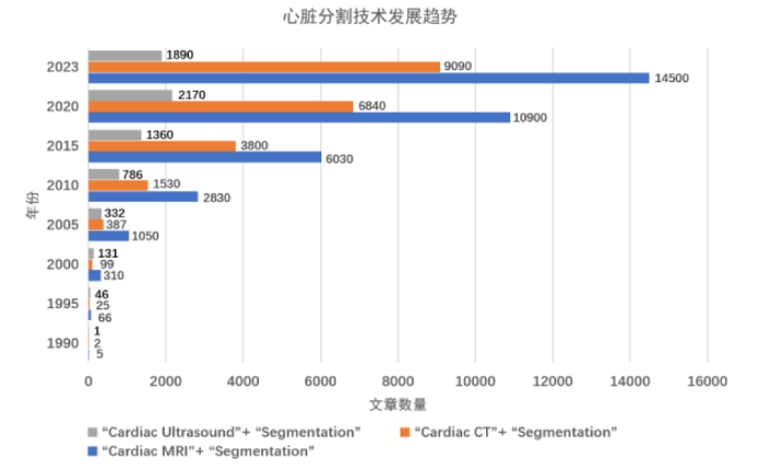


