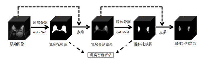
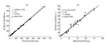
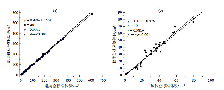
| 1 |
BOYD N F , GUO H , MARTIN L J , et al. Mammographic density and the risk and detection of breast cancer[J]. New Eng J Med, 2007, 356 (1): 227- 236.
|
| 2 |
KLIFA C , CARBALLIDO-GAMIO J , WILMES L , et al. Magnetic resonance imaging for secondary assessment of breast density in a high-risk cohort[J]. Magn Reson Imaging, 2010, 28 (1): 8- 15.
doi: 10.1016/j.mri.2009.05.040
|
| 3 |
WU S D , WEINSTEIN S P , CONANT E F , et al. Automated chest wall line detection for whole-breast segmentation in sagittal breast MR images[J]. Med Phys, 2013, 40 (4): 042301.
doi: 10.1118/1.4793255
|
| 4 |
NIE K , CHEN J H , CHAN S , et al. Development of a quantitative method for analysis of breast density based on three-dimensional automated segmentation of breast in 3-D MR images[J]. Med Phys, 2008, 35 (12): 5253- 5262.
doi: 10.1118/1.3002306
|
| 5 |
GUBERN-MÉRIDA A , KALLENBERG M , MANN R M , et al. Breast segmentation and density estimation in breast MRI: A fully automatic framework[J]. IEEE J Biomed Health, 2015, 19 (1): 349- 357.
doi: 10.1109/JBHI.2014.2311163
|
| 6 |
IVANOVSKA T , LAQUA R , WANG L , et al. A level set based framework for quantitative evaluation of breast tissue density from MRI data[J]. PLoS One, 2014, 9 (11): e112709.
doi: 10.1371/journal.pone.0112709
|
| 7 |
WU S D , WEINSTEIN S P , CONANT E F , et al. Automated fibroglandular tissue segmentation and volumetric density estimation in breast MRI using an atlas-aided fuzzy C-means method[J]. Med Phys, 2013, 40 (12): 122302.
doi: 10.1118/1.4829496
|
| 8 |
KOREZ R, LIKAR B, PERNUŠ F, et al. Model-based segmentation of vertebral bodies from MR images with 3D CNNs[C]//Medical Image Computing and Computer-Assisted Intervention-MICCAI 2016. Cham, Switzerland: Springer International Publishing, 2016: 433-441.
|
| 9 |
MOESKOPS P, WOLTERINK J M, VAN DER VELDEN B H M, et al. Deep learning for multi-task medical image segmentation in multiple modalities[C]//Medical Image Computing and Computer-Assisted Intervention-MICCAI 2016. Cham, Switzerland: Springer International Publishing, 2016: 478-486.
|
| 10 |
RODRIGUEZ-RUIZ A, TEUWEN J, CHUNG K, et al. Pectoral muscle segmentation in breast tomosynthesis with deep learning[C]//Medical Imaging 2018: Computer-Aided Diagnosis. Bellingham, WA: SPIE, 2018: 564-570.
|
| 11 |
ZHANG J , GAO Y Z , PARK S H , et al. Structured learning for 3-D perivascular space segmentation using vascular features[J]. IEEE T Biomed Eng, 2017, 64 (12): 2803- 2812.
doi: 10.1109/TBME.2016.2638918
|
| 12 |
CHRIST P, ETTLINGER F, GRÜN F, et al. Automatic liver and tumor segmentation of CT and MRI volumes using cascaded fully convolutional neural networks[EB/OL]. (2017-2-23)[2020-12-19]. https://arxiv.org/pdf/1505.04597v1.
|
| 13 |
RONNEBERGER O, FISCHER P, BROX T. U-Net: Convolutional networks for biomedical image segmentation[C]//Medical Image Computing and Computer-Assisted Intervention-MICCAI 2015. Cham, Switzerland: Springer International Publishing, 2015: 234-241.
|
| 14 |
ZHAO S Y , WANG Y J . Classification of Alzheimer's disease patients based on magnetic resonance images and an improved UNet++ model[J]. Chinese J Magn Reson, 2020, 37 (3): 321- 331.
|
|
赵尚义, 王远军. 基于磁共振图像和改进的UNet++模型区分阿尔茨海默症患者和健康人群[J]. 波谱学杂志, 2020, 37 (3): 321- 331.
|
| 15 |
KALLENBERG M , PETERSEN K , NIELSEN M , et al. Unsupervised deep learning applied to breast density segmentation and mammographic risk scoring[J]. IEEE Trans Med Imaging, 2016, 35 (5): 1322- 1331.
doi: 10.1109/TMI.2016.2532122
|
| 16 |
LIU P , ZHONG Y M , WANG L J . Automatic segmentation of right ventricle in cine cardiac magnetic resonance image based on a dense and multi-scale u-net method[J]. Chinese J Magn Reson, 2020, 37 (4): 456- 468.
|
|
刘鹏, 钟玉敏, 王丽嘉. 基于密集多尺度U-net网络的电影心脏磁共振图像右心室自动分割[J]. 波谱学杂志, 2020, 37 (4): 456- 468.
|
| 17 |
XIAO L , LOU Y K , ZHOU H Y . A U-Net network-based rapid construction of knee models for specific absorption rate estimation[J]. Chinese J Magn Reson, 2020, 37 (2): 144- 151.
|
|
肖亮, 娄煜堃, 周航宇. 用于SAR估计的基于U-Net网络的快速膝关节模型重建[J]. 波谱学杂志, 2020, 37 (2): 144- 151.
|
| 18 |
ZHANG Y , CHEN J H , CHANG K T , et al. Automatic breast and fibroglandular tissue segmentation in breast MRI using deep learning by a fully-convolutional residual neural network U-Net[J]. Acad Radiol, 2019, 26 (11): 1526- 1535.
doi: 10.1016/j.acra.2019.01.012
|
| 19 |
DALMIŞ M U , LITJENS G , HOLLAND K , et al. Using deep learning to segment breast and fibroglandular tissue in MRI volumes[J]. Med Phys, 2017, 44 (12): 533- 546.
|
| 20 |
PIANTADOSI G, SANSONE M, SANSONE C. Breast segmentation in MRI via U-Net deep convolutional neural networks[C]//Proceedings of 2018 International Conference on Pattern Recognition (ICPR). Piscataway, NJ: IEEE press, 2018: 3917-3922.
|
| 21 |
JIANG L , HU X X , XIAO Q , et al. Fully automated segmentation of whole breast using dynamic programming in dynamic contrast enhanced MR images[J]. Med Phys, 2017, 44 (6): 2400- 2414.
doi: 10.1002/mp.12254
|
| 22 |
ISENSEE F, PETERSEN J, KLEIN A, et al. nnU-Net: Self-adapting framework for U-Net-based medical image segmentation[EB/OL]. (2018-9-27)[2020-12-19]. https://arxiv.org/abs/1809.10486v1
|
| 23 |
HELLER N , ISENSEE F , MAIER-HEIN K H , et al. The state of the art in kidney and kidney tumor segmentation in contrast-enhanced CT imaging: Results of the KiTS19 challenge[J]. Med Image Anal, 2021, 67, 101821.
doi: 10.1016/j.media.2020.101821
|
| 24 |
MA J, WANG Y X, AN X L, et al. Toward data-efficient learning: A benchmark for COVID-19 CT lung and infection segmentation[J]. Med Phys, 2020. https://doi.org/10.1002/mp.14676.
|
| 25 |
PAUL S H . Graphics gems Ⅳ[M]. San Francisco: Margan Kaufmann, 1994, 474- 485.
|
| 26 |
YUSHKEVICH P A , PIVEN J , HAZLETT H C , et al. User-guided 3D active contour segmentation of anatomical structures: Significantly improved efficiency and reliability[J]. NeuroImage, 2006, 31 (3): 1116- 1128.
doi: 10.1016/j.neuroimage.2006.01.015
|
| 27 |
LÓPEZ-LINARES ROMÁN K , GARCÍA OCAÑA M I , LETE URZELAI N , et al. Medical image segmentation using deep learning[J]. Intelligent Systems Reference Library, 2020, 171, 17- 31.
|

 )
)