)
)
)
)
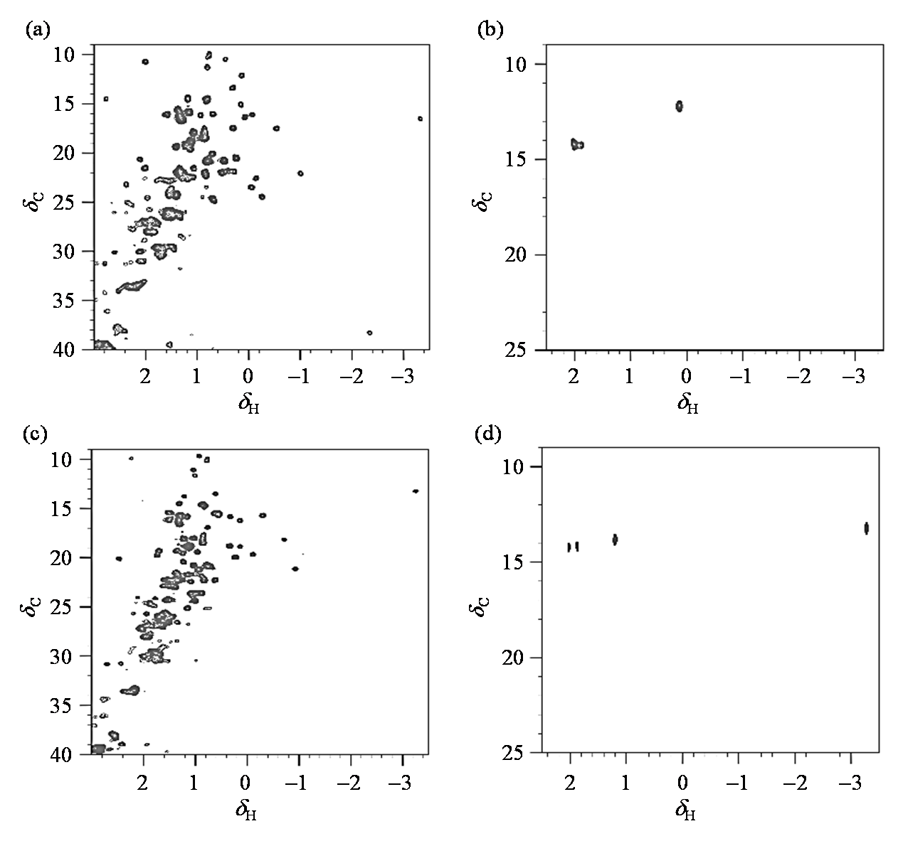
图2. 不同状态下,13C全标记和Met甲基13C标记的细胞色素c的1H-13C HSQC谱图. (a) 0.1 mmol/L 13C全标记细胞色素c,氧化态;(b) 0.1 mmol/L Met甲基选择性13C标记细胞色素c,氧化态;(c) 0.1 mmol/L 13C全标记细胞色素c,还原态;(d) 0.1 mmol/L Met甲基选择性13C标记细胞色素c,还原态. (a)、(c)中Met-6相对信号强度较弱,在(b)、(d)中由于选择性标记了Met末端甲基,所以Met-6的信号明显
Fig. 2. 1H-13C HSQC spectra of 13C full-labeled and methyl-13C methionine labeled cytochrome c under different states. (a) 0.1 mmol/L 13C full-labeled cytochrome c, oxidative state; (b) 0.1 mmol/L methyl- 13C labeling methione labeled cytochrome c, oxidative state; (c) 0.1 mmol/L 13C full-labeled cytochrome c, reductive state; (d) 0.1 mmol/L methyl- 13C methionine labeled cytochrome c, reductive state. (a)、(c) the signal intensity of Met-6 is weak. (b)、(d) the signal of Met-6 is obvious due to the selective labeling of methionine terminal methyl group