基于级联网络的膝关节图像分割与模型构建
Knee Joint Image Segmentation and Model Construction Based on Cascaded Network

基于级联网络的膝关节图像分割与模型构建 |
| 马岩,邢藏菊,肖亮 |
|
Knee Joint Image Segmentation and Model Construction Based on Cascaded Network |
| Yan MA,Cang-ju XING,Liang XIAO |
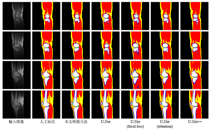
| 图4 使用5种自动分割方法得到的最后分割结果与人工标注结果的对比.从上到下,为一位志愿者图像集中4张连续的等间隔切片,其中,脂肪、肌肉、松质骨、皮质骨、软骨、半月板分别标记为黄色、红色、白色、蓝色、绿色与粉色 |
| Fig.4 The comparison between the final segmentation results obtained by using the 5 automatic segmentation methods and the manual segmentation. From top to bottom, 4 consecutive equally spaced slices are collected for a volunteer image. Among them, fat, muscle, cancellous bone, cortical bone, cartilage, and meniscus are marked as yellow, red, white, blue, green, and pink, respectively |

|