基于级联网络的膝关节图像分割与模型构建
Knee Joint Image Segmentation and Model Construction Based on Cascaded Network

基于级联网络的膝关节图像分割与模型构建 |
| 马岩,邢藏菊,肖亮 |
|
Knee Joint Image Segmentation and Model Construction Based on Cascaded Network |
| Yan MA,Cang-ju XING,Liang XIAO |
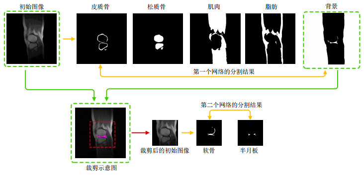
| 图3 第一行分别为初始图像与对应的皮质骨、松质骨、肌肉、脂肪与背景的分割结果,本文所提方法通过提取背景中的空洞并计算其质心得到软骨与半月板的位置信息,以质心为中心进行裁剪,得到包含软骨与半月板的子图,即第二个网络的输入图像.第二行分别展示了裁剪示意图与利用第二个网络得到的软骨与半月板的分割结果 |
| Fig.3 The first row is the initial image and the corresponding segmentation results of cortical bone, cancellous bone, muscle, fat, and background. Extract the cavity in the background and calculate its centroid to obtain the position information of cartilage and meniscus. Cut out with the centroid as the center to obtain a sub-image containing cartilage and meniscus, which is the input image of the second network. The second row respectively shows the cut-out schematic diagram and the segmentation results of cartilage and meniscus with the second network |

|