T2/FLAIR错配征的定量参数在评价较低级别胶质瘤分子分型的诊断价值
The Diagnostic Value of Quantitative Parameters of T2/FLAIR Mismatch Sign in Evaluating the Molecular Typing of Lower-grade Gliomas
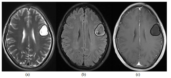
图1. 整体T2/FLAIR错配征的ROI勾画,左侧额叶弥漫性星形细胞瘤IDHMUT/1p/19q+,WHO Ⅱ级.(a)轴位T2WI,病变呈高信号,边界清晰,周围脑组织未见明显水肿;(b)轴位FLAIR,除边缘高信号环外,大部分瘤区与T2WI比呈相对低信号;(c) 轴位T1增强,病变未见明显强化
Fig.1. The ROI of the T2/FLAIR mismatch sign of the entire tumor. Diffuse astrocytoma of the left frontal lobe with IDHMUT/1p/19q+, WHO grade Ⅱ. (a) Axial T2WI, the lesion showed high signal, clear boundary, and no obvious edema in the surrounding brain tissue; (b) Axial FLAIR, except for the edge high signal ring, most of the tumor area has relatively lower signal intensities compared with T2WI; (c) Axial T1 enhancement, no obvious enhancement of the lesion

