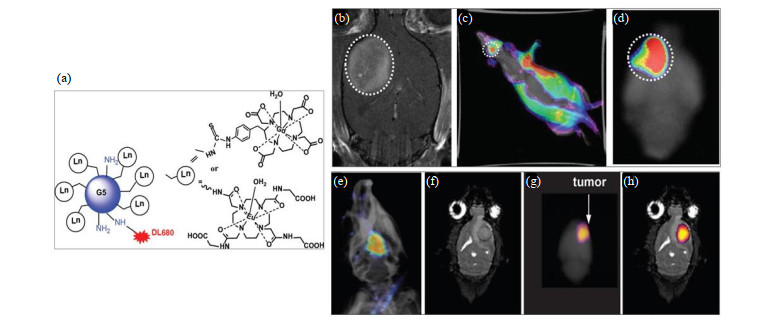
图4. G5树枝状大分子的结构及在胶质瘤动物模型中的检测.(a) Dylight680(DL680)与Gd-DOTA或Eu-DOTA-Gly4装载到G5树枝状聚合物结构中.(b) Gd-G5-DL680,注射剂量为0.03 mmol Gd/kg.分别在白光和过滤激发下获得体内光学图像,发射滤光片设置为750 nm,用来分辨神经胶质瘤中的荧光.(c)大鼠脑的离体荧光成像清楚地显示Gd-G5-DL680在肿瘤内的选择性积累.(d)肿瘤以白色虚线圆圈表示.(e)覆盖在X射线图像上的大鼠头部的体内荧光图像显示脑中U87肿瘤中存在Eu-DOTA-Gly4-G5-DL680纳米颗粒.(f)冠状位磁共振图像显示U87肿瘤的位置.(g)全脑的离体荧光图像也检测到大脑中的纳米颗粒.(h)离体荧光图像叠加在磁共振图像上,以显示纳米颗粒位于U87神经胶质瘤中[
Fig.4. The structure of G5 dendrimer and detection in animal model of glioma. (a) Dylight680 (DL680) was conjugated with Gd-DOTA or Eu-DOTA-Gly4 preloaded G5 dendrimer. (b) The agent was Gd-G5-DL680 and injected at a dose of 0.03 mmol Gd/kg. In vivo optical image obtained under simultaneous white light and filtered excitation detected with the emission filter set at 750 nm demonstrating fluorescence in the glioma. (c) Ex vivo fluorescence imaging of rat brain clearly shows the selective accumulation of the Gd-G5-DL680 within the tumor. (d) Tumor is indicated as dotted white circle. (e) The in vivo fluorescent image of the rat head overlayed on an X-ray image shows the presence of Eu-DOTA-Gly4-G5-DL680 nanoparticle in the U87 tumor in the brain. (f) The coronal MR image shows the location of the U87 tumor. (g) The ex vivo fluorescence image of whole brain also detected the nanoparticle in the brain. (h) The ex vivo fluorescence image was also overlayed on the MR image to show that the nanoparticle was located in the U87 glioma[

